. 2019 Feb 5;12(567):eaau2922.
doi: 10.1126/scisignal.aau2922.

Targeting NOTCH activation in small cell lung cancer through LSD1 inhibition

Affiliations

Targeting NOTCH activation in small cell lung cancer through LSD1 inhibition

Arnaud Augert et al. Sci Signal. .

Abstract

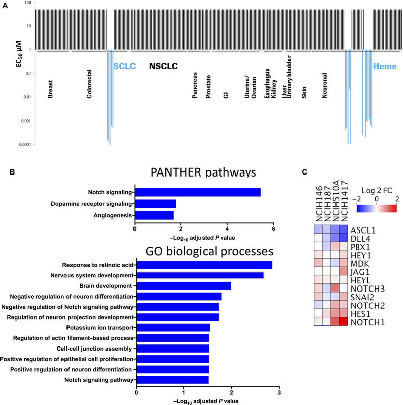
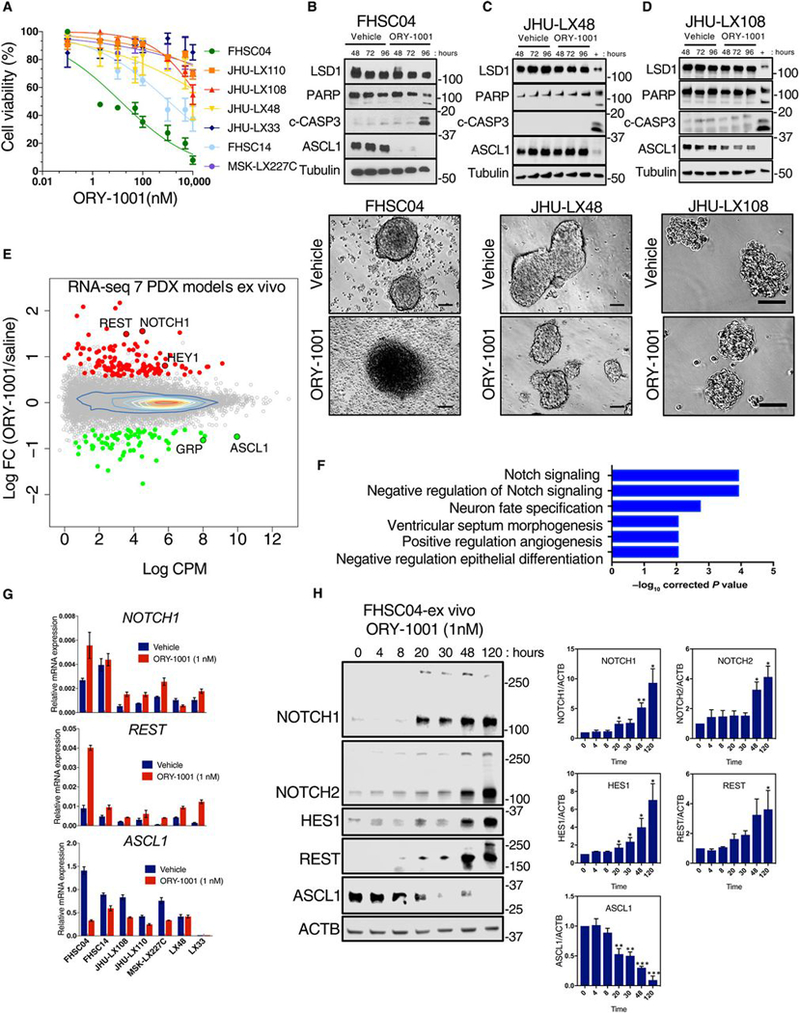
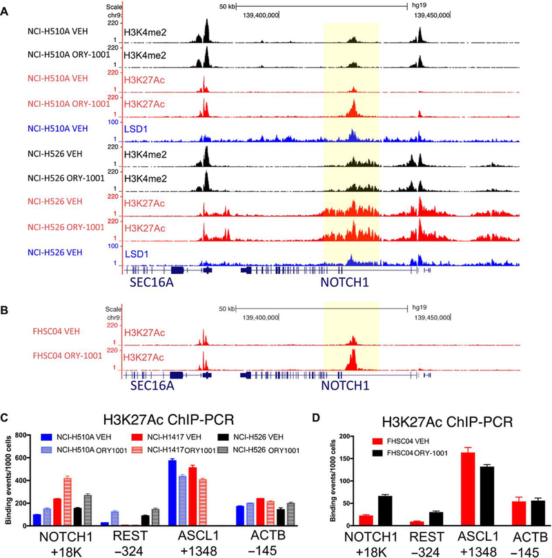
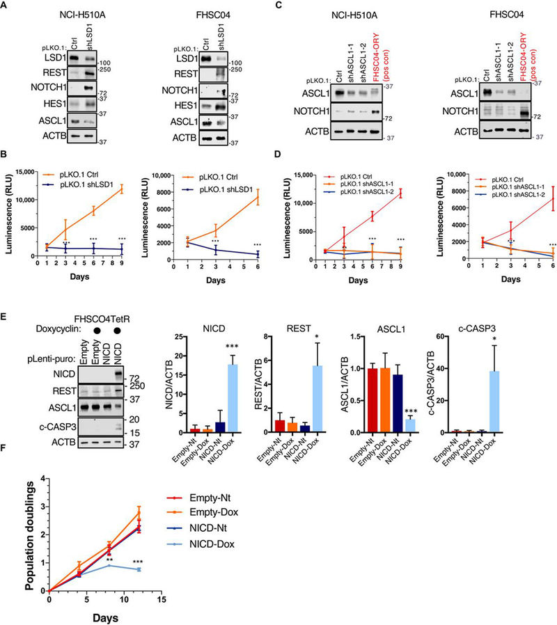
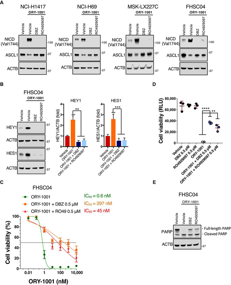
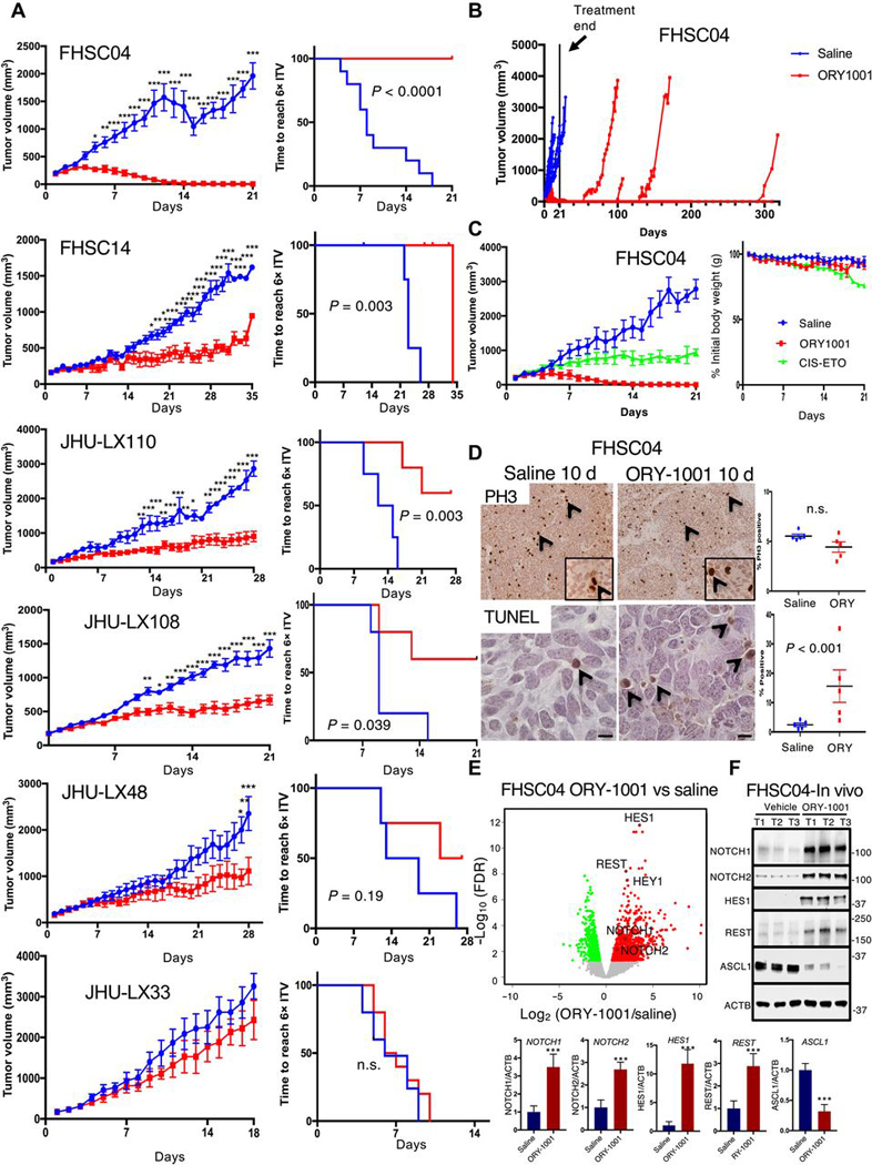
Small cell lung cancer (SCLC) is a recalcitrant, aggressive neuroendocrine-type cancer for which little change to first-line standard-of-care treatment has occurred within the last few decades. Unlike nonsmall cell lung cancer (NSCLC), SCLC harbors few actionable mutations for therapeutic intervention. Lysine-specific histone demethylase 1A (LSD1 also known as KDM1A) inhibitors were previously shown to have selective activity in SCLC models, but the underlying mechanism was elusive. Here, we found that exposure to the selective LSD1 inhibitor ORY-1001 activated the NOTCH pathway, resulting in the suppression of the transcription factor ASCL1 and the repression of SCLC tumorigenesis. Our analyses revealed that LSD1 bound to the NOTCH1 locus, thereby suppressing NOTCH1 expression and downstream signaling. Reactivation of NOTCH signaling with the LSD1 inhibitor reduced the expression of ASCL1 and neuroendocrine cell lineage genes. Knockdown studies confirmed the pharmacological inhibitor-based results. In vivo, sensitivity to LSD1 inhibition in SCLC patient-derived xenograft (PDX) models correlated with the extent of consequential NOTCH pathway activation and repression of a neuroendocrine phenotype. Complete and durable tumor regression occurred with ORY-1001-induced NOTCH activation in a chemoresistant PDX model. Our findings reveal how LSD1 inhibitors function in this tumor and support their potential as a new and targeted therapy for SCLC.


Conflict of interest statement

Competing Interests: DM received research support from Roche that partially supported costs of this research. F. Milletti, WC and F. Mack are employees of Roche. CMR has consulted with Roche and Celgene regarding anti-cancer drug development. The other authors declare that they have no conflicts of interest.

Figures

Fig. 1.
Fig. 1.. ORY-1001 exhibits anti-proliferative activity in a subset of SCLC cell lines.
(A) Anti-proliferative activity of ORY-1001 (in concentrations of up to 10μM) was screened across a panel of 275 cell lines in a 4-day cell viability assay (CellTiter-Glo). Half-maximal effective concentration (EC50) values in responsive cell lines ranged from 0.1–23nM. (B) RNA-Seq analyses performed using DeSeq2(47) in SCLC cell lines (NCI-H146, NCI-H187, NCI-H510A, and NCI-H1417) treated with 10nM ORY-1001 for 96 hours. 1400 differentially expressed genes (table S2) with FDR<0.05 were used for enrichment analysis with the online tool Enrichr (14), assigned according to the PANTHER and Gene Ontology Biological Processes databases. For the PANTHER pathways listed, all significant pathways with FDR<0.05 are shown. For GO Biological Processes, a subset of significant processes with FDR<0.05 are shown, including Notch signaling (GO:0007219). (C) Heat map showing differentially expressed genes in Notch pathway (GO:0007219), showing log2 fold change in ORY-1001–treated relative to vehicle-treated cells.
Fig. 2.
Fig. 2.. LSD1 inhibition with ORY-1001 results in the activation of NOTCH signaling.
(A) Dose response curves of 7 PDX model-derived cells cultured and treated ex vivo with ORY-1001 over 120 hours. Viability was assessed with the CellTiter-Glo assay and calculated relative to the vehicle control. Data are mean ± S.E.M. from at least n=3 biological replicates. (B) Immunoblots (above) and photomicrographs (below) for FHSC04 cells treated ex vivo with ORY-1001. Protein abundance was assessed at 48, 72 and 96 hours of 1nM ORY-1001 treatment and photomicrographs taken at 96 hours. Blots are representative of two independent experiments. c-CASP3, cleaved caspase 3. Scale bar, 100μM. (C and D) As described in (B) for JHU-LX48 (C) and JHU-LX108 (D) cells. As a positive control for cleaved caspase 3 and PARP, lysates from FHSC04 cells treated 96 hours with 1nM ORY-1001 (also represented in B) was run on the same blot (“+” lane). Blots are representative of two independent experiments. (E) RNA-Seq MA plot showing differentially expressed genes across the 7 PDX models treated ex vivo with 1nM ORY-1001. Red, upregulated genes; green, downregulated genes; each with log2 fold change >0.585 and FDR <0.05 as identified using edgeR. (F) Gene Ontology (GO) analysis using GOseq (19) showing the top 6 significant GO terms in the genes differentially expressed with FDR <0.05 (see Methods; table S4 for full list). (G) Abundance of NOTCH1, REST and ASCL1 transcripts in the PDX models ex vivo assessed using real time PCR, with expression plotted relative to that of GAPDH (n=3 independent experiments). (H) Immunoblot showing kinetics of changes in NOTCH1, NOTCH2, REST and ASCL1 protein abundance in the drug-sensitive FHSC04 model treated ex vivo with1 nM ORY-1001 over 120 hours. Data are mean ± SD from three independent experiments. * P<0.05, ** P<0.01, and *** P<0.005 by a two-tailed unpaired Student’s t-test.
Fig. 3.
Fig. 3.. Differential histone acetylation of LSD1-regulated genes.
(A) Read-density tracks, visualized using UCSC genome browser, of normalized ChIP-Seq for H3K4me2 (black), H3K27Ac (red) and LSD1 (blue) in NCI-H510A and NCI-H526 cell lines. Cells were treated with vehicle (VEH) or 1nM ORY-1001 for 96 hours. Shaded area highlights region in NOTCH1 with LSD1 binding and increased H3K27Ac upon ORY-1001 treatment in the NCI-H510A cell line. (B) Read-density tracks of normalized ChIP-Seq for H3K27Ac in cells derived from FHSC04 PDXs, a markedly ORY-1001–responsive model. Cells were treated ex vivo with vehicle (VEH) or 1nM ORY-1001 for 48 hours. Shaded area highlights region in NOTCH1 with increased H3K27Ac. (C and D) ChIP-PCR validation of H3K27Ac changes in NOTCH1, REST, ASCL1 and as a control, ACTB in vehicle in SCLC cell lines treated for 96 hours (C) or FHSC04 cells treated for 48 hours (D) with vehicle or 1nM ORY-1001 for 96 hours. Data are mean ± SD from n=3 experiments.
Fig. 4.
Fig. 4.. Genetic perturbation of LSD1, ASCL1 and NOTCH1 in SCLC cells.
(A) Immunoblot analysis of the indicated proteins in response to lentiviral LSD1 knockdown in NCI-H510A SCLC cell line and FHSC04 PDX model ex vivo. ACTB is used as a loading control. Blots are representative of two independent experiments. (B) Proliferation analysis using CellTiter-Glo in response to LSD1 knockdown, analyzed as summarized below. (C) Immunoblot analysis of the indicated proteins in response to ASCL1 knockdown in NCI-H510A SCLC cell line and FHSC04 PDX model ex vivo. An ORY-1001 treated FHSC04 protein extract is used as a positive control for NOTCH1 and REST. Blots are representative of two independent experiments. (D) Proliferation analysis in response to ASCL1 knockdown, as in (B). (E) Immunoblot analysis of the indicated proteins in FHSC04TetR cells in response to an inducible expression of N1ICD (1μg/μl doxycycline for 96 hours). (F) Growth curve analysis of FHSC04TetR in response to N1ICD expression. All data are mean ± SD from n=3 experiments. *P<0.05, **P<0.01, and ***P<0.005 by a two-tailed unpaired Student’s t-test.
Fig. 5.
Fig. 5.. Pharmacological inhibition of NOTCH signaling partially rescues effects of ORY-1001.
(A) Immunoblot analysis of the indicated proteins in response to ORY-1001 (1nM) with or without the GSIs RO4929097 or DBZ (0.5μM) for 48 hours in SCLC cell lines NCI-H1417 and NCI-H69 as well as ex vivo PDX models MSK-LX227C and FHSC04. ACTB is used as a loading control. Blots are representative of two independent experiments. (B) Immunoblot analysis of HEY1 and HES1 abundance in adherent FHSC04 cells treated as in (A). Data are mean ± SD from n=3 experiments. **P<0.01 and ***P<0.005 by a two-tailed unpaired Student’s t-test. (C) Dose response curves in adherent FHSC04 cells treated ex vivo for 120 hours. Viability was quantified relative to vehicle control using the CellTiter-Glo assay. Data are mean ± SD from n=4 biological replicates, each with 3 technical replicates. (D) An extension of (C), showing the viability of FHSC04 cells in response to ORY-1001 at the 10nM dose, alone and in the presence of RO4929097 or DBZ (0.5μM), with comparison each single GSI alone and vehicle. Data are mean ± SD from n=4 experiments, each with 3 technical replicates. **P<0.01 and ****P<0.0001 by a two-tailed unpaired Student’s t-test. (E) Immunoblot analysis of cleaved PARP in FHSC04 cells in response to 96 hours’ treatment with ORY-1001 (10 nM) alone or with RO4929097 or DBZ (0.5μM). Blots are representative of two independent experiments.
Fig. 6.
Fig. 6.. ORY-1001 efficacy in PDX models of SCLC.
(A) Tumor growth inhibition curves over the treatment period (left) and Kaplan-Meier curves (right) showing time to reach 6X initial tumor volume (ITV) across 6 PDX models of SCLC treated with ORY-1001 (400μg/kg once weekly) or with saline. Mice were treated with saline or ORY-1001 once tumors reached 150mm3. Data are mean ± S.E.M.; n=10 animals total per treatment group for FHSC04 (pooled from two experiments, each with n=5 per group); for all others, n=5–6 per group. Tumor growth curves: *P<0.05, **P<0.005, and ***P<0.0005 by two-way ANOVA with Sidak’s post-test. Kaplan-Meier curves show P-value from log-rank test; n.s. not significant. (B) Tumor volume plots for mice in FHSC04 model treated with saline or ORY-1001 depicted in (A) extending beyond treatment end at 21 days (solid line). Mouse tumor volumes are individually plotted for response and durability of tumor regressions. (C) Repeat of ORY-1001 treatment in FHSC04 with comparison to a cisplatin-etoposide combination therapy (CIS-ETO) (left), n=5 mice per treatment group. Change in body weight is shown to right as CIS-ETO treatment led to reductions in body weight not seen with ORY-1001. Cisplatin was dosed at 5mg/kg once per week, and etoposide at 10mg/kg every 3 days. (D) TUNEL and PH3 immunohistochemistry analysis to assess proliferation and cell death in FHSC04 PDXs collected 10 days after treatment initiation. n=5 tumors per group; P-value, (n.s., not significant) assessed by Student’s t-test. (E) Volcano plot showing differentially expressed genes in FHSC04 PDXs after 10 days’ treatment with ORY-1001 as identified using EdgeR. N=5 control and 5 ORY-1001–treated mice. Red, upregulated genes; green, downregulated genes; FDR<0.01. (F) Western blot analyses and quantifications (below) of ASCL1 and NOTCH-pathway protein abundance in FHSC04 PDX in vivo. Data are mean ± S.E.M. from n = 3 mice per group; tumors harvested 10 days after treatment initiation. ***P<0.005 by two-tailed unpaired Student’s t-test.

References

    1. Gazdar AF, Bunn PA, Minna JD, Small-cell lung cancer: what we know, what we need to know and the path forward. Nature reviews. Cancer 17, 725–737 (2017); published online EpubDec (10.1038/nrc.2017.87). - DOI - PubMed
    1. George J, Lim JS, Jang SJ, Cun Y, Ozretic L, Kong G, Leenders F, Lu X, Fernandez-Cuesta L, Bosco G, Muller C, Dahmen I, Jahchan NS, Park KS, Yang D, Karnezis AN, Vaka D, Torres A, Wang MS, Korbel JO, Menon R, Chun SM, Kim D, Wilkerson M, Hayes N, Engelmann D, Putzer B, Bos M, Michels S, Vlasic I, Seidel D, Pinther B, Schaub P, Becker C, Altmuller J, Yokota J, Kohno T, Iwakawa R, Tsuta K, Noguchi M, Muley T, Hoffmann H, Schnabel PA, Petersen I, Chen Y, Soltermann A, Tischler V, Choi CM, Kim YH, Massion PP, Zou Y, Jovanovic D, Kontic M, Wright GM, Russell PA, Solomon B, Koch I, Lindner M, Muscarella LA, la Torre A, Field JK, Jakopovic M, Knezevic J, Castanos-Velez E, Roz L, Pastorino U, Brustugun OT, Lund-Iversen M, Thunnissen E, Kohler J, Schuler M, Botling J, Sandelin M, Sanchez-Cespedes M, Salvesen HB, Achter V, Lang U, Bogus M, Schneider PM, Zander T, Ansen S, Hallek M, Wolf J, Vingron M, Yatabe Y, Travis WD, Nurnberg P, Reinhardt C, Perner S, Heukamp L, Buttner R, Haas SA, Brambilla E, Peifer M, Sage J, Thomas RK, Comprehensive genomic profiles of small cell lung cancer. Nature 524, 47–53 (2015); published online EpubAug 6 (10.1038/nature14664). - DOI - PMC - PubMed
    1. Rudin CM, Durinck S, Stawiski EW, Poirier JT, Modrusan Z, Shames DS, Bergbower EA, Guan Y, Shin J, Guillory J, Rivers CS, Foo CK, Bhatt D, Stinson J, Gnad F, Haverty PM, Gentleman R, Chaudhuri S, Janakiraman V, Jaiswal BS, Parikh C, Yuan W, Zhang Z, Koeppen H, Wu TD, Stern HM, Yauch RL, Huffman KE, Paskulin DD, Illei PB, Varella-Garcia M, Gazdar AF, de Sauvage FJ, Bourgon R, Minna JD, Brock MV, Seshagiri S, Comprehensive genomic analysis identifies SOX2 as a frequently amplified gene in small-cell lung cancer. Nature genetics, (2012); published online EpubSep 2 (10.1038/ng.2405). - DOI - PMC - PubMed
    1. Augert A, Zhang Q, Bates B, Cui M, Wang X, Wildey G, Dowlati A, MacPherson D, Small Cell Lung Cancer Exhibits Frequent Inactivating Mutations in the Histone Methyltransferase KMT2D/MLL2: CALGB 151111 (Alliance). Journal of thoracic oncology : official publication of the International Association for the Study of Lung Cancer 12, 704–713 (2017); published online EpubApr (10.1016/j.jtho.2016.12.011). - DOI - PMC - PubMed
    1. Borromeo MD, Savage TK, Kollipara RK, He M, Augustyn A, Osborne JK, Girard L, Minna JD, Gazdar AF, Cobb MH, Johnson JE, ASCL1 and NEUROD1 Reveal Heterogeneity in Pulmonary Neuroendocrine Tumors and Regulate Distinct Genetic Programs. Cell reports, (2016); published online EpubJul 20 (10.1016/j.celrep.2016.06.081). - DOI - PMC - PubMed

Publication types

MeSH terms