. 2019 Feb 5;10(1):424.
doi: 10.1038/s41467-018-08278-4.

BMP9 stimulates joint regeneration at digit amputation wounds in mice

Affiliations

BMP9 stimulates joint regeneration at digit amputation wounds in mice

Ling Yu et al. Nat Commun. .

Abstract

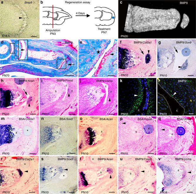
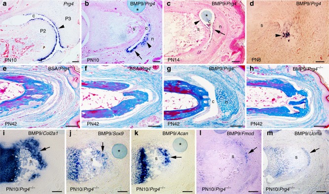
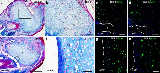
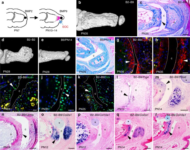
A major goal of regenerative medicine is to stimulate tissue regeneration after traumatic injury. We previously discovered that treating digit amputation wounds with BMP2 in neonatal mice stimulates endochondral ossification to regenerate the stump bone. Here we show that treating the amputation wound with BMP9 stimulates regeneration of a synovial joint that forms an articulation with the stump bone. Regenerated structures include a skeletal element lined with articular cartilage and a synovial cavity, and we demonstrate that this response requires the Prg4 gene. Combining BMP2 and BMP9 treatments in sequence stimulates the regeneration of bone and joint. These studies provide evidence that treatment of growth factors can be used to engineer a regeneration response from a non-regenerating amputation wound.


Conflict of interest statement

K.M., L.Y. and M.Y. disclose patent no. 9,833,481 B2 (4104-1) on BMP9-induced articular cartilage regeneration. The authors declare no competing interests.

Figures

Fig. 1
Fig. 1
Bone morphogenetic protein 9 (BMP9) stimulates regeneration of joint structures. a Bmp9 transcripts are localized to digit joints undergoing cavitation (arrowhead) in E18.5 embryos. b P2 level amputation of neonatal digits is used to test for induced regeneration. Following amputation (red line) at postnatal day 3 (PN3), epidermal closure is completed 4 days later (PN7), and an agarose microcarrier bead (blue dot) is implanted between the wound epidermis and the stump. c Microcomputed tomography (µCT) rendering of a BMP9-treated digit showing regeneration of a skeletal element articulating with the stump after 4 weeks (PN35). d Mallory trichrome staining of BMP9-treated digit 9 weeks after BMP9 treatment (PN70). Regenerated skeletal element (arrow) forms a joint-like structure with the digit stump. Articular cartilage of the P1/P2 joint is shown at higher magnification in the inset. e Higher magnification of the inset in (d) shows a cavity (c) separating a layer of articular cartilage (arrowhead) lining the regenerated skeletal element (n) and the stump bone (s). Small patches of articular cartilage (star) form on the stump bone. fj In situ hybridization of cavity (arrow)-forming BMP9-treated digits showing localization of transcripts to the regenerated chondrogenic nodule but not the digit stump (s) 72 h after bead (*) implantation. f Col2a1. g Sox9. h Acan. i Fmod. j Ucma. k, l Immunofluorescence staining of Doublecortin (Dcx) expression. k Immunostaining shows Dcx+ cells (arrowhead) lining the synovial cavity of an uninjured joint at PN10. l Isolated superficial cells of the BMP9-induced nodule express Dcx (arrowhead) 72 h after treatment (PN10). The cavity is outlined with dashed white lines. Dcx+ cells are also observed internal to the superficial layer. mq In situ hybridization of bovine serum albumin (BSA)-treated control digits 72 h after bead implantation showing no expression associated with the amputation wound distal to the stump. m Col2a1. n Sox9. o Acan. p Fmod. q Ucma. rv In situ hybridization of non-cavity-forming BMP9-treated digits showing localization of transcripts to the distal stump 72 h after bead implantation. r Col2a1. s Sox9. t Acan. u Fmod. v Ucma. Right is distal, top is dorsal. Scale bars: a, fj, mv = 100 µm; d = 250 µm; e, k = 50 µm; l = 25 µm
Fig. 2
Fig. 2
Proteoglycan 4 (Prg4) is required for bone morphogenetic protein 9 (BMP9)-induced cavitation. ad In situ hybridization for Prg4 expression. a Prg4 is expressed by cells lining the P2/P3 synovial cavity (arrowhead) and forming the superficial layer of the articular cartilage of the neonatal joint at PN10. b In BMP9-treated cavity-forming digits, Prg4 is expressed by cells lining the induced cavity (arrow) and by cells forming the chondrogenic nodule (arrowhead) 72 h after treatment. c At 7 days after treatment with a BMP9 bead, Prg4 is expressed by cells lining the cavity on both the stump side (arrowhead) and the nodule side (arrow). d At 24 h after BMP9 treatment, Prg4 transcripts are found localized in clusters of cells in the wound bed (arrowhead) associated with the initiation of the cavitation response. e, f Control bovine serum albumin (BSA) bead implantation does not induce an articulation response in PRG4+/− (e) or PRG4−/− (f) digit amputations. g BMP9 treatment of PRG4+/- digit amputations induces an articulation response that includes a distal nodule and cavity. h No regeneration response is observed after BMP9 treatment in PRG4−/− mutants. im In situ hybridization of chondrogenic genes expressed at the distal stump in non-cavity-forming PRG4−/− mutant digits 72 h after BMP9 treatment. i Col2a1. j Sox9. k Acan. l Fmod. m Ucma. Distal is to the right and dorsal is to the top; n nodule, c cavity, s stump, * indicates bead. Scale bars: ad, im = 100 µm; eh = 200 µm
Fig. 3
Fig. 3
Bone morphogenetic protein 9 (BMP9) stimulates stump chondrogenesis and cavitation in adult digits. a Mallory trichrome staining of an adult BMP9-treated digit harvested at 7 days post implant (DPI) showing induced chondrogenesis is contiguous with the bone stump. b Higher magnification of the inset in (a) of BMP9-induced chondrocytes at 7 DPI. c, d Immunofluorescence staining for the cartilage marker proteins Sox9 and Acan illustrate chondrogenesis contiguous with the stump bone (dashed white lines) at 7 DPI (n nodule, c cavity, s stump). e Mallory trichrome staining of an adult BMP9-treated digit showing cavity and chondrogenic nodule formation at 14 DPI. f Higher magnification of the inset in (e) showing the chondrogenic and cavitation response at 14 DPI. g, h Immunohistochemistry of the 14 DPI cavity-forming BMP9-treated digit. Dashed white lines outline the cavity. g Cells lining the cavity and within the nodule express the cartilage marker protein Sox9. h Acan immunostaining is restricted to cells within the central region of the nodule. Distal is to the right and dorsal is to the top. Scale bars: a, e = 500 µm; b, c, d = 100 µm; fh = 50 µm
Fig. 4
Fig. 4
Sequential bone morphogenetic protein 2/9 (BMP2/BMP9) treatment stimulates bone and joint regeneration. a Diagram illustrating sequential BMP2 and BMP9 (B2–B9) treatment. A BMP2 bead is implanted at PN7. A BMP9 bead is implanted distal to the induced endochondral ossification center (EOC) at PN10 or PN14. bm All B2–B9 samples are from the 3-day interval study. b microcomputed tomography (µCT) rendering of a B2–B9-regenerate at PN35 identifies a distal articulation. c Mallory trichrome staining of a B2–B9 regenerate showing cartilage capping the stump and surrounding the nodule (n nodule, c cavity, s stump). d µCT rendering of a PN28 regenerate treated with BMP2 at PN7 and PN10 displaying excessive bone growth and no joint formation. e PN28 µCT rendering of a regenerate treated with BMP9 at PN14. Thus, 50% of the digits formed a nodule that articulated with the stump. f Mallory trichrome staining of digit shown in (e). The regenerate formed a cavity and a cartilage-lined nodule but the stump bone lacks a chondrogenic cell layer. gk Immunofluorescence staining for marker proteins expressed by cells lining both sides of regenerated and control joints. The joint cavity is identified by dashed white lines. Red blood cell autofluorescence (yellow) identifies ossifying regions. g ColII (arrowhead) of the B2–B9 induced cavity and h a control digit joint cavity. i Acan (arrowhead) of the B2–B9-induced cavity and j a control digit joint cavity. k Dcx (arrowhead) of the B2–B9-induced cavity. lr In situ hybridization of B2–B9 regenerates (* indicates bead). l Prg4 transcripts are expressed by cells of the regenerated cavity (arrowhead) 96 h after BMP9 treatment. m, n Fmod (arrowhead in m) and Ucma (arrowhead in n) transcripts are expressed in cells of the distal digit stump and the nodule 96 h following BMP9 treatment. o, p Cells of the distal stump (arrowhead) and the regenerated nodule express Col2a1 (o), whereas Col10a1 is not expressed (p) 48 h after BMP9 treatment. q, r Cells of the distal stump (arrowhead) and the regenerated nodule express Col2a1 (q) but only cells of the stump express Col10a1 (r) 96 h after BMP9 treatment. Right is distal, dorsal is top. Scale bars: c = 200 µm; gk = 50 µm; f, lr = 100 µm

References

    1. Dawson LA, et al. Analogous cellular contribution and healing mechanisms following digit amputation and phalangeal fracture in mice. Regeneration (Oxf.) 2016;3:39–51. doi: 10.1002/reg2.51. - DOI - PMC - PubMed
    1. Turner NJ, Johnson SA, Badylak SF. A histomorphologic study of the normal healing response following digit amputation in C57bl/6 and MRL/MpJ mice. Arch. Histol. Cytol. 2010;73:103–111. doi: 10.1679/aohc.73.103. - DOI - PubMed
    1. Dolan CP, Dawson LA, Muneoka K. Digit tip regeneration: merging regeneration biology with regenerative medicine. Stem Cells Transl. Med. 2018;7:262–270. doi: 10.1002/sctm.17-0236. - DOI - PMC - PubMed
    1. Yu L, Han M, Yan M, Lee J, Muneoka K. BMP2 induces segment-specific skeletal regeneration from digit and limb amputations by establishing a new endochondral ossification center. Dev. Biol. 2012;372:263–273. doi: 10.1016/j.ydbio.2012.09.021. - DOI - PMC - PubMed
    1. Dawson LA, et al. The periosteal requirement and temporal dynamics of BMP2-induced middle phalanx regeneration in the adult mouse. Regeneration (Oxf.) 2017;4:140–150. doi: 10.1002/reg2.81. - DOI - PMC - PubMed

Publication types

MeSH terms