. 2019 Feb;26(2):129-136.
doi: 10.1038/s41594-018-0181-y. Epub 2019 Feb 4.

Dynamics and selective remodeling of the DNA-binding domains of RPA

Affiliations

Dynamics and selective remodeling of the DNA-binding domains of RPA

Nilisha Pokhrel et al. Nat Struct Mol Biol. 2019 Feb.

Abstract

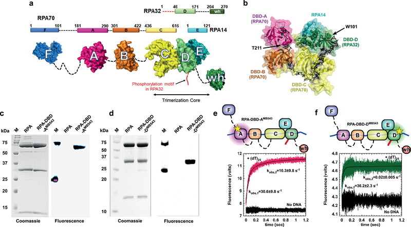
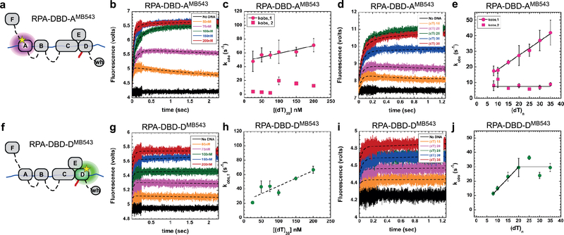
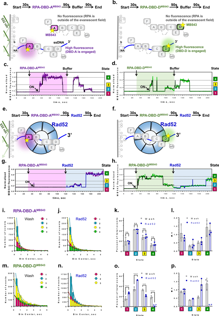
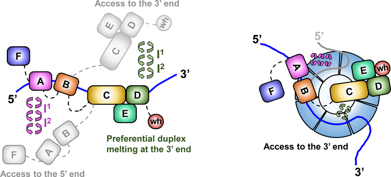
Replication protein A (RPA) coordinates important DNA metabolic events by stabilizing single-stranded DNA (ssDNA) intermediates, activating the DNA-damage response and handing off ssDNA to the appropriate downstream players. Six DNA-binding domains (DBDs) in RPA promote high-affinity binding to ssDNA yet also allow RPA displacement by lower affinity proteins. We generated fluorescent versions of Saccharomyces cerevisiae RPA and visualized the conformational dynamics of individual DBDs in the context of the full-length protein. We show that both DBD-A and DBD-D rapidly bind to and dissociate from ssDNA while RPA remains bound to ssDNA. The recombination mediator protein Rad52 selectively modulates the dynamics of DBD-D. These findings reveal how RPA-interacting proteins with lower ssDNA binding affinities can access the occluded ssDNA and remodel individual DBDs to replace RPA.


Conflict of interest statement

Competing Financial Interests Statements:

The authors declare no competing financial interests.

Figures

Figure 1.
Figure 1.. Non-canonical amino acid-based fluorescent RPAs report on individual DBD dynamics.
a) The residue numbers for the three RPA subunits and their respective DNA binding domains (DBDs A-F) are denoted. The winged-helix (wh) domain in RPA32 and DBD-F in RPA70 mediate interactions with RPA-interacting proteins (RIPs). The N-terminus of RPA32 that is phosphorylated is shown in red. Crystal structures of the ordered domains are shown as surface representations with intervening disordered linkers as dotted lines (black). DBD-C, DBD-D and RPA14 interact to form the trimerization core. b) Crystal structure of the DNA binding domains of U. maydis RPA bound to ssDNA (PDB ID:4GNX). Residues T211 in DBD-A and W101 in DBD-D are sites where 4-azidophenylalanine (4AZP) is incorporated (residue numbering in Saccharomyces cerevisiae RPA). The bound ssDNA is shown as sticks (black). c & d) Coomassie and fluorescence imaging of RPA complexes labeled with MB543 at either DBD-A or DBD-D. Only the fluorescently-labeled domains are visualized upon fluorescence imaging suggesting site-specific labeling of each domain, respectively. e & f) RPA-DBD-AMB543 and RPA-DBD-DMB543 binding to ssDNA was analyzed by monitoring the change in MB543 fluorescence. Robust change in fluorescence depicts engagement of specific DBDs onto ssDNA. Data were fit and analyzed as described in Methods. Values depicted in panels e & f represent the mean and s.e. from n=3 independent experiments. Uncropped gel images of panels c & d are shown in Supplementary Dataset 1.
Figure 2.
Figure 2.. DNA binding dynamics of individual DBDs.
a & f) Cartoons depicting RPA-DBD-AMB543 or RPA-DBD-DMB543 binding to ssDNA and producing a change in fluorescence. Stopped flow experiments done with b & c) increasing concentrations of [(dT)35] ssDNA or d & e) with ssDNA of increasing length, captures the observed rates in fluorescence change for RPA-DBD-AMB543. g – j) Stopped flow analysis of RPA-DBD-DMB543 ssDNA binding dynamics. The data for RPA-DBD-AMB543 are best fit using a two-step model whereas the data for RPA-DBD-DMB543 fit to a one-step process, suggesting distinct DNA context dependent changes in their dynamics. Error bars in panels c, e, h & j represent the mean and s.e. from n=3 independent experiments.
Figure 3.
Figure 3.. Single-molecule analysis quantifies the conformational dynamics of DBDs and the effect of the recombination mediator Rad52.
a & b) Experimental scheme for visualizing conformational dynamics of DBD-A and DBD-D. Binding of fluorescently labeled RPA to a surface-tethered ssDNA (purple line) brings the MB543 fluorophore within the evanescent field of TIRFM. NA – neutravidin, b – biotin. c & d) Representative fluorescence trajectories for individual RPA-DBD-AMB543and RPA-DBD-DMB543 molecules, (purple and green lines, respectively). Black lines are the results of ebFRET fitting. Additional examples of representative trajectories are presented in Supplementary Datasets 2 & 3. e & f) Experimental scheme for visualizing the effect of Rad52 on the conformational dynamics of DBD-A and DBD-D. g & h) Representative fluorescence trajectories depicting conformational dynamics of the individual RPA-DBD-AMB543and RPA-DBD-DMB543 molecules upon addition of Rad52. Additional trajectories are shown in the Supplementary Datasets 5 & 6. i-j) Dwell time histograms for the four fluorescent states obtained by the ebFRET fitting of RPA-DBD-AMB543 trajectories from three independent experiments after buffer wash (i), and Rad52 wash (j). The trajectories were reduced from 120 sec to 210 sec. Solid lines represent single-exponential fit. The data are summarized in Supplementary Table 1. k) Fractional visitation to each state available to RPA-DBD-AMB543 alone (grey) and in the presence of Rad52 (blue). The 2-way ANOVA analysis suggests no significant differences between the visitation frequencies in the presence and absence of Rad52 (p>0.1). l) Stability of each state available to RPA-DBD-AMB543 alone (grey) and in the presence of Rad52 (blue). The data on Y axis are the lifetimes for the respective dwell time distributions. m - p) The same analysis was carried out for RPA-DBD-DMB543. Only three fluorescence states were detected in the presence of Rad52. NP – not present. Statistical analysis is performed by ANOVA (***, and **** correspond to p=0.0001 and p<0.0001, respectively).
Figure 4.
Figure 4.. Dynamics of RPA DBDs and modulation by Rad52.
a) Sequential and directional arrangement of the DBDs allows RPA to occlude 20–30 nt of ssDNA (∼20 nt under our experimental conditions; Supplemental Fig 1). When RPA is in a stoichiometric complex with ssDNA, or when the ssDNA is in excess, the individual DBDs of RPA exist in a variety of distinct dynamic conformational DNA bound states. Such conformational flexibility allows access to either the 5′ or the 3′ segment of the DNA to other proteins that function in downstream processes. The circular arrows represent the transitions between multiple fluorescence states we observe in the single molecule experiments and which are implied by the bulk stopped flow experiments. Note that while we illustrate the changes in the conformation of the RPA-ssDNA complex as movement of the DBDs, the same microscopically bound states may arise from ssDNA dissociating and moving away from the respective DBDs. b) The DBDs are also selectively modulated by RPA-interacting proteins (RIPs) such as Rad52. In this case, only the DNA binding dynamics of DBD-D, and possibly the trimerization core, is influenced by Rad52. In the ternary RPA-ssDNA-Rad52 complex, the ssDNA is shared by RPA and Rad52, which also interact with one another. The ability of the DBD-D and other RPA elements contacting the ssDNA near the 3′ end of the occluded sequence is constrained. Such selective DBD modulation may promote loading of Rad51 onto the 3′ end of the ssDNA during homologous recombination.

References

    1. Chen R & Wold MS Replication protein A: single-stranded DNA’s first responder: dynamic DNA-interactions allow replication protein A to direct single-strand DNA intermediates into different pathways for synthesis or repair. Bioessays 36, 1156–61 (2014). - PMC - PubMed
    1. Wold MS Replication protein A: a heterotrimeric, single-stranded DNA-binding protein required for eukaryotic DNA metabolism. Annu Rev Biochem 66, 61–92 (1997). - PubMed
    1. Fanning E, Klimovich V & Nager AR A dynamic model for replication protein A (RPA) function in DNA processing pathways. Nucleic Acids Res 34, 4126–37 (2006). - PMC - PubMed
    1. Nguyen B et al. Diffusion of human replication protein A along single-stranded DNA. J Mol Biol 426, 3246–3261 (2014). - PMC - PubMed
    1. Gibb B et al. Concentration-dependent exchange of replication protein A on single-stranded DNA revealed by single-molecule imaging. PLoS One 9, e87922 (2014). - PMC - PubMed

Methods-only References.

    1. Pokhrel N et al. Monitoring Replication Protein A (RPA) dynamics in homologous recombination through site-specific incorporation of non-canonical amino acids. Nucleic Acids Res 45, 9413–9426 (2017). - PMC - PubMed
    1. Gibb B et al. Protein dynamics during presynaptic-complex assembly on individual single-stranded DNA molecules. Nat Struct Mol Biol 21, 893–900 (2014). - PMC - PubMed
    1. Kumaran S, Kozlov AG & Lohman TM Saccharomyces cerevisiae replication protein A binds to single-stranded DNA in multiple salt-dependent modes. Biochemistry 45, 11958–73 (2006). - PMC - PubMed
    1. Hengel SR et al. Small-molecule inhibitors identify the RAD52-ssDNA interaction as critical for recovery from replication stress and for survival of BRCA2 deficient cells. Elife 5(2016). - PMC - PubMed
    1. Grimme JM & Spies M FRET-based assays to monitor DNA binding and annealing by Rad52 recombination mediator protein. Methods Mol Biol 745, 463–83 (2011). - PubMed

Publication types

MeSH terms