. 2018 Dec 12;8(3):1548241.
doi: 10.1080/2162402X.2018.1548241. eCollection 2019.

Inhibition of Hedgehog signaling reprograms the dysfunctional immune microenvironment in breast cancer

Affiliations

Inhibition of Hedgehog signaling reprograms the dysfunctional immune microenvironment in breast cancer

Ann Hanna et al. Oncoimmunology. .

Abstract

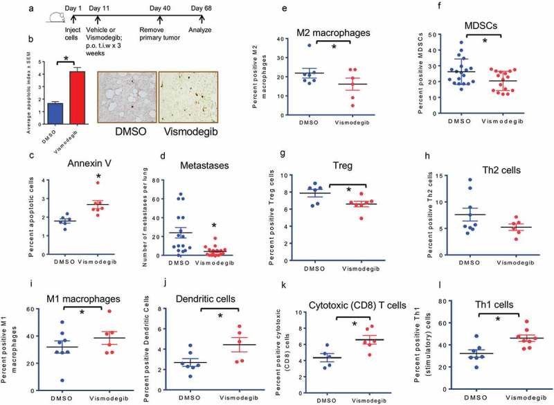
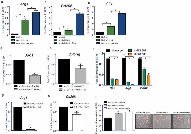
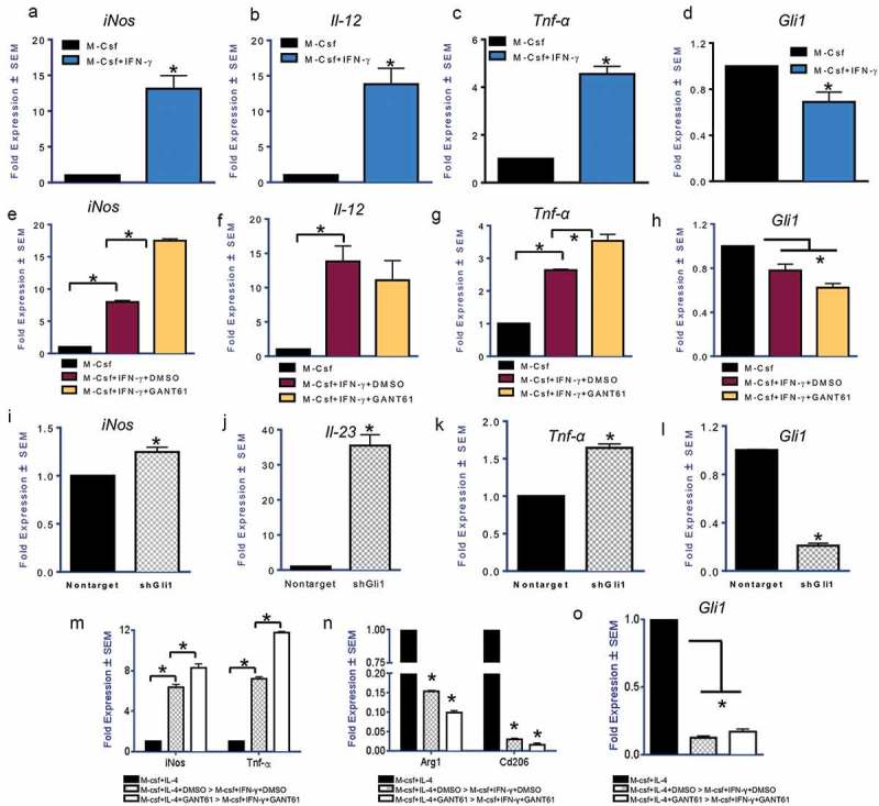
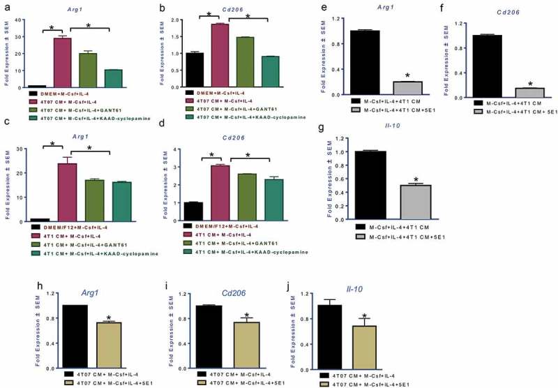
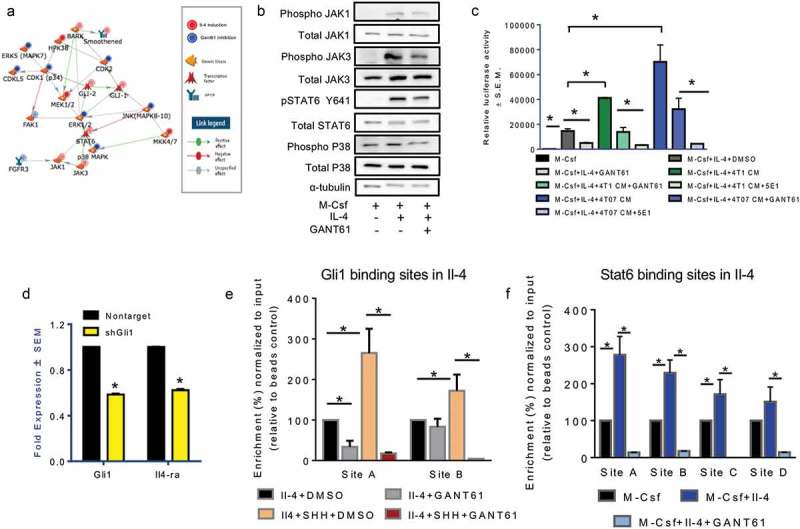
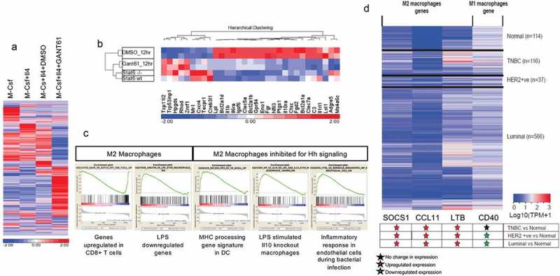
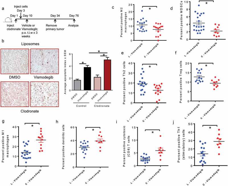
Host responses to tumor cells include tumor suppressing or promoting mechanisms. We sought to detail the effect of Hedgehog (Hh) pathway inhibition on the composition of the mammary tumor immune portfolio. We hypothesized that Hh signaling mediates a crosstalk between breast cancer cells and macrophages that dictates alternative polarization of macrophages and consequently supports a tumor-promoting microenvironment. We used an immunocompetent, syngeneic mouse mammary cancer model to inhibit Hh signaling with the pharmacological inhibitor, Vismodegib. Using molecular and functional assays, we identified that Hedgehog (Hh) signaling mediates a molecular crosstalk between mammary cancer cells and macrophages that culminates in alternative polarization of macrophages. We carried out an unbiased kinomics and genomics assessment to unravel changes in global kinomic and gene signatures impacted by Hh signaling. Our investigations reveal that in an immunocompetent mammary cancer model, the administration of Vismodegib led to changes in the portfolio of tumor-infiltrating immune cells. This was characterized by a marked reduction in immune-suppressive innate and adaptive cells concomitant with an enrichment of cytotoxic immune cells. Breast cancer cells induce M2 polarization of macrophages via a crosstalk mediated by Hh ligands that alters critical kinomic and genomic signatures. Macrophage depletion improved the benefit of Hedgehog inhibition on eliciting an immunogenic, pro-inflammatory profile. We define a novel role for Hh signaling in disabling anti-tumor immunity. Inhibition of Hh signaling presents with dual advantages of tumor cell-targeting as well as re-educating a dysfunctional tumor microenvironment.

Keywords: Hedgehog signaling; breast cancer; metastasis; tumor associated macrophages; tumor microenvironment.


Figures

Figure 1.
Figure 1.
Inhibiting Hedgehog signaling in vivo blunts the inhibitory immune response and elicits an inflammatory immune response(a) Schematic of Hh inhibition strategy used in vivo. (b) Tumor sections from DMSO and Vismodegib-treated mice were assessed for apoptosis by TUNEL staining. Tumors from Vismodegib-treated mice show significantly greater levels of apoptotic cells relative to vehicle-treated mice (p < 0.0001). (c)Tumors from Vismodegib-treated mice show significantly greater levels of late apoptotic cells relative to vehicle-treated mice (p = 0.0055). Tumor cells from DMSO and Vismodegib-treated mice were analyzed by flow cytometry analysis for late apoptotic cells as indicated by PI and Annexin V following the exclusion of lineage positive immune cells (CD45, CD31, and Ter119) and inclusion of CD24+ ve epithelial cells. (d) Vismodegib-treated mice show significantly decreased pulmonary metastasis. Lung metastasis in mice was assessed by microscopic counting of lungs fixed and stained in Bouin’s solution post tumor resection (p = 0.0009). (E-L) Following resection and processing of the primary tumor, non-viable cells were excluded and immune cell infiltrates were assayed based on the following markers: (e) M2 macrophages: CD11b, Arg1, and CD206 positive cells (p = 0.04). (f) Myeloid-derived suppressor cells: CD11b, LY6G and LY6C double positive cells (p = 0.02). (g) Regulatory T cells: CD3, CD4, FOXP3, and CD25 positive cells (p = 0.03). (h) Type II helper T cells: CD3, CD4, and GATA3 positive cells (p = 0.1). (i) M1 macrophages: CD11b, F4/80 and CD80 positive cells (p = 0.042). (j) Dendritic cells: CD11c and MHC II positive cells (p = 0.04). (k) Cytotoxic CD8 T cells: CD3, CD8, and Granzyme B positive cells (p = 0.015). (l) Activated Type I helper T cells: CD3, CD4, and IFN-γ positive cells (p = 0.0078). *p< statistically significant difference relative to DMSO-treated mice.
Figure 2.
Figure 2.
Hh signaling potentiates alternative activation of macrophages.The addition of recombinant Sonic Hedgehog ligand (100 nM) for 24 hours notably upregulates (a) Arg1 (p < 0.0001 and p = 0.0008) and (b) Cd206 (p < 0.0001 and p = 0.001). Shh ligand treatment was done in serum-free conditions. (c) Gli1 expression (p = 0.0007 and p < 0.0001) is an indication of activated Hh signaling. Gli inhibitor, GANT61 (10 μM for 24 hours) reduces the expression of (d) Arg1 (p < 0.0001) and (e) Cd206 (p = 0.0082). Transcript levels of Arg1 (p = 0.0023 for sh802 and p = 0.0032 for sh803) and Cd206 (p = 0.0087 for sh802 and p = 0.0015 for sh803) are significantly decreased in polarized RAW 264.7 cells transduced with an RFP expressing short hairpin RNA targeting Gli1 (p < 0.0001 for both shRNAs) (f). The expression of (g) Arg1 (p < 0.0001) and (h) Cd206 (p = 0.016) was significantly decreased in M2 polarized macrophages treated with the SMO inhibitor, BMS-833923 (2.5 μM). (i) Phagocytosis of bacterial particles was assessed using fluorescence microscopy in M2 stimulated macrophages inhibited for Hh signaling with BMS-833923 (2.5 μM) (p < 0.0001) or Vismodegib (20 μM) (p = 0.0006). Inhibition of Hh signaling significantly upregulates phagocytic activity. *statistically significant difference.
Figure 3.
Figure 3.
Inhibition of Hedgehog signaling facilitates classical activation of macrophages (a-d) Transcript levels of iNos (p = 0.029), Il-12 (p = 0.022), and Tnf-α (p = 0.0085), in macrophages treated with IFN-γ (50 ng/ml for 24 hours) are significantly increased with a concomitant reduction in the levels of Gli1 (p = 0.048). (e-h) Treatment of inflammatory macrophages with GANT61 (10 μM) causes an upregulation in the expression of iNos (p = 0.0002) and Tnf-α (p = 0.008). Il-12 expression did not change significantly. Gli1 transcript levels are reduced (p = 0.03) indicative of reduced potency of Hh signaling. (i-l) Stable silencing of Gli1 (p = 0.0008) in RAW 264.7 cells is consistent with an increase in the expression of iNos (p = 0.009), Il-23 (p = 0.0089), and Tnf-α (p = 0.0020) when assayed in M2 polarizing conditions. (m-o) Macrophages initially polarized to be immunosuppressive can be reversed to assume an inflammatory phenotype when treated with IFN-γ (50 ng/ml). Inhibition of Gli with GANT61 (p = 0.004) permits a further increase in expression of iNos (p = 0.007 and 0.023) and TNF-α (p = 0.008 and 0.005) with a concomitant decrease in Arg1 (p = 0.002 and 0.01) and Cd206 (p = 0.0007 and 0.002). *statistically significant difference.
Figure 4.
Figure 4.
Mammary carcinoma cells upregulate alternative polarization of macrophages by eliciting activation of Hedgehog signaling Transcript levels of Arg1 (a (p = 0.0008), c (p = 0.01)) and Cd206 (b (p = 0.002), d (p = 0.002)) are significantly upregulated in presence of conditioned medium (CM) from 4T07 and 4T1 invasive mammary carcinoma cells. Treatment with GANT61 (10 μM) or KAAD-Cyclopamine (20 μM) significantly decreased the immunosuppressive polarization induced by the CM (Arg1 in 4T07 cells: p = 0.0034; Arg1 in 4T1 cells: p = 0.022; Cd206 in 4T07 cells: p = 0.0013; Cd206 in 4T1 cells: p = 0.006). The addition of Hh ligand squelching antibody, 5E1 (2.5 μg/ml), significantly decreases the effect of the 4T1 and 4T07 CM on its ability to induce a potent M2 phenotype marked by Arg1 (e, h), Cd206 (f, i), and Il-10 (g, j) (p < 0.0001 for all comparisons). *statistically significant difference.
Figure 5.
Figure 5.
Hedgehog signaling mediates molecular mechanisms that dictate alternative macrophage polarization and its blockade promotes a pro-inflammatory, immunogenic profile. (a) Network model generated from kinomics assessment of M2 polarized macrophages treated with DMSO (vehicle control) or GANT61 (10 μM). Phosphorylated tyrosine and serine/threonine residues were assessed using PamGene PamStation Kinomic Array platform. (b) Inhibition of Hh signaling decreases phosphorylation of JAK1, JAK3, STAT6, and p38 kinases. Lysates from M2-polarized RAW 264.7 cells treated with DMSO (vehicle control) or GANT61 (10 μM) were assessed for total JAK1, JAK3, STAT6, p38 and pJAK1, pJAK3, pSTAT6, and p-p38 expression using western blot analysis. α-tubulin was used as a loading control. (c) Inhibiting Hh signaling significantly decreases the activity of the STAT6 luciferase reporter (p = 0.0007). CM from 4T1 (p = 0.0002) and 4T07 cells (p = 0.0022) potently activates STAT6 activity; squelching of Hh ligands with 5E1 antibody (4T1CM: p < 0.0001; 4T07 CM: p = 0.001) or inhibition with GANT61 (4T1CM: p = 0.02; 4T07 CM: p = 0.0015) significantly decreased the ability of the CM to activate STAT6. RAW 264.7 cells were transfected with the p4X STAT6 plasmid, and cultured to induce M2 polarization in presence or absence of vehicle control, DMSO, or GANT61 (10 μM) or CM from 4T1 and 4T07 cells with or without 5E1 (2.5 μg/ml) or GANT61 (10 μM). *p < 0.05. (d) The transcript levels of Il-4Ra (p < 0.00010) are significantly decreased in macrophages silenced for Gli1 (p < 0.0001) and polarized toward the M2 phenotype. (e) ChIP was performed in macrophages treated with M-Csf and Il-4 and DMSO or GANT61 (10 μM) in the presence and absence of SHH (100 nM) using an anti-GLI1 antibody followed by qPCR with primers specific for two putative Gli1 binding sites on the IL-4 promoter. (f) Stat6 was ChIP-ed in macrophages treated with M-Csf and Il-4 and DMSO or GANT61 (10 μM) followed by qPCR with primers specific for four putative Stat6 binding sites on the IL-4 promoter. *statistically significant difference.
Figure 6.
Figure 6.
Inhibiting Hedgehog signaling upregulates an inflammatory gene expression signature (a) Heat map of the gene signature of macrophages derived from RAW 264.7 cells treated with M-Csf (M0 state), M-Csf and IL-4 (M2 macrophages), M-Csf, IL-4, and DMSO (M2 and vehicle control), and M-Csf, IL-4, and 20 μM GANT61 (M2 inhibited for Hh signaling). GANT61-treated macrophages demonstrate clear differences from vehicle controls. Log2-transformed RPKM-normalized intensities were used for Z-normalization with color indicating above (in red) or below (in blue) average. (b) GANT61-treated macrophages demonstrate similarity to Stat6 (-/-) macrophages, although Stat6 (-/-) and Stat6 (WT) show fewer differences among these genes. (c) Gene set enrichment analysis of RNAseq data depicts that the molecular gene signature of M2 macrophages is characteristically immunosuppressive. Inhibition of Hh/Gli signaling reveals an overlap with inflammatory signature in bacterial infections and antigen presentation by dendritic cells. (d) Heatmap showing expression level of M1 and M2 macrophage genes in normal (n = 114), HER2 Positive (n = 37), Luminal (n = 566), and triple negative breast cancer [TNBC] (n = 116) samples from TCGA breast invasive carcinoma [BRCA] dataset. Transcript per million (TPM) value for each sample is obtained by multiplying RSEM scaled_estimate by 106.The significance of differential expression is estimated via student’s T-test.
Figure 7.
Figure 7.
Macrophage depletion improves the benefit of inhibiting Hedgehog signaling on eliciting a pro-inflammatory, immunogenic response (a) Schematic of macrophages depletion using liposomal clodronate followed by Hh inhibition strategy used in vivo. (b) Tumor sections from DMSO and Vismodegib-treated mice were assessed for apoptosis by TUNEL staining. Tumors from Vismodegib-treated mice in the control liposomes (L) group show significantly greater levels of apoptotic cells relative to vehicle-treated mice (p < 0.0001). Tumors from Vismodegib-treated mice depleted for macrophages with clodronate (C) display a significantly higher apoptotic index than their vehicle treated counterparts (p < 0.0001). Following resection and processing of the primary tumor, non-viable cells were excluded and immune cell infiltrates were assayed based on the following markers: (c) M2 macrophages: CD11b, LY6C-ve, F4/80 + ve, Arg1, and CD206 positive cells (p = 0.0151). (d) Myeloid-derived suppressor cells: CD11b, LY6G and LY6C double positive cells (p = 0.0133). (e) Type II helper T cells: CD3, CD4, and GATA3 positive cells (p = 0.0184). (f) Regulatory T cells: CD3, CD4, and FOXP3, and CD25 positive cells (p = 0.0056). (g) M1 macrophages: F4/80, CD80 and CD86 positive cells (p < 0.0001). (h) Dendritic cells: CD11c, MHC II and CD86 positive cells (p = 0.0085). (i) Cytotoxic CD8 T cells: CD3, CD8, and Granzyme B positive cells (0.0005). (j) Activated Type I helper T cells: CD3, CD4, and IFN-γ positive cells (p = 0.0021). *p< statistically significant difference relative to DMSO-treated mice.

References

    1. Mao Y, Keller ET, Garfield DH, Shen K, Wang J.. Stromal cells in tumor microenvironment and breast cancer. Cancer Metastasis Rev. 2013;32(1–2):303–315. DOI: 10.1007/s10555-012-9415-3. - DOI - PMC - PubMed
    1. Turley SJ, Cremasco V, Astarita JL.. Immunological hallmarks of stromal cells in the tumour microenvironment. Nat Rev Immunol. 2015;15(11):669–682. DOI: 10.1038/nri3902. - DOI - PubMed
    1. Varol C, Mildner A, Jung S. Macrophages: development and tissue specialization. Annu Rev Immunol. 2015;33:643–675. DOI: 10.1146/annurev-immunol-032414-112220. - DOI - PubMed
    1. Obeid E, Nanda R, Fu YX, Olopade OI. The role of tumor-associated macrophages in breast cancer progression (review). Int J Oncol. 2013;43(1):5–12. DOI: 10.3892/ijo.2013.1938. - DOI - PMC - PubMed
    1. Das A, Sinha M, Datta S, Abas M, Chaffee S, Sen CK, Roy S. Monocyte and macrophage plasticity in tissue repair and regeneration. Am J Pathol. 2015;185(10):2596–2606. DOI: 10.1016/j.ajpath.2015.06.001. - DOI - PMC - PubMed

Publication types

LinkOut - more resources