

Chest wall lipoblastoma in a 3 year-old boy
- PMID: 30723667
- PMCID: PMC6350110
- DOI: 10.1016/j.rmcr.2019.01.013
Chest wall lipoblastoma in a 3 year-old boy
Abstract
Background: Lipoblastoma is a rare, benign, fatty tissue tumor that occurs in infancy and early childhood. The most common tumor locations are the extremities and the torso. The location of this tumor in the chest wall and an intrathoracic extension is uncommon.
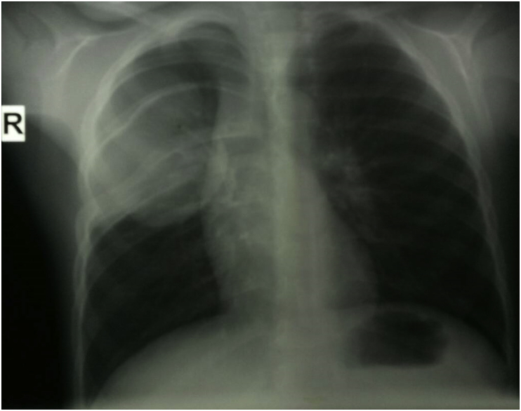
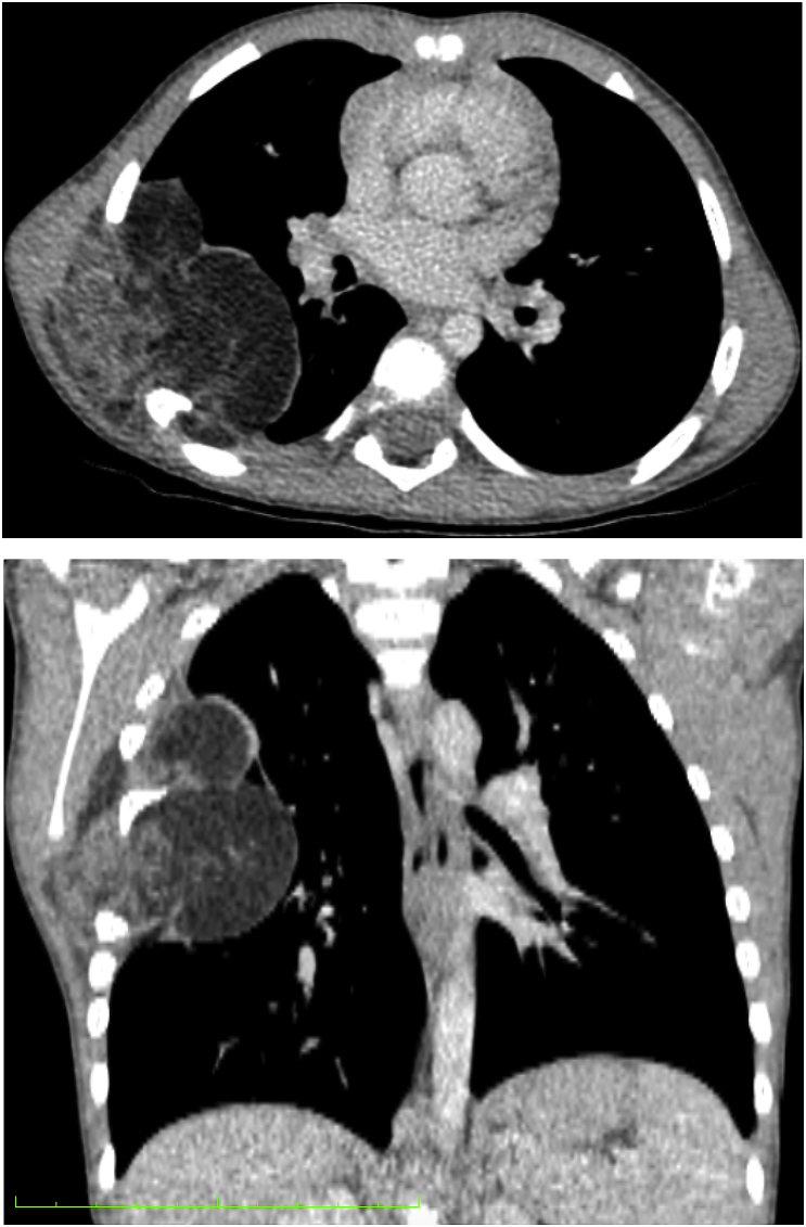
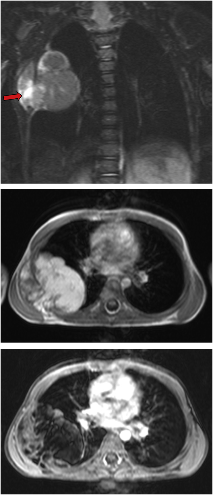
Case report: We present a case of a 3-year-old boy with anterior chest wall lipoblastoma with an intrathoracic extension. Computed tomography was suggestive of lipoblastoma. The mass was completely excised through a right posterolateral thoracotomy. The histologic examination of the lesion confirmed the diagnosis of lipoblastoma.
Conclusion: Although extremely rare, chest wall lipoblastoma should be included in the differential diagnosis of thoracic mass in childhood.
Keywords: Chest wall; Children; Lipoblastoma; Thoracic mass.
Figures
References
-
- Benato C., Falezza G., Lonardoni A., Magnanelli G., Ricci M., Gilioli E., Calabr F. Acute respiratory distress caused by a giant mediastinal lipoblastoma in a 16-month-old boy. Ann. Thorac. Surg. 2011;92(6):e119–e120. - PubMed
-
- Harrer J., Hammon G., Wagner T., Bolkenius M. Lipoblastoma and lipoblastomatosis: a report of two cases and review of the literature. Eur. J. Pediatr. Surg. 2001;11(5):342–349. - PubMed
-
- Salem R., Zohd M., Njim L., Maazoun K., Jellali M.A., Zrig A., Mnari W., Harzallah W., Nouri A., Zakhama A., Golli M. Lipoblastoma: a rare lesion in the differential diagnosis of childhood mediastinal tumors. J. Pediatr. Surg. 2011;46(5):e21–e23. - PubMed
-
- Moholkar S., Sebire N.J., Roebuck D.J. Radiological-pathological correlation in lipoblastoma and lipoblastomatosis. Pediatr. Radiol. 2006;36(8):851–856. - PubMed
-
- Glazer H.S., Wick M.R., Anderson D.J., Semenkovich J.W., Molina P.L., Siegel M.J., Sagel S.S. CT of fatty thoracic masses. Am. J. Roentgenol. 1992;159(6):1181–1187. - PubMed
Publication types
LinkOut - more resources
Full Text Sources
