

Review: Bioengineering approach for the repair and regeneration of peripheral nerve
- PMID: 30723843
- PMCID: PMC6351356
- DOI: 10.1016/j.bioactmat.2018.09.001
Review: Bioengineering approach for the repair and regeneration of peripheral nerve
Erratum in
-
Erratum regarding missing Declaration of Competing Interest statements in previously published articles.Bioact Mater. 2020 Dec 4;6(6):1789-1790. doi: 10.1016/j.bioactmat.2020.11.009. eCollection 2021 Jun. Bioact Mater. 2020. PMID: 33336111 Free PMC article.
Abstract
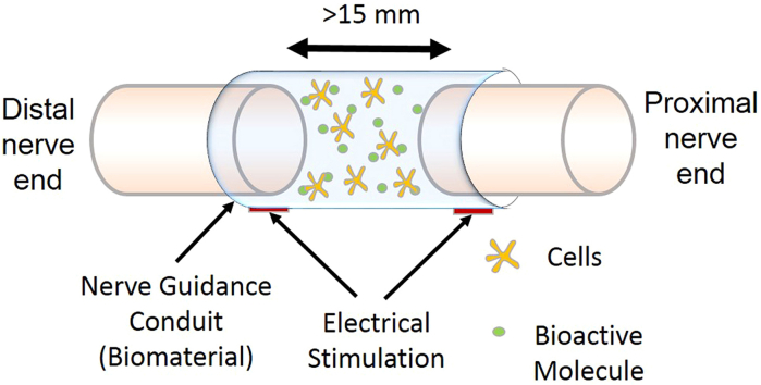
Complex craniofacial surgeries of damaged tissues have several limitations, which present complications and challenges when trying to replicate facial function and structure. Traditional treatment techniques have shown suitable nerve function regeneration with various drawbacks. As technology continues to advance, new methods have been explored in order to regenerate damaged nerves in an effort to more efficiently and effectively regain original function and structure. This article will summarize recent bioengineering strategies involving biodegradable composite scaffolds, bioactive factors, and external stimuli alone or in combination to support peripheral nerve regeneration. Particular emphasis is made on the contributions of growth factors and electrical stimulation on the regenerative process.
Keywords: Composite materials; Electrical stimulation; Growth factor; Peripheral nerve regeneration.
Figures
References
-
- Birch R. 2010. The Peripheral Nervous System: Gross Anatomy; pp. 1–41.
-
- Burnett M.G., Zager E.L. Pathophysiology of peripheral nerve injury: a brief review. Neurosurg. Focus. 2004;16:1–7. - PubMed
Publication types
Grants and funding
LinkOut - more resources
Full Text Sources
