

Paraspinal Muscle DTI Metrics Predict Muscle Strength
- PMID: 30723976
- PMCID: PMC6767405
- DOI: 10.1002/jmri.26679
Paraspinal Muscle DTI Metrics Predict Muscle Strength
Abstract
Background: The paraspinal muscles play an important role in the onset and progression of lower back pain. It would be of clinical interest to identify imaging biomarkers of the paraspinal musculature that are related to muscle function and strength. Diffusion tensor imaging (DTI) enables the microstructural examination of muscle tissue and its pathological changes.
Purpose: To investigate associations of DTI parameters of the lumbar paraspinal muscles with isometric strength measurements in healthy volunteers.
Study type: Prospective.
Subjects: Twenty-one healthy subjects (12 male, 9 female; age = 30.1 ± 5.6 years; body mass index [BMI] = 27.5 ± 2.6 kg/m2 ) were recruited.
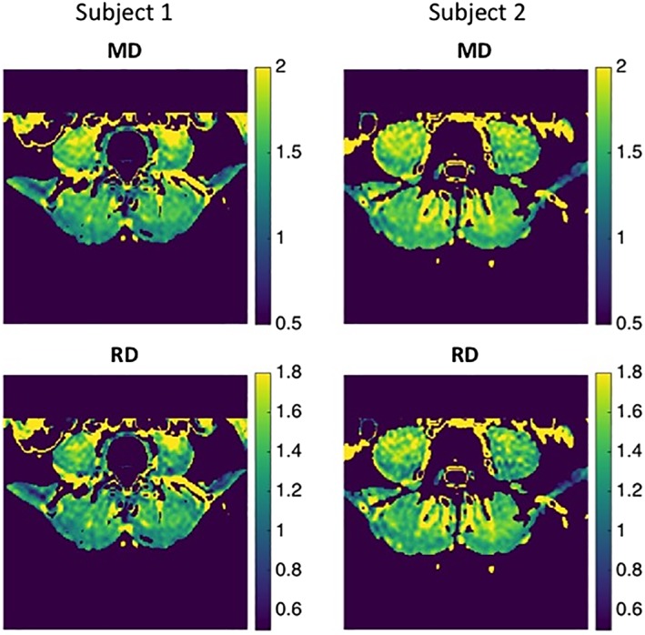
Field strength/sequence: 3 T/single-shot echo planar imaging (ss-EPI) DTI in 24 directions; six-echo 3D spoiled gradient echo sequence for chemical shift encoding-based water-fat separation.
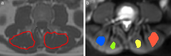
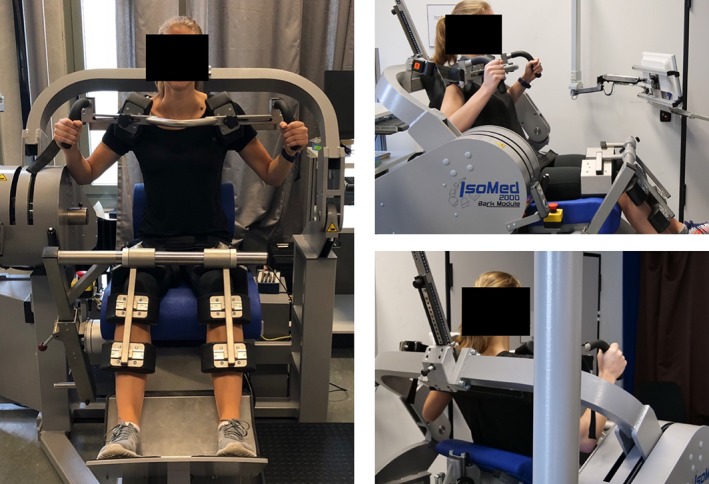
Assessment: Paraspinal muscles at the lumbar spine were examined. Erector spinae muscles were segmented bilaterally; cross-sectional area (CSA), proton density fat fraction (PDFF), and DTI parameters were calculated. Muscle flexion and extension maximum isometric torque values [Nm] at the back were measured with an isokinetic dynamometer and the ratio of extension to flexion strength (E/F) calculated.
Statistical tests: Pearson correlation coefficients; multivariate regression models.
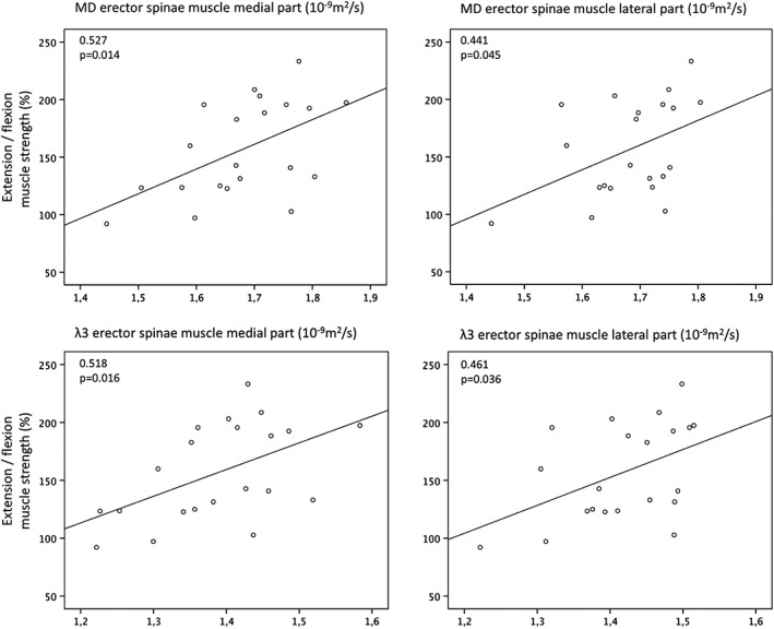
Results: Significant positive correlations were found between the ratio of extension to flexion (E/F) strength and mean diffusivity (MD) (P = 0.019), RD (P = 0.02) and the eigenvalues (λ1: P = 0.026, λ2: P = 0.033, λ3: P = 0.014). In multivariate regression models λ3 of the erector spinae muscle λ3 and gender remained statistically significant predictors of E/F (R2adj = 0.42, P = 0.003).
Data conclusion: DTI allowed the identification of muscle microstructure differences related to back muscle function that were not reflected by CSA and PDFF. DTI may potentially track subtle changes of back muscle tissue composition.
Level of evidence: 3 Technical Efficacy: Stage 2 J. Magn. Reson. Imaging 2019;50:816-823.
Keywords: diffusion tensor imaging; lumbar spine; muscle microstructure; muscle strength; paraspinal musculature.
© 2019 The Authors. Journal of Magnetic Resonance Imaging published by Wiley Periodicals, Inc. on behalf of International Society for Magnetic Resonance in Medicine.
Figures
References
-
- Fehlings MG, Tetreault L, Nater A, et al. The aging of the global population: The changing epidemiology of disease and spinal disorders. Neurosurgery 2015;77(Suppl 4):S1–5. - PubMed
-
- Lotz JC, Haughton V, Boden SD, et al. New treatments and imaging strategies in degenerative disease of the intervertebral disks. Radiology 2012;264:6–19. - PubMed
-
- Dahlqvist JR, Vissing CR, Thomsen C, Vissing J. Severe paraspinal muscle involvement in facioscapulohumeral muscular dystrophy. Neurology 2014;83:1178–1183. - PubMed
Publication types
MeSH terms
Grants and funding
LinkOut - more resources
Full Text Sources
Research Materials
Miscellaneous
