

Autoimmune glial fibrillary acidic protein astrocytopathy
- PMID: 30724768
- PMCID: PMC6522205
- DOI: 10.1097/WCO.0000000000000676
Autoimmune glial fibrillary acidic protein astrocytopathy
Abstract
Purpose of review: To describe a recently characterized autoimmune, inflammatory central nervous system (CNS) disorder known as autoimmune glial fibrillary acidic protein (GFAP) astrocytopathy.
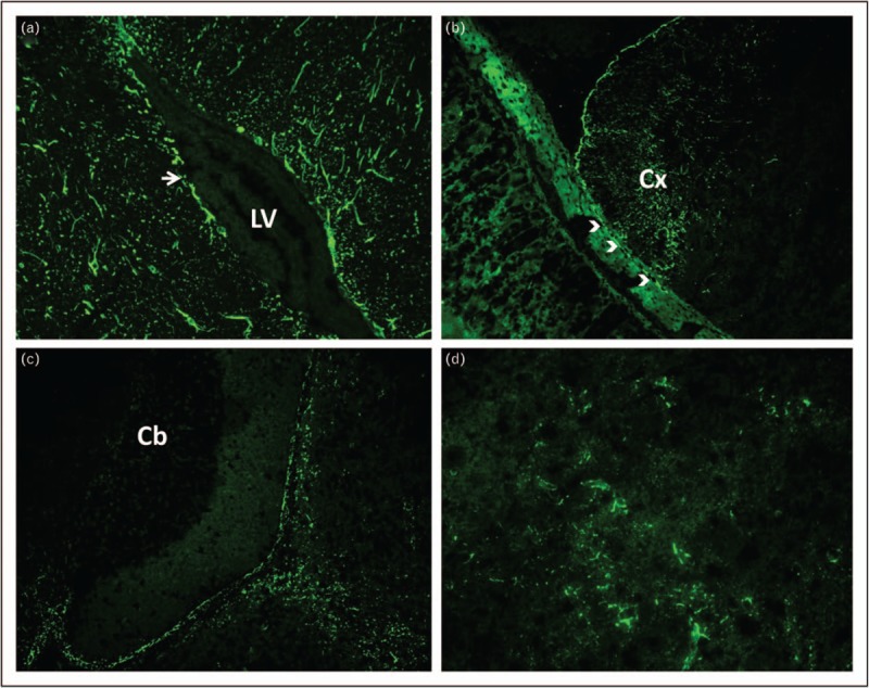
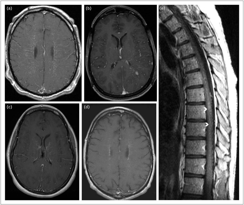
Recent findings: Affected patients present with symptoms of one or more of meningitis (headache and neck ache), encephalitis (delirium, tremor, seizures, or psychiatric symptoms), and myelitis (sensory symptoms and weakness). Optic disc papillitis (blurred vision) is common. CNS inflammation is evident in characteristic T1 postgadolinium enhancement of GFAP-enriched CNS regions, and lymphocytic cerebrospinal fluid (CSF) white cell count elevation. CSF is more reliable than serum for GFAP-immunoglobulin G (IgG) testing. Ovarian teratoma commonly coexists, particularly among patients with accompanying N-methyl-D-aspartate receptor or aquaporin-4 autoimmunity. Parainfectious autoimmunity is suspected in some other patients, though the culprit organism is rarely verified. Pathophysiologic relevance of T cells is underscored by neuropathology and cases of dysregulated T-cell function (HIV or checkpoint inhibitor cancer therapy). Corticosteroid-responsiveness is a hallmark of the disease. Relapses occur in approximately 20% of patients, necessitating transition to a steroid-sparing drug. Reported outcomes vary, though in the authors' experience, early and sustained intervention usually portends recovery.
Summary: Autoimmune GFAP astrocytopathy is a treatable autoimmune CNS disease diagnosable by GFAP-IgG testing in CSF. This disease presents opportunities to explore novel mechanisms of CNS autoimmunity and inflammation.
Figures
References
-
- Fang B, McKeon A, Hinson SR, et al. Autoimmune glial fibrillary acidic protein astrocytopathy: a novel meningoencephalomyelitis. JAMA Neurol 2016; 73:1297–1307. - PubMed
-
The original study describing autoimmune GFAP astrocytopathy.
-
- Long Y, Liang J, Xu H, et al. Autoimmune glial fibrillary acidic protein astrocytopathy in Chinese patients: a retrospective study. Eur J Neurol 2018; 25:477–483. - PubMed
-
This article contains a large experience and includes some pathological data.
-
- Aksamit AJ, Parisi J. Chronic microglial encephalomyelitis. Ann Neurol 2012; 72:S110.
-
- Flanagan EP, Hinson SR, Lennon VA, et al. Glial fibrillary acidic protein immunoglobulin G as biomarker of autoimmune astrocytopathy: analysis of 102 patients. Ann Neurol 2017; 81:298–309. - PubMed
-
This article is the largest and most detailed experience of autoimmune GFAP astrocytopathy published to date.
-
- Shibuya M, Matsuki N, Fujiwara K, et al. Autoantibodies against glial fibrillary acidic protein (GFAP) in cerebrospinal fluids from Pug dogs with necrotizing meningoencephalitis. J Vet Med Sci 2007; 69:241–245. - PubMed
Publication types
MeSH terms
Substances
LinkOut - more resources
Full Text Sources
Research Materials
Miscellaneous
