Review
. 2019 Feb 6;10(1):12.
doi: 10.1186/s13244-019-0694-x.

Thyroglossal duct pathology and mimics

Affiliations
Review

Thyroglossal duct pathology and mimics

Swapnil Patel et al. Insights Imaging. .

Abstract

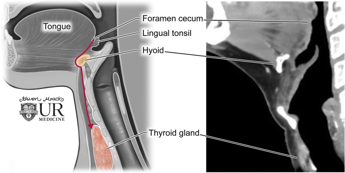
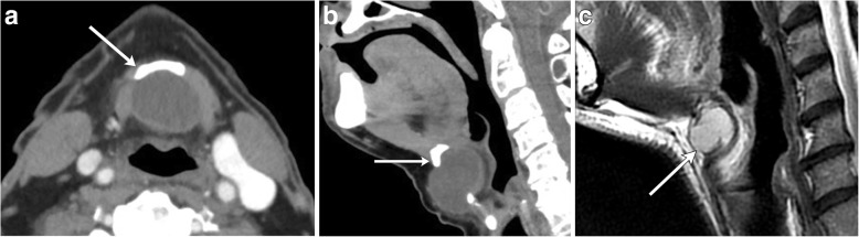
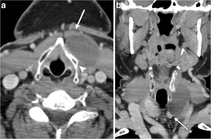
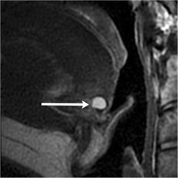
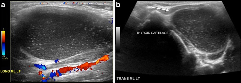
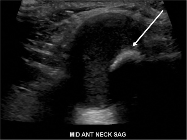
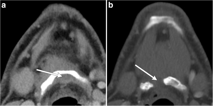
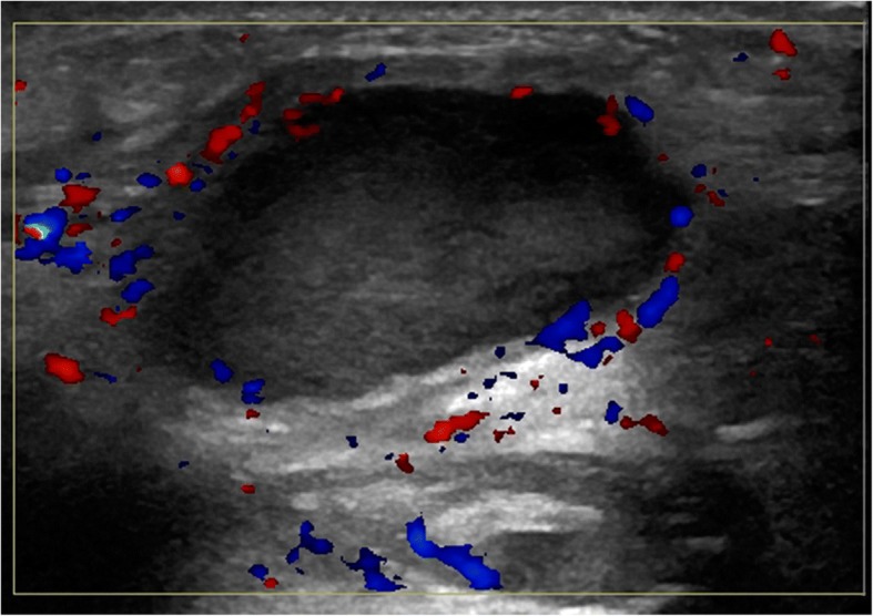
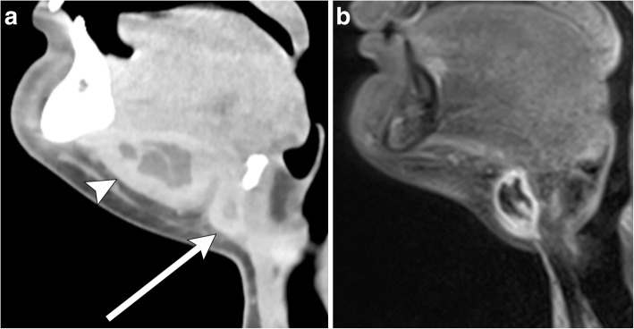
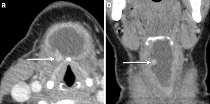
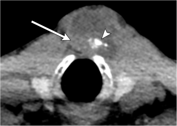
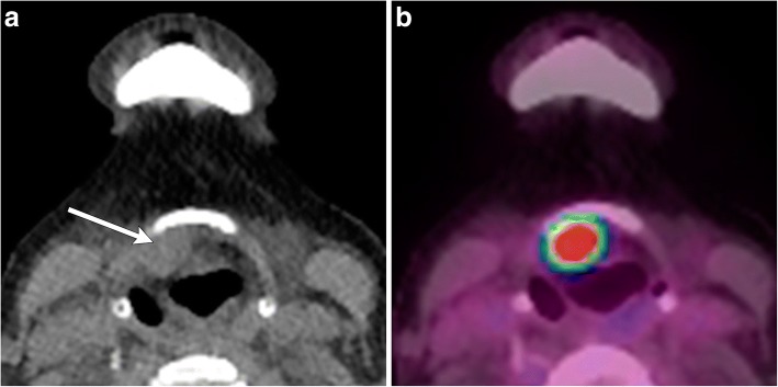
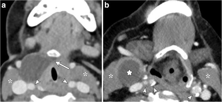
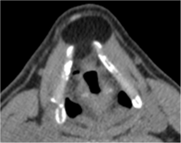
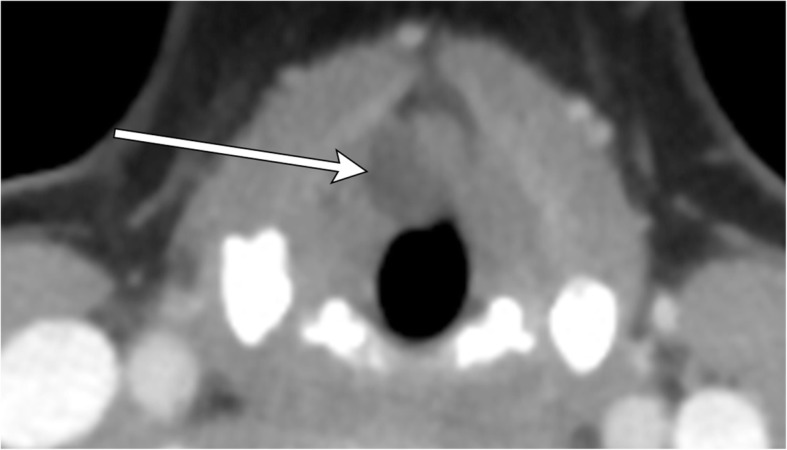
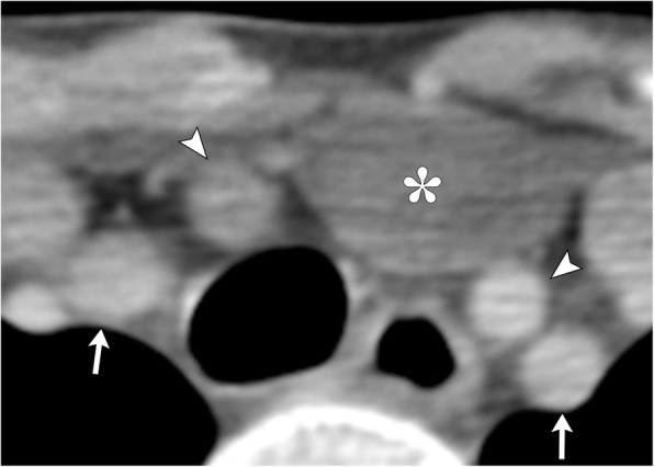
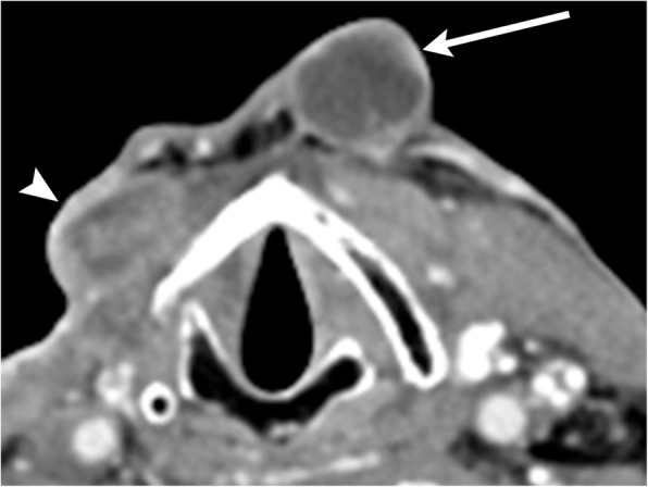
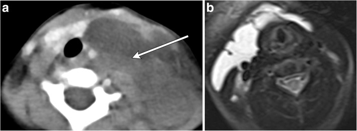
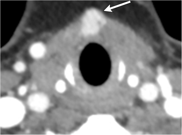
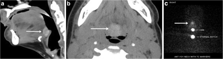
Congenital anterior neck masses comprise a rare group of lesions typically diagnosed in childhood. Most commonly, lesions are anomalies of the thyroglossal duct, namely the thyroglossal duct cyst, along with ectopic thyroid tissue. Although usually suspected based on clinical examination, imaging can confirm the diagnosis, assess the extent, and evaluate for associated complications. Imaging characteristics on ultrasound, CT, and MRI may at times be equivocal; differential considerations include branchial cleft cyst, dermoid/epidermoid, laryngocele, thymic cyst, lymphatic malformation, and metastatic disease. Thus, understanding of the embryologic course of thyroid development is crucial with recognition of critical landmarks such as the foramen cecum, hyoid bone, thyroid cartilage, and strap musculature to aid in the diagnosis of an anterior neck mass.

Keywords: Cystic lesions; Ectopic thyroid; Neck mass; Thyroglossal duct.


Conflict of interest statement

Competing interests

The authors declare that they have no competing interests.

Publisher’s Note

Springer Nature remains neutral with regard to jurisdictional claims in published maps and institutional affiliations.

Figures

Fig. 1
Fig. 1
Thyroglossal duct anatomy. Illustration of the anatomic course of the thyroglossal duct during embryological development (left). Corresponding anatomy on a sagittal contrast-enhanced neck CT (right)
Fig. 2
Fig. 2
Midline thyroglossal duct cysts. Axial (a) and sagittal (b) contrast-enhanced CT images of the neck demonstrate the close relationship of a thyroglossal duct cyst (non-enhancing cystic structure within the midline neck) with the hyoid bone (arrows). c Sagittal T2-weighted MR image of a different patient reveals a fluid signal lesion extending posterior to the hyoid bone (arrow)
Fig. 3
Fig. 3
Paramidline thyroglossal duct cyst. a Axial contrast-enhanced CT images of the infrahyoid neck demonstrate a well-circumscribed homogenous low attenuation mass located between the thyroid cartilage (arrowhead) and strap muscle (arrow). b Coronal contrast-enhanced CT image shows the paramidline location and extension to upper pole of the left thyroid lobe (arrow)
Fig. 4
Fig. 4
Lingual thyroglossal duct cyst. Sagittal T2-weighted MR image demonstrates a fluid signal lesion at the foramen cecum (arrow)
Fig. 5
Fig. 5
Thyroglossal duct cyst containing debris. a Long axis grayscale ultrasound image with color Doppler demonstrates a well-circumscribed hypoechoic structure containing tiny hyperechoic foci compatible with debris. Note the lack of vascularity within the lesion and posterior through transmission. b Grayscale ultrasound image illustrates the paramidline location adjacent to the thyroid cartilage and deep to the strap muscle (arrow)
Fig. 6
Fig. 6
Infrahyoid thyroglossal duct cyst. Long axis grayscale ultrasound image shows a midline cyst in contact with and extending posterior to the hyoid bone (arrow)
Fig. 7
Fig. 7
Sistrunk procedure. a Axial contrast-enhanced CT image demonstrates a thyroglossal duct cyst wrapping around the hyoid bone (arrow). b Post-operative axial contrast-enhanced CT image demonstrates removal of the thyroglossal duct cyst and a partial hyoidectomy (arrow)
Fig. 8
Fig. 8
Infected thyroglossal duct cyst. A 23-year-old male presents with several days of fever, as well as a warm anterior neck mass. Long axis ultrasound image with color Doppler reveals a thick-walled hypoechoic structure containing low level echoes with peripheral hyperemia in the paramidline anterior neck
Fig. 9
Fig. 9
Infected thyroglossal duct cyst with abscess. a Sagittal contrast-enhanced CT image of the neck shows a thick-walled cyst adjacent to the hyoid bone (arrow). There is a communicating peripherally enhancing fluid collection along the floor of the mouth, consistent with abscess (arrowhead). Note stranding of the adjacent fat. b Sagittal post-contrast T1-weighted MR imaging of the neck demonstrates interval resolution of the abscess and underlying thyroglossal duct cyst with residual inflammatory changes. Note the relative increased thickness of the wall of the infected thyroglossal duct cyst compared to one that is not infected (see Fig. 2)
Fig. 10
Fig. 10
Thyroglossal ductal cyst with malignancy. Axial (a) and coronal (b) contrast-enhanced CT images of the neck show an irregularly thick-walled thyroglossal duct cyst with enhancing mural nodule (arrows). Resection revealed papillary carcinoma
Fig. 11
Fig. 11
Thyroglossal duct cyst with malignancy. Axial CT image demonstrates a thyroglossal duct cyst, which is predominantly cystic, but also has soft tissue attenuation (arrow) and calcifications (arrowhead) at the posterior aspect. Resection revealed papillary carcinoma
Fig. 12
Fig. 12
Malignancy of thyroglossal duct remnant. a Axial non-contrast CT image demonstrates a soft tissue mass adjacent to the posterior margin of the hyoid bone (arrow). b Axial fused PET/CT image demonstrates FDG avidity in this lesion. Pathology proven papillary carcinoma
Fig. 13
Fig. 13
Thyroglossal duct cyst and branchial cleft cyst. a Axial contrast-enhanced CT image shows a pathology proven thyroglossal duct cyst located anterior to the carotid vessels (arrowheads) and anteromedially to the sternocleidomastoid muscle (asterisks) with a tail-like extension medially to the hyoid bone (arrow). b Axial contrast-enhanced CT image demonstrates a second branchial cleft cyst (star) in a similar location within the right neck without hyoid extension
Fig. 14
Fig. 14
Dermoid. Axial non-contrast CT image illustrates a neck mass located anterior to the thyroid cartilage and superficial to the strap muscles. The lesion demonstrates the same attenuation as adjacent subcutaneous fat
Fig. 15
Fig. 15
Internal laryngocele. Axial contrast-enhanced CT image shows a non-enhancing fluid attenuating structure in right paraglottic region with mass effect on false vocal cord (arrow)
Fig. 16
Fig. 16
Thymic Cyst. Axial contrast-enhanced CT image demonstrates a cystic mass (asterisk) in the left neck base between the right and left common carotid arteries (arrowhead), extending into the superior mediastinum. Bilateral subclavian arteries are also visualized (arrows)
Fig. 17
Fig. 17
Necrotic metastatic lymphadenopathy (primary later found to be tongue base squamous cell carcinoma). Axial contrast-enhanced CT image illustrates an irregularly thick-walled cystic mass anterior to the left thyroid cartilage (arrow). Careful search in other areas of the neck demonstrate another similar appearing cystic lymph node adjacent to the right thyroid cartilage (arrowhead)
Fig. 18
Fig. 18
Lymphangioma. a Axial contrast-enhanced CT image demonstrates a non-enhancing multilocular, trans-spatial fluid-attenuation mass centered in the left neck base with mass effect and rightward displacement of trachea and esophagus. Note the fluid-fluid levels indicative of prior hemorrhage (arrow), distinguishing this lesion from a thyroglossal duct cyst. b Axial T2-weighted MR image in a different patient demonstrates a trans-spatial multiseptated cystic mass extending from the right lateral infrahyoid neck across the anterior aspect of the thyroid cartilage
Fig. 19
Fig. 19
Ectopic thyroid tissue. Axial contrast-enhanced CT image demonstrates a focal high attenuating soft tissue mass in the midline anterior neck along the anterior margin of the thyroid cartilage (arrow). Invaginated by strap musculature, this mass resembles the thyroid gland in attenuation and occurs in expected course of the thyroglossal duct tract
Fig. 20
Fig. 20
Lingual thyroid. a Sagittal contrast-enhanced CT image of the neck demonstrates a homogenously enhancing lingual thyroid at the base of the tongue encroaching on the valleculae (arrow). b Axial non-contrast CT shows the inherently high attenuating midline mass at the base of the tongue (arrow). c I-123 thyroid scan confirms the diagnosis, expected iodine uptake of the lingual thyroid (arrow)

References

    1. Zander DA, Smoker WR. Imaging of ectopic thyroid tissue and thyroglossal duct cysts. Radiographics. 2014;34:37–50. doi: 10.1148/rg.341135055. - DOI - PubMed
    1. Branstetter B, Weissman J, Kennedy T, Whitaker M (2000) The CT appearance of thyroglossal duct carcinoma. AJNR Am J Neuroradiol 21:1547–1550 - PMC - PubMed
    1. Imhof H, Czerny C, Hörmann M, Krestan C (2004) Tumors and tumor-like lesions of the neck: from childhood to adult. Eur Radiol 14(Suppl 4):L155–L165. 10.1007/s00330-003-2035-0 - PubMed
    1. Koeller KK, Alamo L, Adair CF, Smirniotopoulos JG (1999) Congenital cystic masses of the neck: radiologic-pathologic correlation. Radiographics 19:121–146 - PubMed
    1. Nakayama S, Kimachi K, Nakayama K, Ikebe T, Ozeki S (2009) Thyroglossal duct cyst occurring in the floor of the mouth: report of 2 cases. J Oral Maxillofac Surg 67:2690–2693 - PubMed

LinkOut - more resources