Clinical Trial
. 2019 Mar 20;37(9):693-702.
doi: 10.1200/JCO.18.01896. Epub 2019 Feb 6.

Durable Tumor Regression and Overall Survival in Patients With Advanced Merkel Cell Carcinoma Receiving Pembrolizumab as First-Line Therapy

Affiliations
Clinical Trial

Durable Tumor Regression and Overall Survival in Patients With Advanced Merkel Cell Carcinoma Receiving Pembrolizumab as First-Line Therapy

Paul Nghiem et al. J Clin Oncol. .

Abstract

Purpose: Merkel cell carcinoma (MCC) is an aggressive skin cancer often caused by the Merkel cell polyomavirus. Clinical trials of programmed cell death-1 pathway inhibitors for advanced MCC (aMCC) demonstrate increased progression-free survival (PFS) compared with historical chemotherapy data. However, response durability and overall survival (OS) data are limited.

Patients and methods: In this multicenter phase II trial (Cancer Immunotherapy Trials Network-09/Keynote-017), 50 adults naïve to systemic therapy for aMCC received pembrolizumab (2 mg/kg every 3 weeks) for up to 2 years. Radiographic responses were assessed centrally per Response Evaluation Criteria in Solid Tumors (RECIST) v1.1.

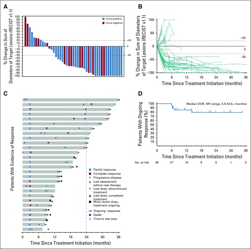
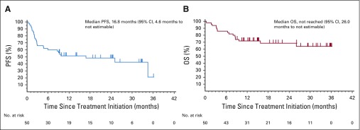
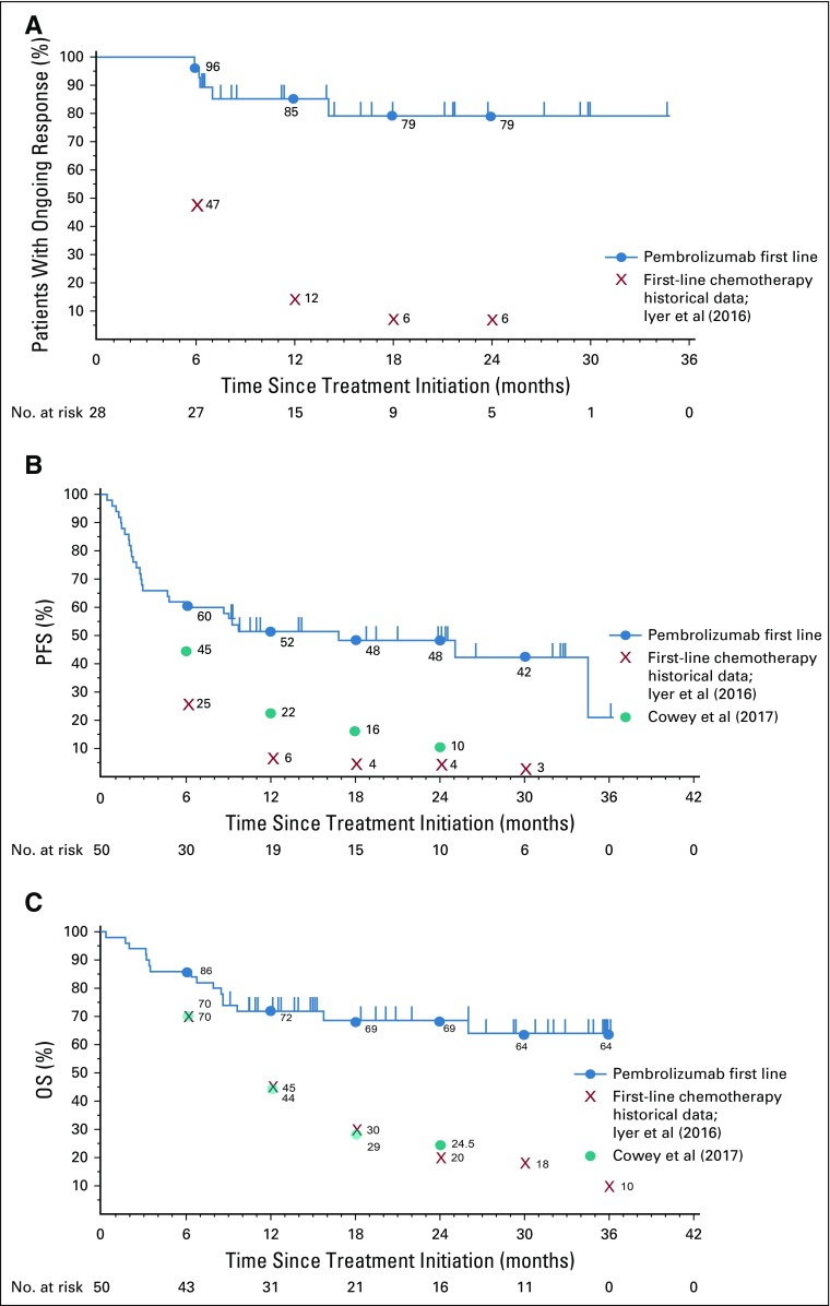
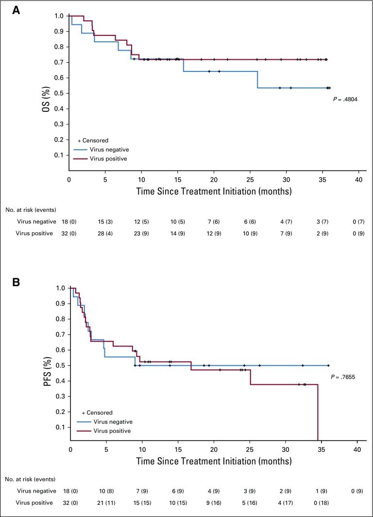
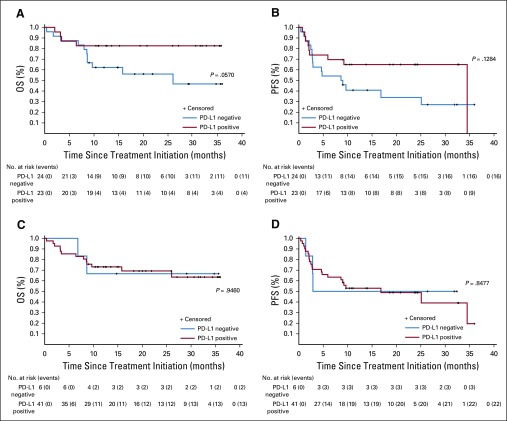
Results: Among 50 patients, the median age was 70.5 years, and 64% had Merkel cell polyomavirus-positive tumors. The objective response rate (ORR) to pembrolizumab was 56% (complete response [24%] plus partial response [32%]; 95% CI, 41.3% to 70.0%), with ORRs of 59% in virus-positive and 53% in virus-negative tumors. Median follow-up time was 14.9 months (range, 0.4 to 36.4+ months). Among 28 responders, median response duration was not reached (range, 5.9 to 34.5+ months). The 24-month PFS rate was 48.3%, and median PFS time was 16.8 months (95% CI, 4.6 months to not estimable). The 24-month OS rate was 68.7%, and median OS time was not reached. Although tumor viral status did not correlate with ORR, PFS, or OS, there was a trend toward improved PFS and OS in patients with programmed death ligand-1-positive tumors. Grade 3 or greater treatment-related adverse events occurred in 14 (28%) of 50 patients and led to treatment discontinuation in seven (14%) of 50 patients, including one treatment-related death.

Conclusion: Here, we present the longest observation to date of patients with aMCC receiving first-line anti-programmed cell death-1 therapy. Pembrolizumab demonstrated durable tumor control, a generally manageable safety profile, and favorable OS compared with historical data from patients treated with first-line chemotherapy.

Trial registration: ClinicalTrials.gov NCT02267603.


Figures

FIG 1.
FIG 1.
Tumor regression in patients with advanced Merkel cell carcinoma (aMCC) receiving pembrolizumab. (A) Waterfall plot showing the maximum change in tumor burden (sum of target lesion measurements) compared with baseline for 45 radiographically evaluable patients. Patients with Merkel cell polyomavirus–positive tumors are indicated by blue bars. Horizontal dotted lines indicate Response Evaluation Criteria in Solid Tumors (RECIST) v1.1 for partial response (30% or greater decrease in sum of target lesion diameters from baseline, assuming no new lesions) and progressive disease (20% or greater increase in sum of target lesion diameters). Five patients are not depicted; three patients did not have measurable target lesions per RECIST v1.1 at baseline, and two patients with clinical disease progression did not undergo repeat imaging of target lesions. (B) Spider plot showing response kinetics in patients with aMCC receiving pembrolizumab. Percent change in sum of target lesion diameters from baseline is depicted for 45 evaluable patients, based on serial measurements performed approximately every 9 to 12 weeks. (C) Swimmer plot showing durability of response among 28 patients with aMCC who experienced a confirmed partial or complete response to pembrolizumab. In this group of patients, median time to response was 2.8 months (range, 1.5 to 9.7 months). Median duration of response was not reached at the time of data analysis (range, 5.9 to 34.5+ months). Dashed vertical line represents maximum duration of therapy per protocol. (D) Kaplan-Meier curve showing duration of response (DOR) among the 28 patients depicted in panel C. Patients without an event were censored (tick mark) at the last disease assessment date. NR, not reached.
FIG 2.
FIG 2.
Response to pembrolizumab in two patients with metastatic Merkel cell carcinoma (MCC). (A) A 92-year-old woman with a history of a virus-negative primary MCC on the scalp in 2016 developed cervical adenopathy and multiple liver lesions. Liver biopsy revealed metastatic MCC. The patient began pembrolizumab in May 2017. She experienced a partial response at 3 months, which was ongoing 13 months after treatment initiation (June 2018). Computed tomography scan images are from baseline and at 3 months (time of response). Yellow arrowheads (left panels) and red circles (right panels) indicate regressing cervical adenopathy and liver metastases, respectively. Treatment-related adverse effects included grade 1 pruritus, anterior uveitis, and creatinine elevation. As of July 2018, the patient’s Eastern Cooperative Oncology Group performance status (ECOG PS) was stable (ECOG PS of 1), and she continued to receive pembrolizumab per protocol. (B) A 72-year-old man developed a virus-negative primary MCC on the scalp in September 2014, which was treated with wide surgical excision. In December 2014, metastatic MCC was identified in the parotid gland, cervical lymph nodes, and numerous sites in the liver (yellow arrowheads). The patient began pembrolizumab therapy in February 2015. After two doses, he experienced a partial response (middle panel), but developed grade 4 immune-mediated hepatitis (peak AST and ALT of 1,223 and 1,281 IU/L, respectively; upper normal limits for AST and ALT are 38 and 48 IU/L, respectively), necessitating discontinuation of pembrolizumab and administration of corticosteroid therapy in March 2015. Hepatic aminotransferases normalized by June 2015. Importantly, the patient’s target lesions continued to shrink with continued observation and no additional cancer therapy and completely resolved by February 2018 (left panel; yellow arrows indicate prior locations of liver metastases). The patient was asymptomatic and without evidence of disease as of August 2018.
FIG 3.
FIG 3.
Progression-free survival (PFS) and overall survival (OS) analyses. (A) Kaplan-Meier curve depicting PFS among 50 patients with advanced Merkel cell carcinoma (aMCC) receiving pembrolizumab. PFS was measured from the time of treatment initiation until either disease progression (Response Evaluation Criteria in Solid Tumors [RECIST] v1.1) or death, whichever occurred first. At 24 months, estimated PFS was 48.3%. Median PFS was 16.8 months (95% CI, 4.6 months to not estimable). (B) Kaplan-Meier curve depicting OS among 50 patients with aMCC receiving pembrolizumab. At 24 months, estimated OS was 68.7%. Median OS was not reached at the time of the analysis (95% CI, 26.0 months to not estimable).
FIG A1.
FIG A1.
Kaplan-Meier curves showing response characteristics of pembrolizumab-treated patients in the current trial, compared with published cohorts of patients with advanced Merkel cell carcinoma (aMCC) treated with chemotherapy. Blue circles represent Kaplan-Meier estimates of pembrolizumab data at the indicated time points. Kaplan-Meier estimates of historical first-line chemotherapy cohort data from Iyer et al (2016) and from Cowey et al (2017) are also indicated. (A) Median duration of response among 28 patients with aMCC who experienced a partial or complete response (RECIST v1.1) to pembrolizumab was not reached (NR) at the time of analysis (range, 5.9 to 34.5+ months, where + indicates an ongoing response). In contrast, Iyer et al reported that only 6% of chemotherapy responses persisted at 18 to 24 months. (B) Median progression-free survival (PFS) was 16.8 months for pembrolizumab (95% CI, 4.6 months to not estimable), as compared with historical data from patients receiving first-line chemotherapy, whose median PFS was 3.1 months as reported by Iyer et al (red X’s) and 4.6 months (95% CI, 3.0 to 7.0 months) as reported by Cowey et al (teal circles). (C) Median overall survival (OS) for pembrolizumab had not been reached at the time of analysis (95% CI, 26.0 months to not estimable). Historical first-line chemotherapy data reported median OS durations of 9.5 months (Iyer et al) and 10.2 months (95% CI, 7.4 to 15.2 months; Cowey et al). Tick marks indicate censored events, defined for PFS as the time to the last assessment before the date of data analysis for patients without disease progression or death and defined for OS as the time to the last known alive date before the date of data analysis for patients without a death.
FIG A2.
FIG A2.
Kaplan-Meier curves showing (A) overall survival (OS) and (B) progression-free survival (PFS) of pembrolizumab-treated patients, according to tumor Merkel cell polyomavirus (MCPyV) status. There was no statistically significant correlation of OS or PFS with MCPyV status. P values are based on log-rank test.
FIG A3.
FIG A3.
Kaplan-Meier curves showing progression-free survival (PFS) and overall survival (OS) of pembrolizumab-treated patients, according to tumor specimen programmed death ligand-1 (PD-L1) expression status. (A) OS and (B) PFS by PD-L1 expression on the surface of tumor cells only. (C) OS and (D) PFS by PD-L1 status on any cell type (tumor cells plus infiltrating immune cells). Patients with PD-L1 expression on tumor cells seemed to have improved OS and PFS, although these trends did not reach statistical significance (P = .057 for OS; P = .128 for PFS). P values are based on log-rank test.

References

    1. Harms KL, Healy MA, Nghiem P, et al. Analysis of prognostic factors from 9387 Merkel cell carcinoma cases forms the basis for the new 8th Edition AJCC Staging System. Ann Surg Oncol. 2016;23:3564–3571. - PMC - PubMed
    1. Paulson KG, Park SY, Vandeven NA, et al. Merkel cell carcinoma: Current US incidence and projected increases based on changing demographics. J Am Acad Dermatol. 2018;78:457–463.e2. - PMC - PubMed
    1. Feng H, Shuda M, Chang Y, et al. Clonal integration of a polyomavirus in human Merkel cell carcinoma. Science. 2008;319:1096–1100. - PMC - PubMed
    1. Becker JC, Stang A, DeCaprio JA, et al. Merkel cell carcinoma. Nat Rev Dis Primers. 2017;3:17077. - PMC - PubMed
    1. Wong SQ, Waldeck K, Vergara IA, et al. UV-associated mutations underlie the etiology of MCV-negative Merkel cell carcinomas. Cancer Res. 2015;75:5228–5234. - PubMed

Publication types

MeSH terms

Associated data