. 2019 Mar 11;20(3):1443-1454.
doi: 10.1021/acs.biomac.9b00018. Epub 2019 Feb 18.

RGD-Functionalized Nanofibers Increase Early GFAP Expression during Neural Differentiation of Mouse Embryonic Stem Cells

RGD-Functionalized Nanofibers Increase Early GFAP Expression during Neural Differentiation of Mouse Embryonic Stem Cells

Diana L Philip et al. Biomacromolecules. .

Abstract

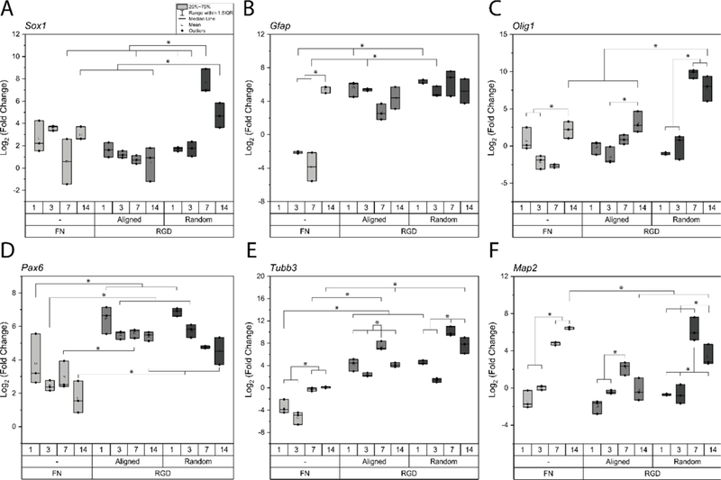
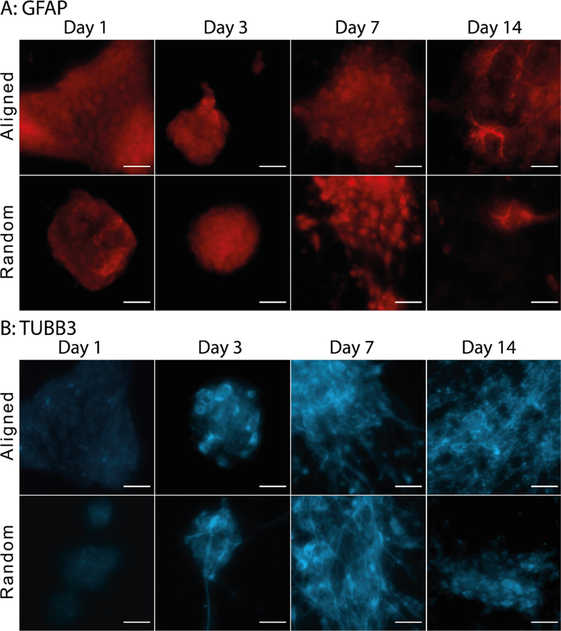
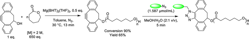
Stem cell differentiation toward a specific lineage is controlled by its microenvironment. Polymer scaffolds have long been investigated to provide microenvironment cues; however, synthetic polymers lack the specific signaling motifs necessary to direct cellular responses on their own. In this study, we fabricated random and aligned poly(ε-caprolactone) nanofiber substrates, surface-functionalized with RGD viastrain-promoted azide-alkyne cycloaddition, that were used to investigate the role of a covalently tethered bioactive peptide (RGD) and nanofiber orientation on neural differentiation of mouse embryonic stem cells. Gene and protein expression showed neural differentiation progression over 14 days, with similar expression on RGD random and aligned nanofibers for neurons and glia over time. The high levels of glial fibrillary acidic protein expression at early time points were indicative of neural progenitors, and occurred earlier than on controls or in previous reports. These results highlight the influence of RGD binding versus topography in differentiation.


Figures

Figure 1:
Figure 1:
(A) Analysis by DMF size exclusion chromatography confirms successful synthesis of high molecular mass DIBO-terminated poly(ε-caprolactone) (Mn = 60,600 Da, Mw = 83,700 Da, Đm = 1.38). Molecular mass was determined against PS standards. (B, D) Nanofibers were fabricated by electrospinning from a solution of DIBO-terminated poly(e-caprolactone) in HFIP (17% w/v) and a voltage of 15 kV. Cover glasses were placed on aluminum foil or in the gaps of metal collector plate for collecting of random or highly aligned nanofibers. (C, E) Analysis of SEM images was performed to estimate topography of nanofibers. NIH ImageJ was used to estimate fiber diameter (ᴓ = 212 ± 63 nm for aligned and 219 ± 36 nm for random fibers) and alignment (Directionality™ plugin, average angle = 0 ± 6° for aligned and −2 ± 111° random nanofiber scaffolds). (F) Post-electrospinning modification with GRGDS or GRGES peptides via strain-promoted azidealkyne cycloaddition. The concentration of GRGDS or GRGES peptides was measured using UV–visible spectrophotometry by comparison of absorbance at 306 nm (peak corresponding to DIBO groups) before (black curve) and after (green curve) post-electrospinning modification.
Figure 2:
Figure 2:
Summary of neural precursor (A), glial (B,C), and neuronal (D-F) gene expression over 14 days of neural differentiation of mouse embryonic stem cells, on fibronectin coated surfaces and RGD functionalized aligned and random nanofibers. Gene expression is represented as log2 (fold change). Statistically significances are highlighted between groups with p<0.05 considered significant.
Figure 3:
Figure 3:
Protein quantification of glial (A, C) and neuronal (B, D) proteins. Cells were considered positive for the respective protein if they possessed the appropriate protein morphology. Prior to morphology assessment, the images were thresholded according to the brightness and contrast settings of control images, which were samples stained with secondary antibodies and nuclei stain only. Protein positive cells were normalized to the total number of cells expressing at least one protein label, and expressed as a percentage. Data is represented as average of single and double labeled protein ± std dev of total labeled proteins. Double labeling of GFAP is noted with SOX1 (days 1 and 3) and OLIG1 (days 7 and 14); TUBB3 with SOX1; OLIG1 with GFAP; and MAP2 with GAP43. * represents statistical difference between respective topography timepoints and groups, with a p<0.05 considered significant.
Figure 4:
Figure 4:
Glial (A) and neuronal (B) protein expression of mESCs for 14 days of neural differentiation. Images have been adjusted to control thresholds to highlight cells expressing positive markers, and have been enhanced for display. At early time points, cells expressed GFAP, however more distinct glial morphology was seen at later time points. Similarly, neuronal expression was also found at early time points, however more distinct neurites were found at later time points of neural differentiation. Scale bar of 20 μm.
Figure 5:
Figure 5:
Neurite extension tracings. TUBB3 from day 7 on (A) aligned and (B) random nanofibers were used for (B, E) neurite tracings and (C, F) directionality measurements. (B) The aligned traces were rotated to orient the aligned fibers (from phase image) at 0°, and directionality of these neurite traces was measured. Gaussian fit (the red curve) was applied to measure neurite orientation. Scale bar of 50 μm.
Scheme 1:
Scheme 1:
DIBO-end-functionalized poly(ε-caprolactone) was synthesized via ring-opening polymerization of ε-caprolactone using DIBO as an initiator and Mg(BHT)2(THF)2 as a catalyst. Surface of DIBO-PCL was modified post-electrospinning with GRGDS or GRGES peptides via strain-promoted azide-alkyne cycloaddition.

References

    1. Song B-W , In Vivo Assessment of Stem Cells for Treating Neurodegenerative Disease: Current Approaches and Future Prospects. Stem Cells Int. 2017, 2017, 5. - PMC - PubMed
    1. Lunn JS; Sakowski SA; Hur J; Feldman EL, Stem cell technology for neurodegenerative diseases. Annals of neurology 2011, 70 (3), 353–361. - PMC - PubMed
    1. Chen R; Hunt JA, Biomimetic materials processing for tissue-engineering processes. J. Mater. Chem. 2007, 17 (38).
    1. Sperling LE; Reis KP; Pozzobon LG; Girardi CS; Pranke P, Influence of random and oriented electrospun fibrous poly(lactic-co-glycolic acid) scaffolds on neural differentiation of mouse embryonic stem cells. J. Biomed. Mater. Res. A 2017, 105 (5), 1333–1345. - PubMed
    1. Sharifi F; Patel BB; Dzuilko AK; Montazami R; Sakaguchi DS; Hashemi N, Polycaprolactone Microfibrous Scaffolds to Navigate Neural Stem Cells. Biomacromolecules 2016, 17 (10), 3287–3297. - PubMed

Publication types

Substances