. 2019 Oct;25(19-20):1426-1437.
doi: 10.1089/ten.TEA.2018.0258. Epub 2019 Aug 20.

Temporal Impact of Substrate Anisotropy on Differentiating Cardiomyocyte Alignment and Functionality

Affiliations

Temporal Impact of Substrate Anisotropy on Differentiating Cardiomyocyte Alignment and Functionality

Alicia C B Allen et al. Tissue Eng Part A. 2019 Oct.

Abstract

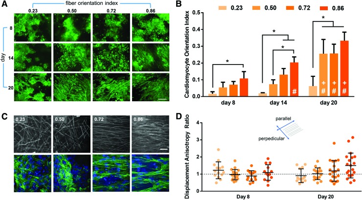
Anisotropic biomaterials can affect cell function by driving cell alignment, which is critical for cardiac engineered tissues. Recent work, however, has shown that pluripotent stem cell-derived cardiomyocytes may self-align over long periods of time. To determine how the degree of biomaterial substrate anisotropy impacts differentiating cardiomyocyte structure and function, we differentiated mouse embryonic stem cells to cardiomyocytes on nonaligned, semialigned, and aligned fibrous substrates and evaluated cell alignment, contractile displacement, and calcium transient synchronicity over time. Although cardiomyocyte gene expression was not affected by fiber alignment, we observed gradient- and threshold-based differences in cardiomyocyte alignment and function. Cardiomyocyte alignment increased with the degree of fiber alignment in a gradient-based manner at early time points and in a threshold-based manner at later time points. Calcium transient synchronization tightly followed cardiomyocyte alignment behavior, allowing highly anisotropic biomaterials to drive calcium transient synchronization within 8 days, while such synchronized cardiomyocyte behavior required 20 days of culture on nonaligned biomaterials. In contrast, cardiomyocyte contractile displacement had no directional preference on day 8 yet became anisotropic in the direction of fiber alignment on aligned fibers by day 20. Biomaterial anisotropy impact on differentiating cardiomyocyte structure and function is temporally dependent. Impact Statement This work demonstrates that biomaterial anisotropy can quickly drive desired pluripotent stem cell-derived cardiomyocyte structure and function. Such an understanding of matrix anisotropy's time-dependent influence on stem cell-derived cardiomyocyte function will have future applications in the development of cardiac cell therapies and in vitro cardiac tissues for drug testing. Furthermore, our work has broader implications concerning biomaterial anisotropy effects on other cell types in which function relies on alignment, such as myocytes and neurons.

Keywords: anisotropy; biomaterials; cardiac differentiation; tissue engineering.


Conflict of interest statement

No competing financial interests exist.

Figures

<b>FIG. 1.</b>
FIG. 1.
Nonaligned, semialigned, and aligned fibers are fabricated by electrospinning onto a rotating collector, with collector rotational speed modulating fiber alignment. (A) Schematic of the electrospinning process. A polymer solution jet is ejected from the charged needle and is deposited as fibers on a rotating grounded collector. (B) SEM images of electrospun fibers showing fiber alignment. Collector rotational speed (RPM) is indicated in top left corner; fiber orientation index is indicated in the top right corner. Scale bar is 10 μm. (C) Electrospun fiber orientation index was calculated to determine the degree of fiber alignment at each collector speed. (D) Electrospun fiber mean diameter was measured for each collector speed. *p < 0.05. RPM, revolutions per minute; SEM, scanning electron microscope. Color images are available online.
<b>FIG. 2.</b>
FIG. 2.
mESCs on electrospun fibers differentiate to cardiomyocytes. (A) Schematic showing the seeding and differentiation timeline. mESC differentiation on fibers was tracked by measuring changes in fluorescence intensity of Nkx2.5 (B) and α-MHC (C) reporter cell lines from days 7 to 20. (D) Effect of fiber alignment on mRNA expression of cardiac progenitor markers Mesp1 and ISL1 on day 8. *p < 0.05 compared to control. (E) Effect of fiber alignment and day of differentiation on mRNA expression of cardiac marker Myh6. *p < 0.05 compared to control; #p < 0.05 compared to day 8. (F) Representative images of cells differentiated for 14 days on fibers. Immunostaining for cardiac marker cTnT confirms presence of cardiomyocytes (scale bar = 100 μm); immunostaining for VIM, SMA, and CD31 shows that fibroblasts are present but smooth muscle cells and endothelial cells are not (scale bar = 200 μm). (G) High-magnification images of cardiomyocytes on aligned fibers stained for α-actinin, which is localized to the Z-disk, show that sarcomeres of neighboring cells are in register. Scale bar is 25 μm. bFGF, basic fibroblast growth factor; BMP, bone morphogenic protein; cTnT, cardiac troponin T; DAPI, 4’,6-Diamidine-2’-phenylindole dihydrochloride; FGF10, fibroblast growth factor 10; LIF, leukemia inhibition factor; mESCs, mouse embryonic stem cells; MHC, myosin heavy chain; mRNA, messenger RNA; n.s., not statistically significant; SMA, smooth muscle actin; VEGF, vascular endothelial growth factor; VIM, vimentin. Color images are available online.
<b>FIG. 3.</b>
FIG. 3.
mESC-derived cardiomyocyte alignment depends on degree of fiber alignment. (A) cTnT+ cardiomyocytes on nonaligned, semialigned, and aligned fibers at days 8, 14, and 20. Scale bar is 200 μm. (B) Cardiomyocyte orientation index was determined to compare the degree of cardiomyocyte alignment over time and versus fiber orientation index. *p < 0.05; #p < 0.05 versus day 8; +p < 0.05 versus day 14. (C) High-magnification images of cTnT+ cardiomyocytes (bottom) and underlying fibers (top) show that cardiomyocytes align in the direction of fiber alignment. Scale bar = 20 μm. (D) Effect of fiber alignment on cardiomyocyte contractile displacement parallel and perpendicular to fiber direction on days 8 and 20. The dashed line indicates a displacement anisotropy ratio of 1, when displacement is equal in the parallel and perpendicular directions. Color images are available online.
<b>FIG. 4.</b>
FIG. 4.
Cardiomyocyte intracellular calcium transients were tracked by staining with Fluo-4, AM, an intracellular calcium-sensitive fluorophore. Representative Fluo-4 signals at day 8 (A) and day 20 (B) for cardiomyocytes derived on nonaligned (0.23 fibers, top) and aligned (0.86 fibers, bottom). Scale bars = 50 μm. (C) Beating rate and (D) synchronicity (as measured by TPA-MAD) at days 8 and 20 for cardiomyocytes derived on nonaligned and aligned fibers. *p < 0.05. TPA-MAD, time of peak arrival median absolute deviation. Color images are available online.

References

    1. Benjamin E.J., Virani S.S., Callaway C.W., et al. . Heart disease and stroke statistics-2018 update: a report from the American Heart Association. Circulation 137, e67, 2018 - PubMed
    1. Bergmann O., Bhardwaj R.D., Bernard S., et al. . Evidence for cardiomyocyte renewal in humans. Science 324, 98, 2009 - PMC - PubMed
    1. Thygesen K., Alpert J.S., Jaffe A.S., Simoons M.L., Chaitman B.R., and White H.D. Third universal definition of myocardial infarction. Circulation 126, 2020, 2012 - PubMed
    1. Fan D., Takawale A., Lee J., and Kassiri Z. Cardiac fibroblasts, fibrosis and extracellular matrix remodeling in heart disease. Fibrogenesis Tissue Repair 5, 15, 2012 - PMC - PubMed
    1. Takahashi K., and Yamanaka S. Induction of pluripotent stem cells from mouse embryonic and adult fibroblast cultures by defined factors. Cell 126, 663, 2006 - PubMed

Publication types

LinkOut - more resources