. 2019 Feb 6;38(1):55.
doi: 10.1186/s13046-019-1074-6.

MicroRNA-7 as a potential therapeutic target for aberrant NF-κB-driven distant metastasis of gastric cancer

Affiliations

MicroRNA-7 as a potential therapeutic target for aberrant NF-κB-driven distant metastasis of gastric cancer

Tingbo Ye et al. J Exp Clin Cancer Res. .

Abstract

Background: Dysregulated miR-7 and aberrant NF-κB activation were reported in various human cancers. However, the expression profile, clinical relevance and dysregulated mechanism of miR-7 and NF-κB RelA/p65 in human gastric cancers (GC) metastasis remain largely unknown. This study is to investigate the expression profile, clinical relevance and dysregulated mechanism of miR-7 and NF-κB RelA/p65 in GC and to explore the potential therapeutic effect of miR-7 to GC distant metastasis.

Methods: TCGA STAD and NCBI GEO database were used to investigate the expression profile of miR-7 and NF-κB RelA/p65 and clinical relevance. Lentivirus-mediated gene delivery was applied to explore the therapeutic effect of miR-7 in GC. Real-time PCR, FACS, IHC, IF, reporter gene assay, IP, pre-miRNA-7 processing and binding assays were performed.

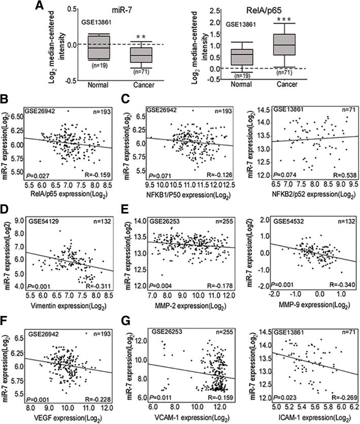
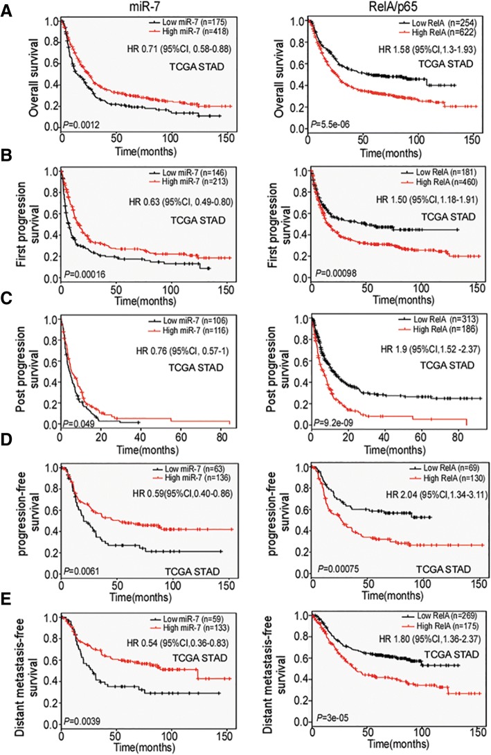
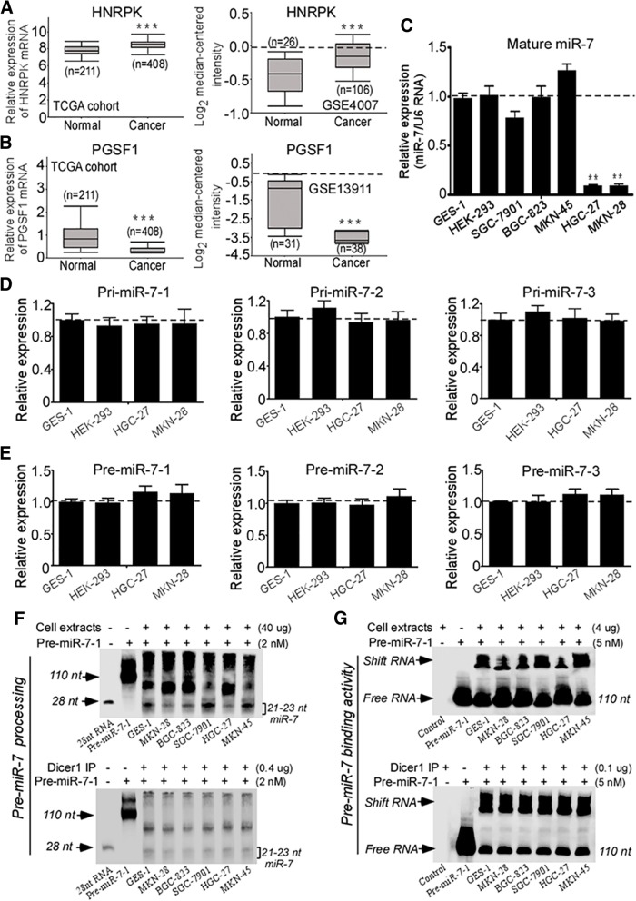
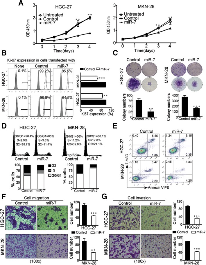
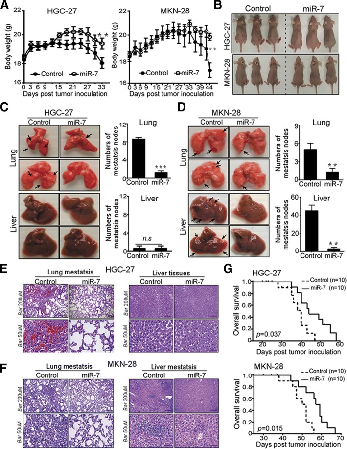
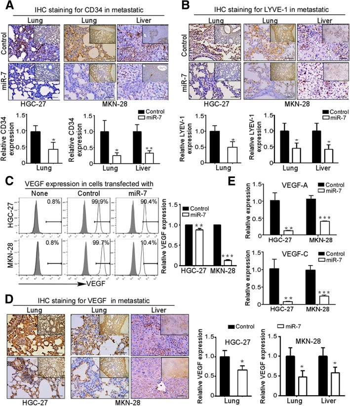
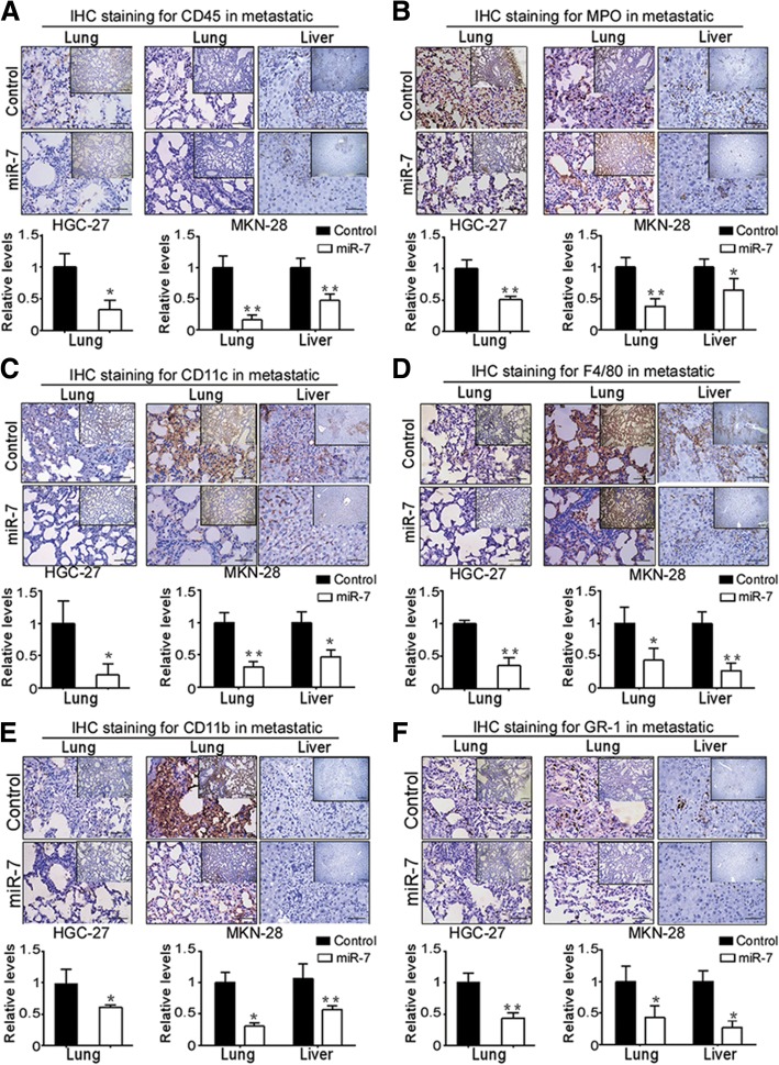
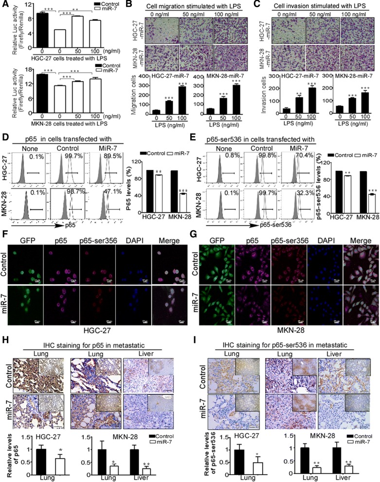
Results: Low miR-7 correlated with high RelA/p65 in GC with a clinical relevance that low miR-7 and high RelA/p65 as prognostic indicators of poor survival outcome of GC patients. Moreover, an impaired pre-miR-7 processing caused by dysregulated Dicer1 expression is associated with downregulated miR-7 in GC cells. Functionally, delivery of miR-7 displays therapeutic effects to GC lung and liver metastasis by alleviating hemangiogenesis, lymphangiogenesis as well as inflammation cells infiltration. Mechanistically, miR-7 suppresses NF-κB transcriptional activity and its downstream metastasis-related molecules Vimentin, ICAM-1, VCAM-1, MMP-2, MMP-9 and VEGF by reducing p65 and p-p65-ser536 expression. Pharmacologic prevention of NF-κB activator LPS obviously restored miR-7-suppressed NF-κB transcriptional activation and significantly reverted miR-7-inhibited cell migration and invasion.

Conclusions: Our data suggest loss of miR-7 in GC promotes p65-mediated aberrant NF-κB activation, facilitating GC metastasis and ultimately resulting in the worse clinical outcome. Thus, miR-7 may act as novel prognostic biomarker and potential therapeutic target for aberrant NF-κB-driven GC distant metastasis.

Keywords: Clinical outcome; Distant metastasis; Gastric Cancer; MiR-7; NF-κB.


Conflict of interest statement

Ethics approval and consent to participate

All animal procedures were approved by the Institutional Animal Care Committee of Chongqing medical University (Chongqing, China).

Consent for publication

None.

Competing interests

The authors declare that they have no competing interests.

Publisher’s Note

Springer Nature remains neutral with regard to jurisdictional claims in published maps and institutional affiliations.

Figures

Fig. 1
Fig. 1
Downregulated miR-7 is correlated with high RelA/p65 in GC. a miR-7 is downregulated whereas RelA/p65 mRNA is increased in GC. Expression of miR-7 and RelA/p65 mRNA was analyzed using the same human GC cohort from the NCBI/GEO/GSE13861 (Normal: normal gastric mucosa; Cancer: gastric cancer). The box plot indicated the log2 transformed mRNA median gene expression level in the tissues. b Correlation of miR-7 with RelA/p65 mRNA expression (n = 193, r = − 0.159, P = 0.027) in GC from NCBI/GEO/GSE26942. c Expression correlation between miR-7 and NFKB1/p50 (n = 193, r = − 0.126, P = 0.071, left panel), NFKB2/p52 mRNA expression (n = 71, r = 0.538, P = 0.074; right panel) in GC from NCBI/GEO /GSE26942 and GSE13861. d Expression correlation between miR-7 and Vimentin (n = 132, r = − 0.311, P = 0.001) mRNA expression in GC from NCBI/GEO/ GSE54129. e-g Expression correlation between miR-7 and MMP-2 (n = 255, r = − 0178, P = 0.001; e, left panel), MMP-9 (n = 132, r = − 0.340, P = 0.001; e, right panel), VEGF (n = 193,r = − 0.228, P = 0.001; f, VCAM-1 (n = 255, left panel, r = − 0.159, P = 0.011; g, left panel) and ICAM-1(n = 71,r = − 0.263, P = 0.023; g right panel,) mRNA expression respectively according to NCBI GEO database(GSE26253, GSE54532, GSE26942, GSE13861). The expression correlation was determined with Pearson’s correlation analysis. ** P < 0.01; *** P < 0.001
Fig. 2
Fig. 2
Low miR-7 and high RelA/p65 expression are correlated with poor prognosis of GC patients. The correlations between miR-7, RelA/p65 and multiply survival rates were determined by Kaplan-Meier analysis from TCGA STAD database. a miR-7 downregulation is related to shorter overall survival (OS) (n = 593, P = 0.0012, left panel). High RelA/p65 indicated poor OS (n = 876, P = 5.5e-06, right panel). b Low miR-7 indicated shorter fist-progression survival (FPS) (n = 359, P = 0.00016, left panel). High RelA/p65 indicated worse FPS (n = 641, P = 0.00098, right panel) according to TCGA cohort. c Low miR-7 indicated poor post-progression survival (PPS) (n = 222, P = 0.049, left panel). High RelA/p65 indicated shorter PPS (n = 499, P = 9.2e-09, right panel). d Low miR-7 is correlated with shorter progression-free survival (PFS) (n = 222, P = 0.049, left panel). High RelA/p65 indicated poor PFS (n = 499, P = 9.2e-09, right panel). e Low miR-7 is correlated with worse distant metastasis-free survival (DMFS) (n = 192, P = 0.0039, left panel). High RelA/p65 indicated shorter DMFS (n = 444, P = 3e-05, right panel). P values were determined by log-rank test
Fig. 3
Fig. 3
Impaired pre-miR-7 processing is responsible for downregulated miR-7 in GC cells. a Upregulated HNRPK (miR-7-1 host gene) mRNA expression in TCGA STAD (n = 619, P < 0.001) and in the NCBI/GEO/GSE4007 database l (n = 132, P < 0.001). b Downregulated PGSF1 (miR-7-3 host gene) mRNA expression of GC in TCGA STAD (n = 619, P < 0.001) and in the NCBI/GEO/GSE13911 database (n = 69, P < 0.001). c Expression of mature miR-7 in five GC cell lines relative to GES-1 and HEK-293 cells. Total RNA was extracted from indicated cells. Expression of mature miR-7 was detected by real-time PCR. U6 RNA was used as an internal control. d-e Expression of pri-miR-7 and pre-miR-7 isoforms in GC cells. Pri-miR-7 isoforms (pri-miR-7-1,2,3) (d) and pre-miR-7 isoforms (pre-miR-7-1,2,3) (e) were quantitated by modified real-time PCR in HGC-27 and MKN-28 cells compared with GES-1 and HEK-293 cells. Human 18S rRNA was used as an internal control. f Pre-miR-7-1 processing assay. Pre-miR-7-1 RNA (2 nM) was incubated in processing buffer with cell extracts (Uper panel) or Dicer1 containing IP complex (Lower panel) respectively. The 110 nt pre-miR-7-1 and mature ~ 21-23 nt miR-7 were detected by RNA gel blot after processing action. g Pre-miR-7-1 binding assay. Pre-miR-7-1 RNA (5 nM) was incubated in binding buffer with cell extracts (Uper panel) or Dicer1 containing IP complex (Lower panel) respectively. RNA gel blot was used to detect shifted binding pre-miR-7-1 RNA and free pre-miR-7-1 RNA (110 nt) after binding action. Data are presented as mean ± SD of three independent experiments. * P < 0.05; ** P < 0.01 between the indicated two groups determined by paired student’s t test
Fig. 4
Fig. 4
Restoration of miR-7 inhibits GC cell viability and invasiveness in vitro. HGC-27 and MKN-28 cells were stably transfected with mature miR-7 or negative control, respectively. a and b Restoration of miR-7 expression decreased Cell viability and inhibited Ki67 expression in GC cells. Cell viability (a) was determined by CCK-8 assay at absorbance 450 nm for 5 consecutive days. Ki67 protein expression in indicated cells (b) were measured by Flow cytometry using specific Ki67 antibodies. Representative images are shown. c Restoration of miR-7 expression suppressed Colony formation in GC cells. Colony formation assay was performed to examine colony formation ability in indicated cells. Colony numbers were counted for 3 weeks after transfection. Representative images of colonies formed are shown. Bar, 100 μm. d and e miR-7 expression promoted significant G0/G1 phase arrest but not cell apoptosis. Cell cycle were measured by flow cytometry analysis after PI staining in indicated cells (d). Cell apoptosis were detected by FACS analysis using Annexin V-PE/7-AAD double staining (e). Representative images of colonies formed and FACS analysis are shown. Data are presented as mean ± SD of three independent experiments. f and g Restoration of miR-7 inhibited GC cell migration and invasion. Cell migration assay (f) and cell invasion assay (g) were performed in 24-well non-coated or Matrigel-coated Transwell Chambers (8-μm pore size). Cell numbers were counted in 5–10 random fields. Data are presented as mean ± SD of three independent experiments. Representative images are shown. ** p < 0.01 and *** p < 0.001 between the indicated two groups determined by paired student’s t test
Fig. 5
Fig. 5
miR-7 inhibited GC distant metastasis and improved overall survival in vivo. HGC-27 and MKN-28 cells were stalely transfected with indicated Lentivirus (MiR-7 and Control). After sorting purification, cells (2 × 106 /mice in 100 μl PBS buffer) were injected tail intravenously into the nu/nu mice (n = 8 mice/group). a and b Restoration of miR-7 in GC cells inhibited body weight loss in experimental metastasis mice. Post-transfection, body weight and the status of nude mice were monitored every 3 days and presented as mean ± SD (a). Representative images of the mice sizes are shown (b). c-f miR-7 markedly suppressed the lung and liver metastasis of GC cells. After indicated time point, lungs and livers from experimental mice were collected and metastatic tumor of the lung and liver surfaces were counted under a dissecting microscope (c and d) and were confirmed by H&E staining on sections from embedded lung and liver tissues (E and F). metastatic tumor was indicated by black arrow. Representative images of metastatic lung and liver tissues and H&E staining are shown. Scale bars indicate 50 μm and 200 μm. g miR-7 markedly improved overall survival (OS) in experimental metastasis mice. Kaplan-Meier analysis was used to analyze OS in established experimental metastasis mice (n = 10mice/group). Log-rank test was used for Kaplan-Meier survival analyses. Data are presented as mean ± SD. *p < 0.05, **p < 0.01 and ***p < 0.001 between the indicated two groups determined by paired student’s t test
Fig. 6
Fig. 6
VEGF driven-hemangiogenesis and lymphangiogenesis are involved in anti-metastasis activation of miR-7 in GC. a and b miR-7 expression inhibited hemangiogenesis and lymphangiogenesis in vivo. Experimental metastasis mice from HGC-27 and MKN-28 cells were established and metastatic lung and liver tissues were collected as described in Materials & Methods. Hemangiogenesis was evaluated in metastatic lung and liver by IHC staining for CD34 hemendothelial marker (a). Lymphangiogenesis was evaluated by detecting Lymphatic vessels density through IHC staining for LYVE-1 (b). Representative IHC images are shown. Data are presented as mean ± SD. c-d miR-7 alleviated VEGF expression in vitro and in vivo. VEGF expression in GC cells were detected by flow cytometry (c). IHC staining was used to evaluate VEGF expression in metastatic lung and liver tissues (d). Representative IHC images are shown. e miR-7 alleviated VEGF-A and VEGF-C expression. VEGF-A and VEGF-C mRNA were detected by Real-Time PCR. Data are presented as mean ± SD. *p < 0.05, **p < 0.01, ***p < 0.001 between the indicated two groups determined by paired student’s t test. Scale bars: (main) 50 μm; (inset) 200 μm
Fig. 7
Fig. 7
miR-7 alleviates inflammation cells infiltration in GC metastasis. Experimental metastasis mice were established as described in Materials & Methods. Inflammation cells was evaluated in embedded metastatic lung and liver tissues by IHC staining using specific antibodies including CD45 (a), MPO (b), CD11c (c), F4/80 (d), CD11b (e) and GR-1 (f). Positive staining cells were counted from 5 high magnification fields (× 400) in metastatic tissues. Relative expression was normalized to 1 compared with control group. Representative IHC images are shown. *p < 0.05, **p < 0.01 and ***p < 0.001 between the indicated two groups determined by paired student’s t test. Scale bars: (main) 50 μm; (inset) 200 μm
Fig. 8
Fig. 8
miR-7 suppresses NF-κB transcriptional activity by reducing p65 and active NF-κB/p65 expression. a miR-7 suppressed NF-κB transcriptional activity while NF-κB activator LPS reversed this activity. NF-κB transcriptional activity was detected by dual-luciferase reporter assay. NF-κB activator LPS was used in indicated groups. The data were presented as fold inductions of the ratio was normalized to Renilla luciferase activity. b-c LPS treatment reverted the abilities of cell migration and cell invasion after miR-7 transfection. Indicated cells were treated with or without 50 ng/ul or 100 ng/ul LPS and then cell migration and cell invasion were analyzed in miR-7-transfected HGC-27 (b) and MKN-28 cells (c). Data are presented as mean ± SD of three independent experiments. Representative images are shown. d-e Restoration of miR-7 decreased total p65 and active p-p65(Ser536) levels in GC cells. p65 and p-p65(Ser536) proteins were detected by FACS analysis in miR-7-transfected HGC-27 (d) and MKN-28 cells (e). Representative images are shown. f-g Cellular distribution of p65 and p-p65(Ser536) in miR-7-transfected GC cells. Immunofluorescence (IF) staining was used to detect cellular distribution of p65 and p-p65(Ser536) in indicated GC cells (GFP). AF555 (Red) or AF647 (Pink)-conjugated secondary antibodies were used to detect primary antibodies. Nuclei were counterstained with DAPI (Blue). Images were captured using a confocal microscope (Scale bars: 25 μm). h-i Restoration of miR-7 reduced p65 and p-p65(Ser536) expression in metastatic tissues. p65 and p-p65(Ser536) were detected by IHC staining in metastatic lung and liver tissues. Representative IHC images are shown. Scale bars: (main) 50 μm; (inset) 200 μm. *p < 0.05, **p < 0.01, ***p < 0.001 between the indicated two groups determined by paired student’s t test

Similar articles

Cited by

References

    1. Jemal A, Bray F, Center MM, Ferlay J, Ward E, Forman D. Global cancer statistics. CA Cancer J Clin. 2011;61:69–90. doi: 10.3322/caac.20107. - DOI - PubMed
    1. N. Cancer Genome Atlas Research Comprehensive molecular characterization of gastric adenocarcinoma. Nature. 2014;513:202–209. doi: 10.1038/nature13480. - DOI - PMC - PubMed
    1. Bartel DP. MicroRNAs: genomics, biogenesis, mechanism. and function Cell. 2004;116:281–297. - PubMed
    1. Yin Y, Li J, Chen S, Zhou T, Si J. MicroRNAs as diagnostic biomarkers in gastric cancer. Int J Mol Sci. 2012;13:12544–12555. doi: 10.3390/ijms131012544. - DOI - PMC - PubMed
    1. Li Z, Cao Y, Jie Z, Liu Y, Li Y, Li J, et al. miR-495 and miR-551a inhibit the migration and invasion of human gastric cancer cells by directly interacting with PRL-3. Cancer Lett. 2012;323:41–47. doi: 10.1016/j.canlet.2012.03.029. - DOI - PubMed

MeSH terms