Review
. 2019 Feb 6;28(151):180065.
doi: 10.1183/16000617.0065-2018. Print 2019 Mar 31.

Tumoral pulmonary hypertension

Affiliations
Review

Tumoral pulmonary hypertension

Laura C Price et al. Eur Respir Rev. .

Abstract

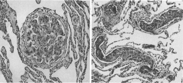
Tumoral pulmonary hypertension (PH) comprises a variety of subtypes in patients with a current or previous malignancy. Tumoral PH principally includes the tumour-related pulmonary microvascular conditions pulmonary tumour microembolism and pulmonary tumour thrombotic microangiopathy. These inter-related conditions are frequently found in post mortem specimens but are notoriously difficult to diagnose ante mortem The outlook for patients remains extremely poor although there is some emerging evidence that pulmonary vasodilators and anti-inflammatory approaches may improve survival. Tumoral PH also includes pulmonary macroembolism and tumours that involve the proximal pulmonary vasculature, such as angiosarcoma; both may mimic pulmonary embolism and chronic thromboembolic PH. Finally, tumoral PH may develop in response to treatments of an underlying malignancy. There is increasing interest in pulmonary arterial hypertension induced by tyrosine kinase inhibitors, such as dasatanib. In addition, radiotherapy and chemotherapeutic agents such as mitomycin-C can cause pulmonary veno-occlusive disease. Tumoral PH should be considered in any patient presenting with unexplained PH, especially if it is poorly responsive to standard approaches or there is a history of malignancy. This article will describe subtypes of tumoral PH, their pathophysiology, investigation and management options in turn.


Conflict of interest statement

Conflict of interest: L.C. Price reports personal fees from Actelion/Johnson and Johnson and has received an educational grant from GSK Pharmaceuticals in the last 2 years. Conflict of interest: M.J. Seckl has nothing to disclose. Conflict of interest: P. Dorfmüller reports personal fees from MSD, Actelion and Roche, outside the submitted work. Conflict of interest: S.J. Wort reports grants and personal fees from Actelion and Bayer, and personal fees from GSK and MSD, outside the submitted work.

Figures

FIGURE 1
FIGURE 1
Neoplastic emboli. Photomicrographs of neoplastic pulmonary emboli. a) Occlusion of a small muscular pulmonary artery by tumour cells from a 55-year-old woman with breast carcinoma. b) Obstruction of multiple pulmonary arterioles by tumour cells from a 36-year-old woman with cervical squamous-cell carcinoma. (Haematoxylin and eosin stain ×120). Reproduced from [4].
FIGURE 2
FIGURE 2
Lung histology from a case of pulmonary tumour thrombotic microangiopathy related to severe pulmonary hypertension. a) Post mortem section showing occlusion of a medium-sized pulmonary arterial lumen by fibrointimal proliferation of fibroblasts and collagen (white arrow) and tumour emboli (black arrow) (Haematoxylin and eosin stain ×4.3). Scale bar=500 μm. b) Post mortem section showing a medium-sized pulmonary artery with two elastic layers, with a normal-sized smooth muscle layer. There is exaggerated luminal occlusion by fibrointimal thickening (white arrow) surrounding nests of tumour emboli (black arrow). The adventitia contains lymphatic tumoral thrombi. Increased alveolar macrophages are seen surrounding the lung (Elastica van Gieson stain ×4.6). Scale bar=500 μm. c) Evidence for fibrointimal proliferation within the lumen of small pulmonary veins (black arrow) and tumour involvement of accompanying lymphatics (white arrow). Inset: veins close to the centrilobular bronchovascular bundles show eccentric fibrointimal remodelling (Elastica van Gieson ×28.4). Scale bar=80 μm. Reproduced from [26].
FIGURE 3
FIGURE 3
Proposed mechanisms for fibrointimal proliferation in pulmonary tumour thrombotic microangiopathy (PTTM). Small nests of carcinomatous cells lodge in the pulmonary vessels, including small pre-capillary arteries (via haematogenous spread) and on the post-capillary side of the pulmonary circulation to pulmonary veins and lymphatics (by lymphatic invasion). Tumour cell and endothelial cell interaction initiates clot formation, and releases further cytokines including vascular endothelial growth factor (VEGF) and platelet-derived growth factor (PDGF). This initiates macrophage recruitment and intimal (endothelial cell and non-endothelial cell like, i.e. myofibroblastic) proliferation. Tissue factor also upregulates VEGF expression on tumour cells, which is angiogenic to intimal cells. PDGF-A and -B are expressed on tumour cells, and anti-phospho-PDGFR-α is expressed on vascular endothelial cell and gastric carcinoma cells. This indicates that PDGF signalling activation of tumour cell growth is present through both autocrine and paracrine mechanisms. The cytokine and adhesive protein osteopontin is expressed on tumour cells in PTTM, and is likely to be a key driver for intimal cell growth. Perivascular CD68-positive macrophages are noted and also reside within intimal layers. Macrophages also stain for CD44, the adhesion molecule which interacts with osteopontin to induce chemotaxis of T-cells and macrophages, propagation of local inflammation and intimal proliferation (through other known macrophage-derived pro-proliferative factors including interleukin (IL)-6). Direct contact with tumour nests is not universal in all vessels where remodelling is present.
FIGURE 4
FIGURE 4
Radiology in pulmonary tumour thrombotic microangiopathy (PTTM). a) Ventilation/perfusion demonstrating sub-segmental defects in lung perfusion in a patient with PTTM. Computed tomography pulmonary angiography did not demonstrate these peripheral lesions (not shown). b) High-resolution computed tomography scanning in PTTM showing widespread ground-glass opacification, micronodules, interlobular septal thickening and small bilateral pleural effusions.
FIGURE 5
FIGURE 5
High-resolution computed tomography (HRCT) of the chest and lung histology (haematoxylin and eosin–saffron staining) from explanted lungs. a) HRCT at diagnosis showed septal lines and centrilobular ground-glass opacities suggestive of pulmonary veno-occlusive disease. b) Improvement of radiological abnormalities after 2 weeks of treatment including diuretics, dobutamine, endothelin receptor antagonist and high-dose corticosteroids. c) Overview of lung parenchyma displaying emphysematous changes and vasculopathy. Note the pulmonary vessels with narrowed lumina (arrows). d) Septal vein with pronounced intimal fibrosis and heavily narrowed lumen. e) Focal capillary congestion in the vicinity of a remodelled vein (bottom). f) Pulmonary artery (centre) with adjacent airway (top left) displaying medial hypertrophy and concentric intimal fibrosis. c) Scale bar=1000 μm. d–f) Scale bar=200 μm. Reproduced from [92].

References

    1. Roberts KE, Hamele-Bena D, Saqi A, et al. . Pulmonary tumor embolism: a review of the literature. Am J Med 2003; 115: 228–232. - PubMed
    1. Kane RD, Hawkins HK, Miller JA, et al. . Microscopic pulmonary tumor emboli associated with dyspnea. Cancer 1975; 36: 1473–1482. - PubMed
    1. Bagshawe KD, Brooks WD. Subacute pulmonary hypertension due to chorionepithelioma. Lancet 1959; 1: 653–658. - PubMed
    1. Shields DJ, Edwards WD. Pulmonary hypertension attributable to neoplastic emboli: an autopsy study of 20 cases and a review of literature. Cardiovasc Pathol 1992; 1: 279–287. - PubMed
    1. Winterbauer RH, Elfenbein IB, Ball WC Jr. Incidence and clinical significance of tumor embolization to the lungs. Am J Med 1968; 45: 271–290. - PubMed

MeSH terms

Substances