. 2019 Apr 3;93(8):e00090-19.
doi: 10.1128/JVI.00090-19. Print 2019 Apr 15.

Differential Antibody-Based Immune Response against Isolated GP1 Receptor-Binding Domains from Lassa and Junín Viruses

Affiliations

Differential Antibody-Based Immune Response against Isolated GP1 Receptor-Binding Domains from Lassa and Junín Viruses

Aliza Borenstein-Katz et al. J Virol. .

Abstract

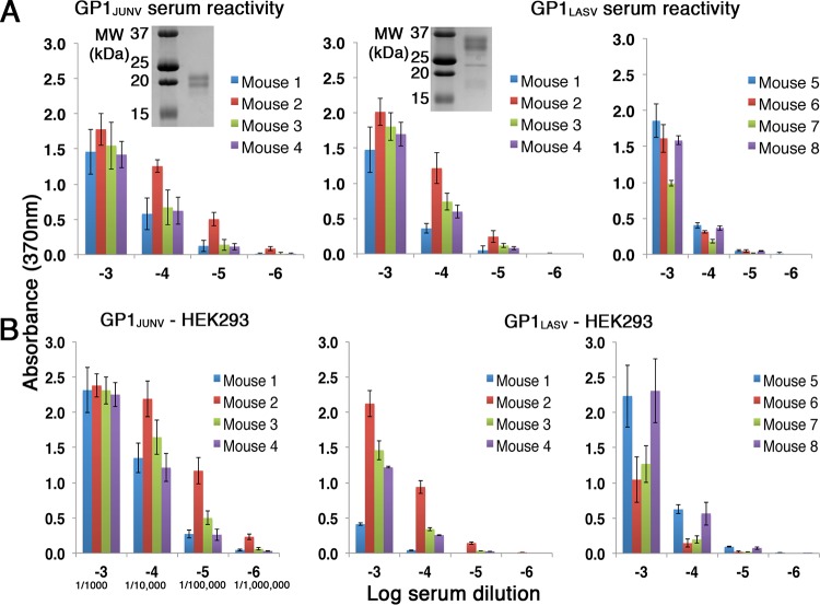
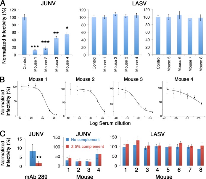
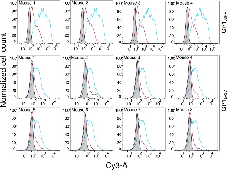
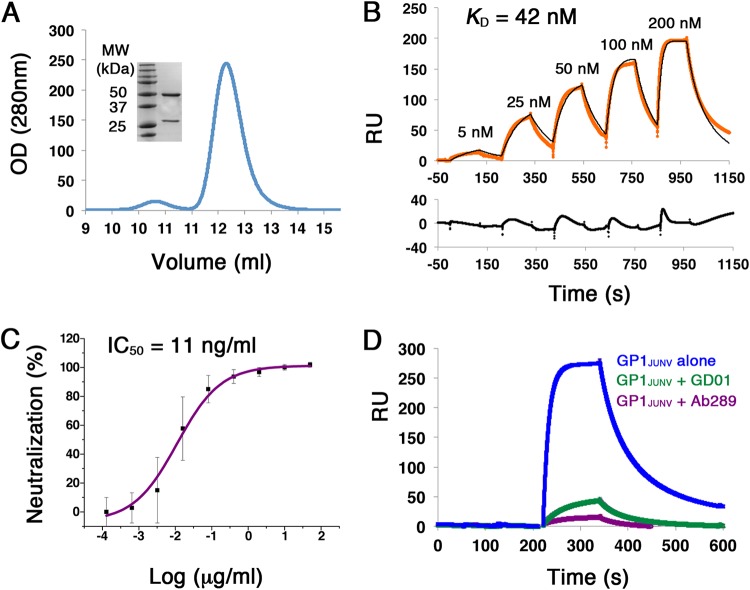
There are two predominant subgroups in the Arenaviridae family of viruses, the Old World and the New World viruses, that use distinct cellular receptors for entry. While New World viruses typically elicit good neutralizing antibody responses, the Old World viruses generally evade such responses. Antibody-based immune responses are directed against the glycoprotein spike complexes that decorate the viruses. A thick coat of glycans reduces the accessibility of antibodies to the surface of spike complexes from Old World viruses, but other mechanisms may further hamper the development of efficient humoral responses. Specifically, it was suggested that the GP1 receptor-binding module of the Old World Lassa virus might help with evasion of the humoral response. Here we investigated the immunogenicity of the GP1 domain from Lassa virus and compared it to that of the GP1 domain from the New World Junín virus. We found striking differences in the ability of antibodies that were developed against these immunogens to target the same GP1 receptor-binding domains in the context of the native spike complexes. Whereas GP1 from Junín virus elicited productive neutralizing responses, GP1 from Lassa virus elicited only nonproductive responses. These differences can be rationalized by the conformational changes that GP1 from Lassa virus but not GP1 from Junín virus undergoes after dissociating from the trimeric spike complex. Hence, shedding of GP1 in the case of Lassa virus can indeed serve as a mechanism to subvert the humoral immune response. Moreover, the realization that a recombinant protein may be used to elicit a productive response against the New World Junín virus may suggest a novel and safe way to design future vaccines.IMPORTANCE Some viruses that belong to the Arenaviridae family, like Lassa and Junín viruses, are notorious human pathogens, which may lead to fatal outcomes when they infect people. It is thus important to develop means to combat these viruses. For developing effective vaccines, it is vital to understand the basic mechanisms that these viruses utilize in order to evade or overcome host immune responses. It was previously noted that the GP1 receptor-binding domain from Lassa virus is shed and accumulates in the serum of infected individuals. This raised the possibility that Lassa virus GP1 may function as an immunological decoy. Here we demonstrate that mice develop nonproductive immune responses against GP1 from Lassa virus, which is in contrast to the effective neutralizing responses that GP1 from Junín virus elicits. Thus, GP1 from Lassa virus is indeed an immunological decoy and GP1 from Junín virus may serve as a constituent of a future vaccine.

Keywords: arenavirus; immune response; vaccines.


Figures

FIG 1
FIG 1
Both GP1JUNV and GP1LASV are immunogenic in mice. (A) ELISA using serial dilutions of sera from mice immunized with GP1JUNV and GP1LASV. Plates were coated with the same immunogen that was produced in insect cells and that was injected into mice. (Left and middle) The response of two groups of 4 mice each that were immunized with GP1JUNV and GP1LASV, respectively. Colors correspond to sera from different mice. (Right) Data for a second group of 4 mice that were immunized with GP1LASV. Bars represent average values from three independent experiments, each consisting of several technical replicates. Error bars represent standard deviations. (Insets) Analyses of the corresponding GP1JUNV and GP1LASV that were used for immunization using SDS-PAGE and Coomassie staining. MW, molecular weight. (B) ELISA using the indicated serum dilution series and plates coated with the GP1 domains produced in HEK293F cells. Bars represent average values from three independent experiments, each consisting of several technical replicates. Error bars represent standard deviations. For convenience, the serum dilutions used are further indicated using a serology standard format in the lower left plot.
FIG 2
FIG 2
GP1JUNV as opposed to GP1LASV elicits a neutralizing response in mice. (A) The infectivity of pseudotyped viruses bearing the spike complexes of JUNV and LASV was tested in the presence of serum samples at 1:250 dilutions from the various GP1JUNV- and GP1LASV-immunized mice. Sera from unimmunized mice were used as controls, and the data were normalized to the values for those sera. Bars represent average values from three independent experiments, each consisting of several technical replicates. Error bars represent standard deviations. Statistically significant reductions in infectivity were determined by a two-tailed Student's t test. *, P < 0.05; **, P < 0.005; ***, P < 0.0005. (B) Dose-response neutralization by GP1JUNV-immunized mouse sera. Graphs show representative results from dose-response experiments using a serum dilution range of 1:250 to 1:8,000 for each of the four mice immunized with GP1JUNV. Mean values from technical replicates are shown, and error bars show standard deviations. Each experiment was repeated at least twice. (C) Complement does not enhance serum neutralization potentials. (Left) Enhanced neutralization of pseudotyped viruses by anti-JUNV Ab289 at 1 μg/ml in the presence of 2.5% complement (red bar) compared to neutralization without the addition of complement (blue bar) as a positive control. The difference in neutralization was significant (two-tailed Student's t test, P < 0.005). Sera from GP1JUNV-immunized mice (middle) and from GP1LASV-immunized mice (right) were tested for neutralization of the corresponding pseudotyped viruses at 1:1,000 and 1:500 dilutions of sera (for GP1JUNV- and GP1LASV-immunized mice, respectively) in the presence or absence of 2.5% complement. None of the differences in neutralization was statistically significant. Results are the averages from three independent experiments, each one of which included several technical replicates.
FIG 3
FIG 3
Reactivity of GP1-immunized mouse sera against GP1 in the context of the full GPC. (A) Syncytium formation assay for cells expressing either GPCJUNV or GPCLASV. Untransfected cells were used as a negative control. Both GPCJUNV and GPCLASV induced cell fusion upon exposure to acidic pH, marked by dark syncytium patches within the yellow boundaries, indicating functional membrane expression of the corresponding GPCs. These are representative images from three independent repeats. (B) Staining of HEK293T cells expressing GPCJUNV on the membrane surface and cytosolic GFP as a transfection control. Images are shown for staining with 1:1,000 dilutions of sera from 4 mice. GFP is shown as an indicator for transfection (green, left column), GPC staining is visualized using a Cy3-conjugated secondary Ab (red, middle column), and a merge between the channels (right column) with DAPI (4′,6-diamidino-2-phenylindole) staining (blue) is shown. (C) Cell staining as described in the legend to panel B using cells expressing GPCLASV that were stained with GP1LASV-immunized mouse sera. Images obtained using a 1:1,000 dilution are shown. Lower serum dilutions up to 1:10 were also tested for GP1LASV-immunized mouse sera and gave similar results (data not shown). Bars, 50 μm (A) and 20 μm (B and C).
FIG 4
FIG 4
Recognition of native unfixed spike complexes by GP1-immunized mouse sera. The results of fluorescent-activated cell sorting analyses of live unfixed cells expressing the GPCJUNV or GPCLASV spike complexes (top row and bottom two rows, respectively) are shown. Each graph shows staining by serum from the indicated mouse in a red curve. Control staining using serum from an unimmunized mouse is shown in gray. For GPCJUNV-bearing cells, staining with purified Ab289 is shown in cyan. For GPCLASV-bearing cells, staining with purified Ab37.7 is shown in cyan.
FIG 5
FIG 5
Structural comparison of soluble GP1LASV with GP1LASV of GPC in the native conformation (GP1LASV-Native). (A) Superpositions of GP1LASV-Native (pink; PDB accession number 5VK2) with soluble GP1LASV (blue; PDB accession number 4ZJF) and GP1MACV (green; PDB accession number 2WFO). The views in the lower row are rotated 180° compared to the views in the upper row. Root mean square deviation (RMSD) values that were calculated using the TM-align server (45) are indicated for each pair of aligned structures. (B) A close-up view of GP1LASV-Native superimposed with GP1LASV showing His131, Asn148, and Tyr150 as sticks. Green dotted lines illustrate the relative C-α atom movements of the indicated residues in the two aligned structures, and the distances are indicated in ångströms. (C) A glycan attached to Asn119 masks a conformation-stable region. Phe117 and His115, which maintain the same conformation in both GP1LASV-Native and GP1LASV, are shown as sticks. Glycans are shown using spheres. The color scheme is the same as that in panel A. (D) The regions that make the α-DG binding site are devoted from glycans. The two regions that form the α-DG binding site are indicated, as are the observed glycans. The color scheme is the same as that described in the legend to panel A.
FIG 6
FIG 6
Isolation and characterization of Ab289 from GP1JUNV-immunized mouse serum. (A) Chromatogram showing the elution profile of Ab289 using SEC. OD, optical density. (Inset) SDS-PAGE analysis of Ab289 stained with Coomassie blue. (B) SPR sensorgram of GP1JUNV as the analyte injected over immobilized Ab289 in a single-cycle kinetic experiment. The recorded sensorgram is shown in orange, and the fitted graph is shown in black. KD was calculated by fitting a 1:1 binding model to the experimental data. A residual plot showing the quality of the fitted models is included below the sensorgram. The binding experiment was repeated twice, and a representative sensorgram is shown. (C) Neutralization IC50 plot of JUNV-pseudotyped viruses by Ab289. The neutralization was tested using a 5-fold dilution series of Ab289. No Ab control was used to determine 100% infectivity. The IC50 was calculated to be 11 ng/ml using a four-parameter sigmoidal function. The indicated values are averages from three independent experiments, each containing several technical replicates. Error bars show standard deviations. (D) Competition SPR experiments. GP1JUNV was injected as the analyte at 200 nM over immobilized Ab289 either alone (blue) or in the presence of 1 μM Fab 289 (purple) or 1 μM Fab GD01 (green) as competitors.

References

    1. Nunberg JH, York J. 2012. The curious case of arenavirus entry, and its inhibition. Viruses 4:83–101. doi:10.3390/v4010083. - DOI - PMC - PubMed
    1. Moraz ML, Kunz S. 2011. Pathogenesis of arenavirus hemorrhagic fevers. Expert Rev Anti Infect Ther 9:49–59. doi:10.1586/eri.10.142. - DOI - PubMed
    1. Geisbert TW, Jahrling PB. 2004. Exotic emerging viral diseases: progress and challenges. Nat Med 10:S110–S121. doi:10.1038/nm1142. - DOI - PubMed
    1. Oldstone MB. 2002. Arenaviruses. II. The molecular pathogenesis of arenavirus infections. Introduction. Curr Top Microbiol Immunol 263:V–XII. - PubMed
    1. Oldstone MB. 2002. Arenaviruses. I. The epidemiology molecular and cell biology of arenaviruses. Introduction. Curr Top Microbiol Immunol 262:V–XII. - PubMed

Publication types

LinkOut - more resources