. 2019 Feb 6;11(478):eaau3776.
doi: 10.1126/scitranslmed.aau3776.

Recurrent group A Streptococcus tonsillitis is an immunosusceptibility disease involving antibody deficiency and aberrant TFH cells

Affiliations

Recurrent group A Streptococcus tonsillitis is an immunosusceptibility disease involving antibody deficiency and aberrant TFH cells

Jennifer M Dan et al. Sci Transl Med. .

Abstract

"Strep throat" is highly prevalent among children, yet it is unknown why only some children develop recurrent tonsillitis (RT), a common indication for tonsillectomy. To gain insights into this classic childhood disease, we performed phenotypic, genotypic, and functional studies on pediatric group A Streptococcus (GAS) RT and non-RT tonsils from two independent cohorts. GAS RT tonsils had smaller germinal centers, with an underrepresentation of GAS-specific CD4+ germinal center T follicular helper (GC-TFH) cells. RT children exhibited reduced antibody responses to an important GAS virulence factor, streptococcal pyrogenic exotoxin A (SpeA). Risk and protective human leukocyte antigen (HLA) class II alleles for RT were identified. Lastly, SpeA induced granzyme B production in GC-TFH cells from RT tonsils with the capacity to kill B cells and the potential to hobble the germinal center response. These observations suggest that RT is a multifactorial disease and that contributors to RT susceptibility include HLA class II differences, aberrant SpeA-activated GC-TFH cells, and lower SpeA antibody titers.


Conflict of interest statement

Competing interests: The authors declare that they have no competing financial interests. S.C. and J.M.D. have an International Patent Application no. PCT/US18/30948, Diagnosis and treatment of infection involving killer T follicular helper cells, methods of preparation, and uses thereof.

Figures

Fig. 1.
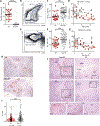
Fig. 1.. RT children have fewer GC-TFH cells in their tonsils.
Immunophenotyping analysis of cohort 1 of patients with and without RT. (A) Number of RT episodes in RT children (n = 23) and non-RT children (n = 11). (B) Flow cytometry of GC-TFH (CXCR5hiPD-1hiCD45RO+CD4+), mTFH (CXCR5+PD-1+CD45RO+CD4+), and non-TFH (CXCR5CD45RO+CD4+) cells. (C) GC-TFH cell frequencies in RT tonsils (n = 26) and non-RT tonsils (n = 39), quantified as percentage of total CD4+ T cells. (D) GC-TFH cells by age. (E) Flow cytometry of BGC cells (CD38+CD20+CD19+), plasma cells (PC; CD38hiCD20+CD19+), and memory (CD27hiCD20+CD19+)/naive (CD27CD20+CD19+) B cells. (F) BGC cell frequencies in RT and non-RT tonsils, quantified as percentage of total B cells. (G) BGC cells by age. (H) Representative Ki67-stained sections from RT and non-RT tonsils. m.(I) Quantitation of GC areas (in μm2) in RT tonsils (n = 21) and non-RT tonsils (n = 16). Each data point represents an individual GC. (J) Staining of BGC cells (Ki67) and GC-TFH cells [programmed cell death protein 1 (PD-1)]. Insets: Enlarged versions of representative GCs stained for Ki67 or PD-1. II and III show PD-1+ GC-TFH cells in representative GCs from a non-RT tonsil and an RT tonsil, respectively. ****P < 0.0001, ***P < 0.001. Statistical significance was determined by Mann-Whitney tests (A to C, E, F, and I) and multivariate analysis of covariance (ANCOVA; D and G). DZ, dark zone; LZ, light zone.
Fig. 2.
Fig. 2.. RT children have lower circulating anti-SpeA IgG titers.
Serological and immunophenotyping analysis of cohort 2 of patients with and without RT. (A) Plasma anti-SLO IgG titers in RT children (n = 23), non-RT children (n = 16), and normal healthy adults (n = 14). LOD, limit of detection; RU, relative units. (B) Plasma anti-SpeA IgG titers in RT children (n = 42), non-RT children (n = 45), and normal healthy adults (n = 17). (C) GC-TFH cell frequencies in RT tonsils (n = 40) and non-RT tonsils (n = 41), quantified as percentage of total CD4+ T cells. (D) GC-TFH cells by age. (E) BGC cell frequencies in RT and non-RT tonsils, quantified as percentage of total B cells. (F) BGC cells by age. *P < 0.05, ***P < 0.001, ****P < 0.0001. Statistical significance was determined by Mann-Whitney test.
Fig. 3.
Fig. 3.. RT tonsils have reduced GAS-specific GC-TFH cells.
(A) Identification of GAS-specific CD4+ T cells (CD45RA) and GAS-specific GC-TFH cells (CD45RACXCR5hiPD-1hi) using OX40+CD25+ AIM (AIM25). Tonsil cells were left unstimulated or stimulated with antibiotic-killed L. lactis (a nonpathogenic Gram-positive bacteria that served as a negative control; 10 μg/ml), heat-inactivated, antibiotic-killed GAS (10 μg/ml), or staphylococcal enterotoxin B [SEB (positive control); 1 μg/ml] for 18 hours. (B) GAS-specific GC-TFH cell frequencies. (C) GAS-specific CD45RACD4+ T cell frequencies, quantified as percentage of total CD4+ T cells, in RT tonsils (n = 31) and non-RT tonsils (n = 35). (D) Fraction of GAS-specific GC-TFH cells (CXCR5hiPD-1hi) among total GAS-specific CD4+ T cell (AIM25+CD45RA) in RT tonsils (n = 31) and non-RT tonsils (n = 35). ****P < 0.0001, *P < 0.05. Statistical significance was determined by paired t test (B) and Mann-Whitney tests (C and D).
Fig. 4.
Fig. 4.. HLA class II associations identified in RT children.
(A) Family history of tonsillectomy (RT = 71 and non-RT = 63). (B) HLA DQB1*06:02 allelic frequency in non-RT children (gray bar, n = 192), RT children (white bar, n = 138), ethnically matched adults from the SD GP (black bar, n = 242), and non-RT children and GP (blue bar, n = 434). NS, not significant. (C) Left: HLA DRB1*01:01 and HLA DRB1*07:01 allelic frequencies in RT tonsils with the lowest quartile of GC activity, defined as the lowest combined frequencies of GC-TFH and BGC cells [green dots, n = 15 (GClo samples); blue dots, n = 46 (GClo samples); red dot, mean of non-RT GC activity]. Right: HLA DRB1*01:01 and HLA DRB1*07:01 allelic frequencies in non-RT tonsils (gray bar, n = 190), GP (black bar, n = 246), and GP and non-RT tonsils (blue bar, n = 436). RT children HLA allele counts (white bar, n = 30). ***P < 0.001, *P < 0.05. Statistical significance was determined by Fisher’s exact test (A to C).
Fig. 5.
Fig. 5.. HLA class II associations identified in RT and non-RT children segregate on the basis of preferential GAS superantigen SpeA binding.
(A) Comparison of AIM+ GC-TFH cells after stimulation with either antibiotic-killed (AK) GAS (10 μg/ml), antibiotic-killed, heat-inactivated (AK HI) GAS (10 μg/ml), or antibiotic-killed SpeA-deficient (AK ΔSpeA) GAS (10 μg/ml); n = 10, including RT and non-RT donors. Heat-inactivation of antibiotic-killed GAS inactivates GAS superantigens. (B) SpeA-responsive GC-TFH cells in tonsils from patients with RT with risk HLA alleles (n = 12) compared to non-RT patients with protective HLA alleles (n = 12). Tonsils were stimulated with SpeA (1 μg/ml) for 18 hours and background-subtracted as determined with unstimulated cells. (C) Histogram flow cytometric quantitation of SpeA binding by HLA allele using HLA cell lines. n = 3 experiments. (D) Proliferation of total CD4+ T cells from peripheral blood mononuclear cells (PBMCs) of HLA DQB1*06:02+ donors cocultured with recombinant SpeA (rSpeA) and a cell line expressing HLA DQB1*06:02 and of CD4+ T cells from PBMCs of HLA DRB1*07:01 + donors cocultured with rSpeA and a cell line expressing HLA DRB1*07:01. n = 4 experiments. **P < 0.01, *P < 0.05 (D). Statistical significance was determined by Mann-Whitney test. CTV, cell trace violet.
Fig. 6.
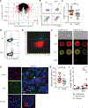
Fig. 6.. SpeA stimulation of GC-TFH cells from RT tonsils induces GzmB.
(A) Volcano plot showing fold change of genes in SpeA-stimulated GC-TFH cells from RT tonsils (n = 5) compared to SpeA-stimulated GC-TFH cells from non-RT tonsils (n = 5). Red dots denote genes with a fold change of <0.5 or >2. (B) Frequency of intracellular GzmB expression in GC-TFH cells by flow cytometry. Tonsil cells were stimulated with SpeA (1 μg/ml) for 24 hours (top). Backgating of the GzmB+ GC-TFH cells among total CD45RA CD4+ T cells (bottom). (C) GzmB+ GC-TFH cells in RT tonsils (n = 20) and non-RT tonsils (n = 17) after SpeA stimulation. (D) Fluorescence-activated cell-sorted GC-TFH cells and autologous B cells were cultured ± SpeA for 5 days and stained for GzmB and perforin expression. n = 3 donors. (E) ImageStream cytometry plot of GzmB+ GC-TFH cells after SpeA stimulation. GC-TFH cells were gated as CXCR5hiPD-1hi of live CD45RACD4+ T cells. n = 1 donor. (F) ImageStream imaging of GC-TFH cells after SpeA stimulation, showing representative GzmB and GzmB+ cells. (G) Confocal microscopy of a GzmB+ CD4+ T cell in a GC in an RT tonsil (*). A GzmB+ CD8+ T cell is also shown for reference (<). m.n = 8 donors. (H) GC-TFH cells (CXCR5hiPD-1hiCD45RACD4+) were cocultured with autologous CTV-labeled B cells (CD19+CD38). Killing was quantified as outlined in Materials and Methods, with controls shown in fig. S7 (J to L). n = 15 and 11 (RT and non-RT donors, respectively). (I) GzmB expression (percentage) by GC-TFH cells from healthy LNs and from RT and non-RT tonsils. GzmB expression after SpeA stimulation of GC-TFH cells from RT tonsils (n = 11), non-RT tonsils (n = 11), or healthy LNs (n = 4). **P < 0.01, *P < 0.05. Statistical significance was determined by Mann-Whitney test (C, H, and I).

Comment in

References

    1. Carapetis JR, Steer AC, Mulholland EK, Weber M, The global burden of group A streptococcal diseases. Lancet Infect. Dis 5, 685–694 (2005). - PubMed
    1. Shulman ST, Bisno AL, Clegg HW, Gerber MA, Kaplan EL, Lee G, Martin JM, Van Beneden C, Clinical practice guideline for the diagnosis and management of group A streptococcal pharyngitis: 2012 update by the Infectious Diseases Society of America. Clin. Infect. Dis 55, 1279–1282 (2012). - PubMed
    1. Ebell MH, Smith MA, Barry HC, Ives K, Carey M, The rational clinical examination. Does this patient have strep throat? JAMA 284, 2912–2918 (2000). - PubMed
    1. Schwartz RH, Kim D, Martin M, Pichichero ME, A reappraisal of the minimum duration of antibiotic treatment before approval of return to school for children with streptococcal pharyngitis. Pediatr. Infect. Dis. J 34, 1302–1304 (2015). - PubMed
    1. Van Brusselen D, Vlieghe E, Schelstraete P, De Meulder F, Vandeputte C, Garmyn K, Laffut W, Van de Voorde P, Streptococcal pharyngitis in children: To treat or not to treat? Eur. J. Pediatr 173, 1275–1283 (2014). - PubMed

Publication types

MeSH terms