. 2019 Feb 19;116(8):3288-3293.
doi: 10.1073/pnas.1809251116. Epub 2019 Feb 6.

Diverse GPCRs exhibit conserved water networks for stabilization and activation

Affiliations

Diverse GPCRs exhibit conserved water networks for stabilization and activation

A J Venkatakrishnan et al. Proc Natl Acad Sci U S A. .

Abstract

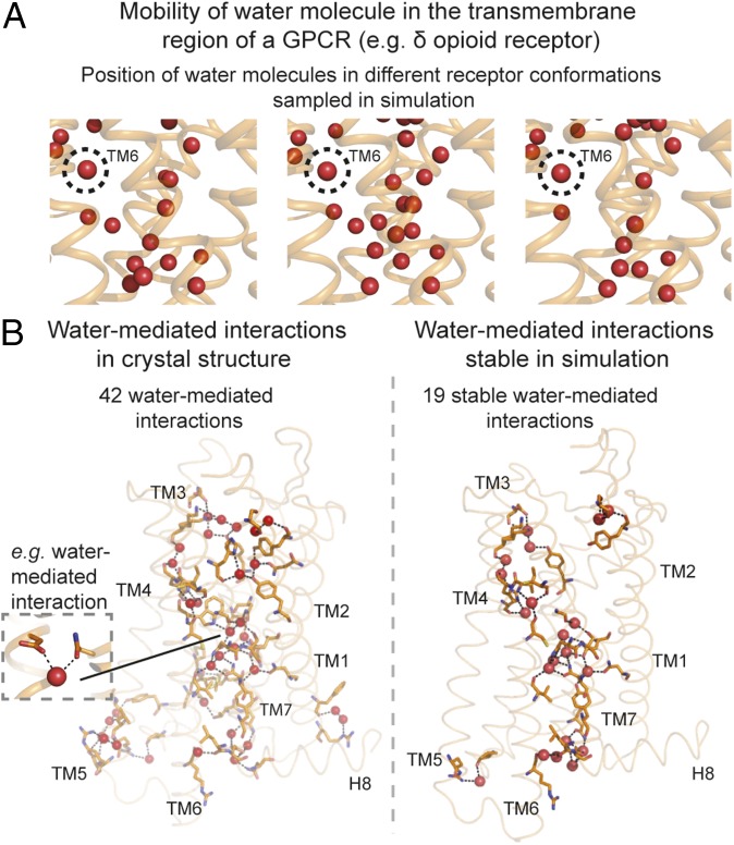
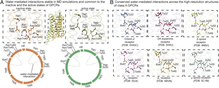
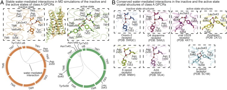
G protein-coupled receptors (GPCRs) have evolved to recognize incredibly diverse extracellular ligands while sharing a common architecture and structurally conserved intracellular signaling partners. It remains unclear how binding of diverse ligands brings about GPCR activation, the common structural change that enables intracellular signaling. Here, we identify highly conserved networks of water-mediated interactions that play a central role in activation. Using atomic-level simulations of diverse GPCRs, we show that most of the water molecules in GPCR crystal structures are highly mobile. Several water molecules near the G protein-coupling interface, however, are stable. These water molecules form two kinds of polar networks that are conserved across diverse GPCRs: (i) a network that is maintained across the inactive and the active states and (ii) a network that rearranges upon activation. Comparative analysis of GPCR crystal structures independently confirms the striking conservation of water-mediated interaction networks. These conserved water-mediated interactions near the G protein-coupling region, along with diverse water-mediated interactions with extracellular ligands, have direct implications for structure-based drug design and GPCR engineering.

Keywords: GPCR dynamics; activation; molecular dynamics simulation; polar network; water molecules.


Conflict of interest statement

Conflict of interest statement: B.K.K. is a cofounder of and consultant for ConfometRx, Inc.

Figures

Fig. 1.
Fig. 1.
Stability of water molecules and water-mediated interactions in GPCRs. (A) Positions of water molecules (red spheres) in one region of a GPCR [the δ-opioid receptor (DOR)] in three simulation snapshots. Receptor transmembrane helices are shown as orange ribbons. Most waters are mobile, but the water at the TM6 kink (circled) is not. (B, Left) Water-mediated interactions present in the crystal structure of DOR. (B, Right) Water-mediated interactions formed over 60% of the time in simulation. Hydrogen bonds forming water-mediated interactions are shown as black dashed lines, and the residues involved in these interactions are shown with orange sticks.
Fig. 2.
Fig. 2.
Conserved water-mediated interactions in inactive and active states of GPCRs. Comparison of the stability of structurally equivalent water-mediated interactions across different GPCRs in (A) inactive states and (B) active states. Four functionally distinct and evolutionarily distant class A GPCRs were compared: A2AR, β2AR, M2R, and MOR. The stability of the water-mediated interactions is estimated based on the frequency of formation in simulations. (A and B, Left) Frames from simulations of four functionally distinct GPCRs. Receptors are shown as ribbons and water molecules are shown as spheres. (A and B, Middle) Heatmap comparing the stability of water-mediated interactions. Columns indicate receptors and rows indicate pairs of structurally equivalent positions, shown using GPCRdb numbers. The cells indicate the measure of stability of water-mediated interactions. Stability is shown as percentage values using corresponding colors. Lighter shades indicate water-mediated interactions that have low stability and darker shades indicate water-mediated interactions that have high stability. Rows with dark cells in all columns indicate interactions that have high stability in all of the receptors being compared. (A and B, Right) Water-mediated interactions that are stable (frequency >60%) across all four receptors mapped onto a GPCR structure. Hydrogen bonds forming water-mediated interactions are shown as black dashed lines, with the residues involved shown as sticks. Inactive state is shown in orange and active state is shown in green.
Fig. 3.
Fig. 3.
State-independent water-mediated interaction network: conserved and stable water-mediated interactions maintained across the inactive and the active states of diverse GPCRs. (A) Stable water-mediated interactions (frequency >60%) present in both the inactive (orange) and the active (green) states of diverse GPCRs. (A, Top) The conserved stable water-mediated interactions mapped onto either an inactive (orange) or active (green) structure. Residues are shown as sticks, water molecules are shown as spheres, and hydrogen bonds forming water-mediated interactions are shown as black dashed lines. (A, Bottom) The conserved stable water-mediated interactions are shown using “flareplots.” In flareplots, the amino acid residues in the transmembrane helices and helix 8 are shown as points on the circle. The water-mediated interactions between the residues are shown as chords connecting the points on the circle. (B) Water-mediated interactions common across the high-resolution structures (resolution of 2.1 Å or better) of diverse GPCRs: inactive Orexin-2 (PDB ID code 5WQC), D4 dopamine (PDB ID code 5WIU), δ-opioid (PDB ID code 4N6H), A2A adenosine (PDB ID code 5IU4), and β1 adrenergic (PDB ID code 4BVN) receptors, and active μ-opioid receptor (PDB ID: 5C1M). Residues are shown as sticks, water molecules are shown as spheres, and hydrogen bonds forming water-mediated interactions are shown as black dashed lines.
Fig. 4.
Fig. 4.
State-dependent water-mediated interaction network: conserved and stable water-mediated interactions maintained exclusively in the inactive states or the active states of diverse GPCRs. (A) Stable water-mediated interactions (frequency >60%) present exclusively in all of the inactive (orange) or all of the active (green) states of diverse GPCRs. (A, Top) The conserved stable water-mediated interactions of inactive states and active states mapped onto inactive (orange) and active (green) structures, respectively. Residues are shown as sticks, water molecules are shown as spheres, and hydrogen bonds forming water-mediated interactions are shown as black dashed lines. (A, Bottom) The conserved stable water-mediated interactions are shown using flareplots. (B) Water-mediated interactions present exclusively in the crystal structures of GPCRs in inactive state or active state. The high-resolution inactive state structures are of the following GPCRs: Orexin-2 (PDB ID code 5WQC), D4 dopamine (PDB ID code 5WIU), δ-opioid (PDB ID code 4N6H), and A2A adenosine (PDB ID code 5IU4) receptors. The active-state crystal structures of the following GPCRs are considered: rhodopsin (PDB ID code 2X72), viral chemokine receptor US28 (PDB ID code 4XT1), and μ-opioid receptor (PDB ID code 5C1M). Residues are shown as sticks, water molecules are shown as spheres, and hydrogen bonds forming water-mediated interactions are shown as black dashed lines.

References

    1. Fredriksson R, Lagerström MC, Lundin L-G, Schiöth HB. The G-protein-coupled receptors in the human genome form five main families. Phylogenetic analysis, paralogon groups, and fingerprints. Mol Pharmacol. 2003;63:1256–1272. - PubMed
    1. Venkatakrishnan AJ, et al. Diverse activation pathways in class A GPCRs converge near the G-protein-coupling region. Nature. 2016;536:484–487. - PMC - PubMed
    1. Venkatakrishnan AJ, et al. Molecular signatures of G-protein-coupled receptors. Nature. 2013;494:185–194. - PubMed
    1. Santos R, et al. A comprehensive map of molecular drug targets. Nat Rev Drug Discov. 2017;16:19–34. - PMC - PubMed
    1. Huang W, et al. Structural insights into µ-opioid receptor activation. Nature. 2015;524:315–321. - PMC - PubMed

Publication types

LinkOut - more resources