. 2019 Feb 6;9(1):1490.
doi: 10.1038/s41598-018-37291-2.

Repeated human deciduous tooth-derived dental pulp cell reprogramming factor transfection yields multipotent intermediate cells with enhanced iPS cell formation capability

Affiliations

Repeated human deciduous tooth-derived dental pulp cell reprogramming factor transfection yields multipotent intermediate cells with enhanced iPS cell formation capability

Miki Soda et al. Sci Rep. .

Abstract

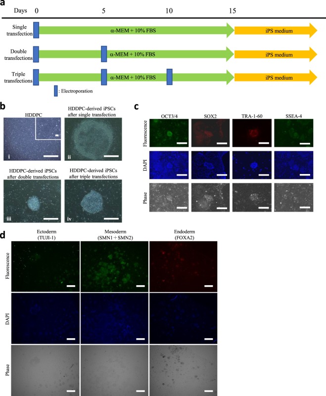
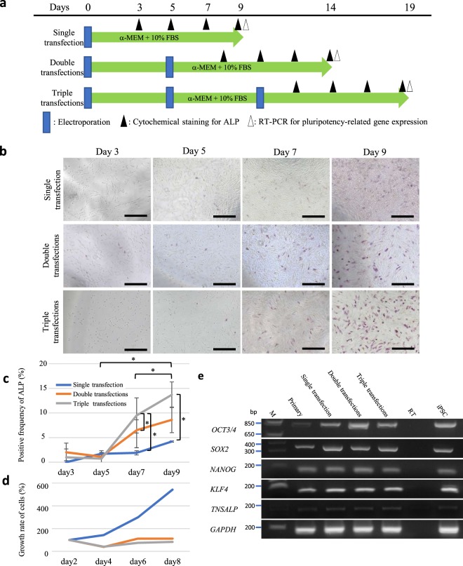
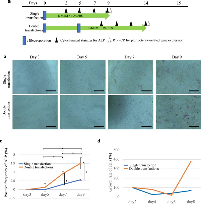
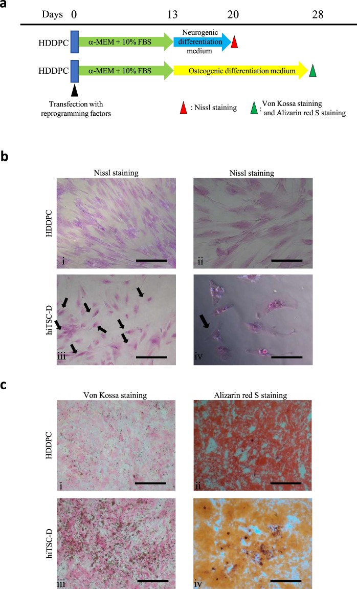
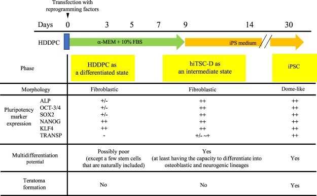
Human tissue-specific stem cells (hTSCs), found throughout the body, can differentiate into several lineages under appropriate conditions in vitro and in vivo. By transfecting terminally differentiated cells with reprogramming factors, we previously produced induced TSCs from the pancreas and hepatocytes that exhibit additional properties than iPSCs, as exemplified by very low tumour formation after xenogenic transplantation. We hypothesised that hTSCs, being partially reprogrammed in a state just prior to iPSC transition, could be isolated from any terminally differentiated cell type through transient reprogramming factor overexpression. Cytochemical staining of human deciduous tooth-derived dental pulp cells (HDDPCs) and human skin-derived fibroblasts following transfection with Yamanaka's factors demonstrated increased ALP activity, a stem cell marker, three weeks after transfection albeit in a small percentage of clones. Repeated transfections (≤3) led to more efficient iPSC generation, with HDDPCs exhibiting greater multipotentiality at two weeks post-transfection than the parental intact HDDPCs. These results indicated the utility of iPSC technology to isolate TSCs from HDDPCs and fibroblasts. Generally, a step-wise loss of pluripotential phenotypes in ESCs/iPSCs occurs during their differentiation process. Our present findings suggest that the reverse phenomenon can also occur upon repeated introduction of reprogramming factors into differentiated cells such as HDDPCs and fibroblasts.


Conflict of interest statement

The authors declare no competing interests.

Figures

Figure 1
Figure 1
Generation of HDDPC-derived iPSCs. (a) Time-line for isolation of iPSCs from HDDPCs via single, double, or triple transfections. (b) Morphology of the HDDPCs (a) and HDDPC-derived iPSC colonies (bd). iPSC colonies obtained after single (i), double (ii), or triple transfections (iii) from the HDDPC lines P02, P03, and P04, respectively. The magnified image is shown in the upper right portion of (i). Bar = 500 μm. (c) Immunocytochemistry of HDDPC (P01)-derived iPSC colonies using antibodies for OCT3/4, SOX2, TRA-1–60, and SSEA-4. Cells were stained by DAPI to visualise the location of nuclei. Bar = 500 μm. (d) Immunocytochemistry of embryoid body-derived outgrowth using antibodies for TUJI-1 (ectodermal marker), SMN1 + SMN2 (mesodermal marker), and FOXA2 (endodermal marker). Cells were stained by DAPI to visualise the location of nuclei. Bar = 500 μm.
Figure 2
Figure 2
Increased pluripotency-related gene expression in HDDPCs after repeated transfection with Yamanaka’s four reprogramming factors. (a) Time-line for assessment of pluripotency-related gene expression in HDDPCs after repeated transfection. (b) Cytochemical evaluation of ALP activity in the HDDPCs after repeated transfections with the reprogramming factors. HDDPCs (P05 line) were transfected with Yamanaka’s four reprogramming factors once, twice, or three times. The treated cells were subjected to cytochemical staining for ALP activity at 3, 5, 7, and 9 days after the final transfection, as shown in (a). Bar = 500 μm. (c,d) Ratio of ALP-positive cells (c) and proliferation curves (d) for HDDPCs when the P05 line was subjected to transfection once, twice, or three times. After the final transfection, cells were seeded onto 4- or 24-well plate, and fixed (for cytochemical staining for ALP activity) or harvested (for counting the cell number) at the days indicated. (e) RT-PCR analysis of HDDPCs after the repeated transfections. HDDPCs (P05 line) were transfected with Yamanaka’s four reprogramming factors once, twice, or three times, and harvested 9 days after the final transfection for RT-PCR analysis for detection of mRNA expression of OCT3/4, SOX2, NANOG, and KLF4, tissue-nonspecific alkaline phosphatase (TNSALP), and glyceraldehyde-3-phosphate dehydrogenase (GAPDH) (as control). Lane 1, HDDPCs (P05 line); lane 2, HDDPCs after the single transfection; lane 3, HDDPCs after the double transfection; lane 4, HDDPCs after the triple transfection; lane 5, no template (as negative control); lane 6, iPSCs (as positive control). M, 100-bp ladder markers.
Figure 3
Figure 3
The changes of ALP activity in human skin-derived fibroblasts after repeated transfection. (a) Time-line for assessment of pluripotency-related gene expression in fibroblasts after repeated transfection. (b) Cytochemical evaluation of ALP activity in the fibroblasts after repeated transfection with the reprogramming factors. Fibroblasts were transfected with Yamanaka’s four reprogramming factors once or twice. The treated cells were subjected to cytochemical staining for ALP activity at 3, 5, 7, and 9 days after the final transfection, as shown in (a). Bar = 500 μm. (c) Ratio of ALP-positive cells and (d) proliferation curves for fibroblasts when the parental fibroblasts were subjected to transfection once or twice. After the final transfection, cells were seeded onto 4- or 24-well plate, and fixed (for cytochemical staining for ALP activity) or harvested (for counting the cell number) at the days indicated.
Figure 4
Figure 4
Microarray analysis. Transcriptome Analysis Console (TAC) Software from Affymetrix was used to analyse global gene expression patterns between HDDPCs and hiTSC-D (a), fibroblasts and hiTSC-F (b), and HDDPCs and fibroblasts (c). (d) Unsupervised hierarchical clustering of gene expression profiles of HDDPCs, hiTSC-D, fibroblasts, and hiTSC-F. Each column represents one biological sample.
Figure 5
Figure 5
Differentiation induction of the intermediate HDDPCs. (a) Time-line for differentiation induction of the HDDPC line P05 (13 days after the single transfection with Yamanaka’s four reprogramming factors) into neurogenic or osteoblastic lineage. (b) Neuronal cell induction 7 days after incubation in neurogenic medium. Nissl staining demonstrated that Nissl bodies are clearly discernible around the nucleus in the transfected cells (arrowed) in (iii), whereas no such bodies are discernible in the untransfected cells in (i). Bar = 500 μm (i) and (iii), 50 μm (ii) and (iv). (c) Osteoblastic cell induction 15 days after incubation in osteogenic medium. Von Kossa staining in (i) and (iii), and staining with Alizarin red-S in (ii) and (iv). Bar = 500 μm.
Figure 6
Figure 6
Summary of the properties of intermediate state cells (hiTSC-D) generated around 9 days after transfection with Yamanaka’s four reprogramming factors.

References

    1. Takahashi K, Yamanaka S. Induction of pluripotent stem cells from mouse embryonic and adult fibroblast cultures by defined factors. Cell. 2006;126:663–676. doi: 10.1016/j.cell.2006.07.024. - DOI - PubMed
    1. Takahashi K, et al. Induction of pluripotent stem cells from adult human fibroblasts by defined factors. Cell. 2007;131:861–872. doi: 10.1016/j.cell.2007.11.019. - DOI - PubMed
    1. Tamaoki, N. et al. Dental pulp cells for induced pluripotent stem cell banking. J. Dent. Res. 89, 773–778, 10.1177/0022034510366846 0022034510366846 [pii] (2010). - PubMed
    1. Inada, E. et al. Alkaline phosphatase and OCT3/4 as useful markers for predicting susceptibility of human deciduous teeth-derived dental pulp cells to reprogramming factor-induced iPS cells. J. Investig. Clin. Dent. 8 Epub ahead of print. 10.1111/jicd.12236 (2017). - PubMed
    1. Yan X, et al. iPS cells reprogrammed from human mesenchymal-like stem/progenitor cells of dental tissue origin. Stem Cells Dev. 2010;19:469–480. doi: 10.1089/scd.2009.0314. - DOI - PMC - PubMed

Publication types

MeSH terms