Review
. 2019 May;15(5):301-316.
doi: 10.1038/s41581-019-0111-1.

Uraemic syndrome of chronic kidney disease: altered remote sensing and signalling

Affiliations
Review

Uraemic syndrome of chronic kidney disease: altered remote sensing and signalling

Sanjay K Nigam et al. Nat Rev Nephrol. 2019 May.

Abstract

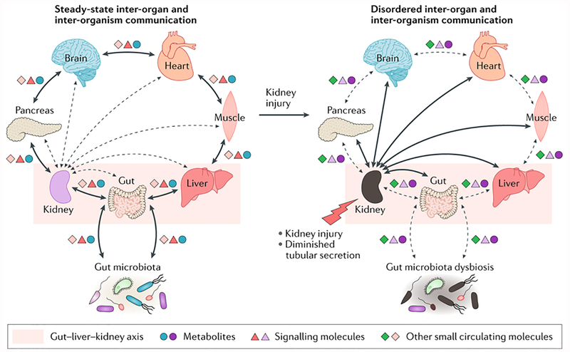
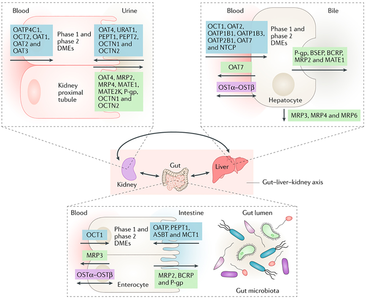
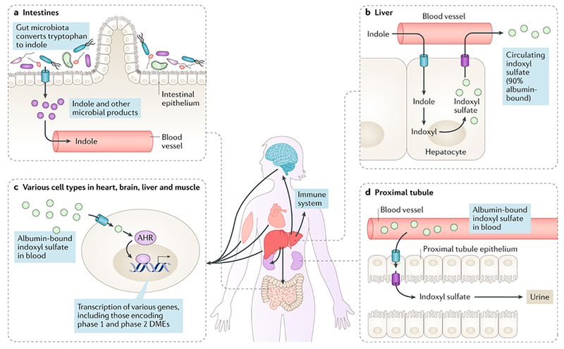
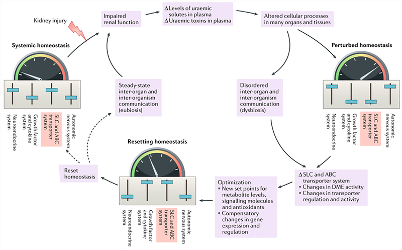
Uraemic syndrome (also known as uremic syndrome) in patients with advanced chronic kidney disease involves the accumulation in plasma of small-molecule uraemic solutes and uraemic toxins (also known as uremic toxins), dysfunction of multiple organs and dysbiosis of the gut microbiota. As such, uraemic syndrome can be viewed as a disease of perturbed inter-organ and inter-organism (host-microbiota) communication. Multiple biological pathways are affected, including those controlled by solute carrier (SLC) and ATP-binding cassette (ABC) transporters and drug-metabolizing enzymes, many of which are also involved in drug absorption, distribution, metabolism and elimination (ADME). The remote sensing and signalling hypothesis identifies SLC and ABC transporter-mediated communication between organs and/or between the host and gut microbiota as key to the homeostasis of metabolites, antioxidants, signalling molecules, microbiota-derived products and dietary components in body tissues and fluid compartments. Thus, this hypothesis provides a useful perspective on the pathobiology of uraemic syndrome. Pathways considered central to drug ADME might be particularly important for the body's attempts to restore homeostasis, including the correction of disturbances due to kidney injury and the accumulation of uraemic solutes and toxins. This Review discusses how the remote sensing and signalling hypothesis helps to provide a systems-level understanding of aspects of uraemia that could lead to novel approaches to its treatment.


Conflict of interest statement

Competing interests

The authors declare no competing interests.

Figures

Figure 1.
Figure 1.. Aberrant inter-organ and inter-organism communication contributes to uremic syndrome.
According to the remote sensing and signaling hypothesis, illustrated here from the perspective of the kidney, steady-state communication between the kidney and other organs and body fluids involves the movement of metabolites, signaling molecules and other small circulating molecules via solute carrier (SLC) and ATP-binding cassette (ABC) transporters. These transporters are expressed in many tissues, including the kidney, liver, pancreas, brain, intestine and muscle. Therefore, injury to the kidney leads to diminished tubular secretion and results in disordered remote communication. Some of these molecules can regulate the expression (via nuclear receptor activation) and/or function of SLC and ABC transporters or phase 1 and phase 2 drug-metabolizing enzymes (DMEs) in distinct cells, tissues and organs, thereby effecting local and global physiological alterations, accompanied by dysbiosis of gut microbiota (including the loss or gain of some bacterial strains). The multi-specificity of some ABC and SLC transporters, together with their different and modifiable tissue expression and/or trafficking, might help to restore homeostasis after organ dysfunction and injury. The differences in color of the symbols representing metabolites, signaling molecules and other small circulating molecules that interact with transporters represent alterations in the concentration and/or identity of these compounds under physiological versus pathological conditions.
Figure 2.
Figure 2.. The gut–liver–kidney axis.
Within the remote sensing and signaling communication network, the gut–liver–kidney axis drives the absorption, distribution, metabolism and excretion of small molecules, including endogenous metabolites, signaling molecules, products of the gut microbiota, nuclear receptors and antioxidants. Products derived from gut microbiota that cross the intestinal barrier are removed from the blood by solute carrier (SLC) and ATP-binding cassette (ABC) transporters on hepatocytes, where they (and other small molecules) are metabolized by phase 1 and phase 2 drug-metabolizing enzymes (DMEs). Ultimately, many of these products are cleared from the body via the kidney in the urine through the action of SLC and ABC transporters on the proximal tubule cells. Some transporters in different tissues are thought to function in both directions. MATE1, multi-drug and toxin extrusion protein 1; MATE2K, 2K splice variant of MATE2; OAT, organic anion transporter; OATP, organic anion-transporting polypeptide; OCT, organic cation transporter; P-gp, P-glycoprotein.
Figure 3.
Figure 3.. The role of indoxyl sulfate in inter-organism and inter-organ remote communication.
a) Indole is created in the lumen of the gut, via the metabolism of tryptophan by the gut microbiota, and absorbed across the gut wall into the blood. b) Circulating indole is taken up by hepatocytes, where it is metabolized first to indoxyl and then to indoxyl sulfate. Indoxyl sulfate is transported back into the circulation where the majority of it circulates bound to albumin and where it is distributed to and interacts with other organs, such as the brain, immune system and muscle, and with the gut microbiota. c) Indoxyl sulfate gains access to tissues and cells, where it signals through the aryl hydrocarbon receptor (AHR), leading to alterations in the expression of a number of genes in these tissues. d) Indoxyl sulfate is ultimately excreted by the kidney via solute carrier (SLC) and ATP-binding cassette (ABC) transporters located in the basolateral (such as organic anion transporter 1 (OAT1) and OAT3) and apical (such as multidrug resistance-associated protein 4 (MRP4)) membranes of proximal tubule cells (influx and efflux transporters are indicated by blue and purple barrels, respectively).
Figure 4.
Figure 4.. A remote sensing and signalling system maintains homeostasis in the steady state and resets homeostasis following perturbations due to kidney dysfunction and microbiota dysbiosis.
Small molecules with informational content are important in transmitting signals to remote tissues and/or organs in the maintenance of homeostasis. In essence, the small-molecule substrates of multi-specific solute carrier (SLC) and ATP-binding cassette (ABC) transporters act in concert with other transporters of limited specificity, as well as with phase 1 and phase 2 drug-metabolizing enzymes (DMEs), as part of a highly flexible inter-organ and inter-organism communication network. Under physiological conditions, this network works in concert with other systems to maintain steady-state homeostasis. Injury to the kidney resulting in diminished kidney function and reduced tubular secretion can lead to the accumulation of uremic solutes and uremic toxins in plasma. The increased levels of these small molecules can lead to alterations in cell functions and processes in multiple tissues and organs, resulting in perturbed homeostasis. The components of this network optimize metabolic pathways, signaling pathways, the control of redox status and other mechanisms necessary for homeostasis in different tissues and body fluid compartments, as well as in different organisms. This system is intertwined with and works in parallel with other homeostatic mediators, such as growth factors, cytokines and the neuroendocrine and vasoregulatory systems. The flexibility of the system enables homeostasis to be restored or reset at a new (presumably compensatory) set point despite the presence or progression of renal disease. Sliders in each picture represent the set points of each of the homeostatic systems, which are altered following organ injury and adjusted during the resetting of homeostasis. The colored scale and arrow represent the homeostatic setting in the various stages.

References

    1. Wu W, Dnyanmote AV & Nigam SK Remote communication through solute carriers and ATP binding cassette drug transporter pathways: an update on the remote sensing and signaling hypothesis. Mol. Pharmacol 79, 795–805 (2011). - PMC - PubMed
    1. Liu X & Dai C Advances in understanding and management of residual renal function in patients with chronic kidney disease. Kidney Dis 2, 187–196 (2017). - PMC - PubMed
    1. Lowenstein J & Grantham JJ Residual renal function: a paradigm shift. Kidney Int 91, 561–565 (2017). - PubMed
    1. Shafi T, Mullangi S, Toth-Manikowski SM, Hwang S & Michels WM Residual kidney function: implications in the era of personalized medicine. Semin. Dial 30, 241–245 (2017). - PubMed
    1. Nigam SK What do drug transporters really do? Nat. Rev. Drug Discov 14, 29–44 (2015). - PMC - PubMed

Publication types

MeSH terms