. 2019 Oct;26(10):2157-2178.
doi: 10.1038/s41418-019-0291-z. Epub 2019 Feb 6.

MINA-1 and WAGO-4 are part of regulatory network coordinating germ cell death and RNAi in C. elegans

Affiliations

MINA-1 and WAGO-4 are part of regulatory network coordinating germ cell death and RNAi in C. elegans

Ataman Sendoel et al. Cell Death Differ. 2019 Oct.

Abstract

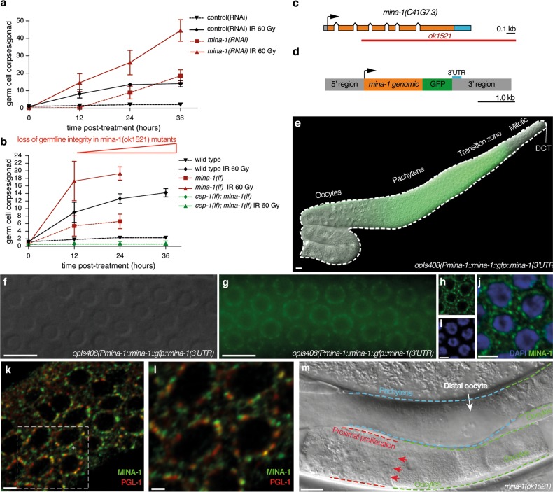
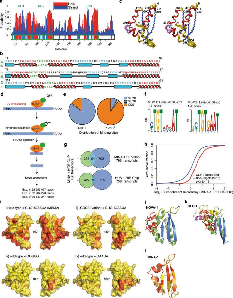
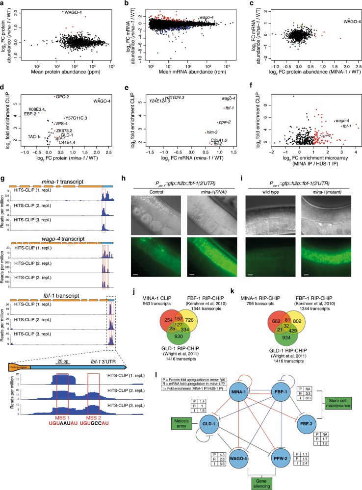
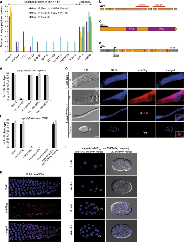
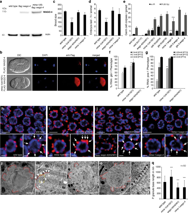
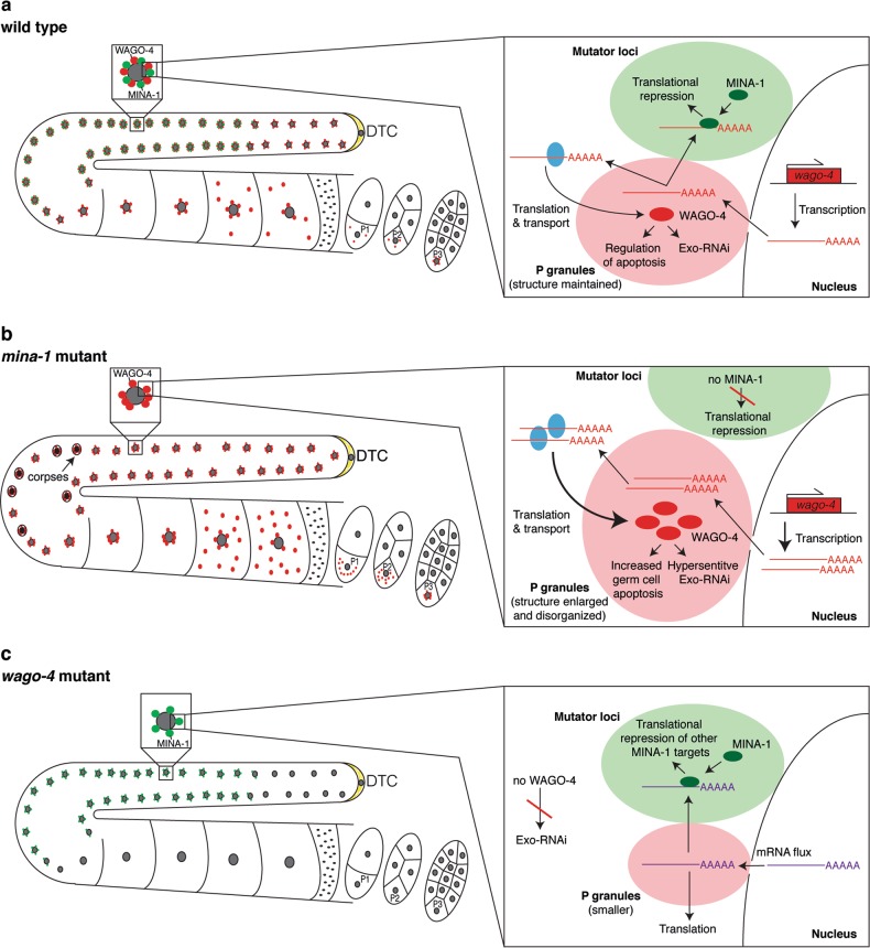
Post-transcriptional control of mRNAs by RNA-binding proteins (RBPs) has a prominent role in the regulation of gene expression. RBPs interact with mRNAs to control their biogenesis, splicing, transport, localization, translation, and stability. Defects in such regulation can lead to a wide range of human diseases from neurological disorders to cancer. Many RBPs are conserved between Caenorhabditis elegans and humans, and several are known to regulate apoptosis in the adult C. elegans germ line. How these RBPs control apoptosis is, however, largely unknown. Here, we identify mina-1(C41G7.3) in a RNA interference-based screen as a novel regulator of apoptosis, which is exclusively expressed in the adult germ line. The absence of MINA-1 causes a dramatic increase in germ cell apoptosis, a reduction in brood size, and an impaired P granules organization and structure. In vivo crosslinking immunoprecipitation experiments revealed that MINA-1 binds a set of mRNAs coding for RBPs associated with germ cell development. Additionally, a system-wide analysis of a mina-1 deletion mutant compared with wild type, including quantitative proteome and transcriptome data, hints to a post-transcriptional regulatory RBP network driven by MINA-1 during germ cell development in C. elegans. In particular, we found that the germline-specific Argonaute WAGO-4 protein levels are increased in mina-1 mutant background. Phenotypic analysis of double mutant mina-1;wago-4 revealed that contemporary loss of MINA-1 and WAGO-4 strongly rescues the phenotypes observed in mina-1 mutant background. To strengthen this functional interaction, we found that upregulation of WAGO-4 in mina-1 mutant animals causes hypersensitivity to exogenous RNAi. Our comprehensive experimental approach allowed us to describe a phenocritical interaction between two RBPs controlling germ cell apoptosis and exogenous RNAi. These findings broaden our understanding of how RBPs can orchestrate different cellular events such as differentiation and death in C. elegans.


Conflict of interest statement

The authors declare that they have no conflict of interest.

Figures

Fig. 1
Fig. 1
MINA-1 is a germline-specific protein controlling germ cell development and apoptosis. a Synchronized L1 animals were raised on RNAi empty vector control or mina-1(RNAi) plates and exposed to IR (60 Gy) as young adults. Germline apoptosis was quantified by DIC microscopy at the indicated time points. Data shown represent the average of three independent experiments ± s.d. (n > 20 animals for each experiment and time point). b Synchronized wild-type, mina-1(ok1521), and cep-1(lg12501);mina-1(ok1521) young adult animals were irradiated and germline apoptosis was quantified at the indicated time points. Data shown represent the average of three independent experiments ± s.d. (n > 20 animals for each experiment and time point). c Schematic representation of the mina-1(C41G7.3) locus. The ok1521 allele deletes exons three to seven of the mina-1 gene. d Schematic representation of the MINA-1::GFP fusion construct used to generate the opIs408 transgene. e MINA-1::GFP localization in a composite germline image of a dissected opIs408(Pmina-1::mina-1::gfp::mina-1(3′-UTR)) animal. GFP and DIC merged. Scale bars, 10 μm. f-j DIC (f) and fluorescence (GFP channel, g) images of germ cells in live opIs408 animals, and confocal microscopy images of germ cells in fixed opIs408 animals (h: α-gfp antibody to detect MINA-1::GFP (green), i: DAPI (DNA, blue), j: merge). Scale bars, 10 μm (f, g), 3 μm (h-j). k, l Confocal microscopy images of germ cells in fixed opIs408 animals using α-PGL-1 antibody (red) as P granule marker and α-gfp antibody to detect MINA-1::GFP (green). l is a higher magnification of area indicated in k. Scale bars, 2 µm (k), 1 µm (l). (Note: Pearson’s correlation of colocalized volume is between –0.4 and –0.6 for different sections). m mina-1(ok1521) mutant animals show additional developmental germline defects, including distal oocytes (gogo phenotype: germ cell, oocyte, germ cell, oocyte; white arrow) and proximal proliferation (red arrows). Scale bar, 20 μm
Fig. 2
Fig. 2
MINA-1 has a KH3 domain with a non-canonical “GNRA” loop essential for RNA binding and binds to target 3′-UTRs mainly via two related binding motifs. a Secondary structure prediction of MINA-1 using the JUFO neural network algorithm. Putative KH domains are indicated by KH1, KH2, and KH3. The probability of α-helical (red bars) and β-strand (blue bars) secondary structure elements is plotted against the sequence of MINA-1. b Sequence and predicted secondary structure elements of the three putative KH domains of MINA-1. Eukaryotic KH domains are characterized by β1-α1-α2-β2-β’-α’ topology and a “GXXG” loop (in green), which is located between helices α1 and α2, which is essential for RNA binding. The predicted α-helices and β-strand are indicated with red and blue colors, respectively. c Stereoview of the 20 lowest-energy conformers representing the solution structure of the KH3 domain of MINA-1 after energy-minimization with AMBER. The sequence boundaries of α-helical (red) and β-stranded (blue) regions are shown; magenta bonds indicate the “GNRA” loop. PDB deposition ID: 6FBL. d Overview of the high-throughput sequencing of RNA isolated by crosslinking immunoprecipitation (HITS-CLIP) of MINA-1::GFP and control. Synchronized young adult opIs408 animals were exposed to UV light to cross-link RNAs and proteins in situ. Cross-linked RNA was co-purified with MINA-1::GFP and subjected to RNA sequencing. Displayed are also the reads achieved in each of the three independent experiments [20, 21]. e Pie charts show the distribution of the binding sites in the 3′-UTR (blue), 5′-UTR (gray), and CDS (orange) of sequenced RNAs of control (total RNA-Seq) and first experiment. f Consensus MINA-1-binding motifs (MBM) identified by HITS-CLIP. MBM1 is present in 300 and MBM2 in 126 of the top MINA-1 500 binding sites, respectively. g Venn diagrams show overlap of transcripts enriched in MINA-1 CLIP (430 transcripts) and in MINA-1 RIP-Chip (796 transcripts) or HUS-1 RIP-Chip (control, 758 transcripts) experiments. Regarding the RIP-Chip experiments, transcripts that were at least twofold enriched (log2 > 1) in either IPs were further considered. The CLIP dataset consists of transcripts enriched compared with RNA-seq of young adults expressing MINA-1::GFP with applying a filter of detection of 1 cpm in all three CLIP replicates. In all, 94 genes overlapped between MINA-1 CLIP and MINA-1 RIP-Chip (P-value = 1.05 × 10–22). h Cumulative fraction analysis of fold change distribution (log2) of CLIP targets and non-targets. P-values were calculated with the Kolmogorov–Smirnov (KS) test. i Surface representation of MINA-1 residues 257–331 showing the combined 1H and 15N chemical shift perturbation (CSP) upon titration of wild-type MINA-1 with (i) CUGUGAAUA-RNA, (iii) CUGUG-RNA, and (iv) GAAUA-RNA and upon titration of the (ii) “GDDA” MINA-1 variant with CUGUGAAUA-RNA. The color coding reflects the gradient of the observed perturbations, ranging from yellow (no CSP) to red (CSP ≥ 0.1). j KH domains typically recognize up to four nucleotides via their canonical nucleic acid-binding surface comprising helices α1 and α2, the GXXG and variable loops and β-strand β2, which is exemplified by the complex of NOVA-1 with UCAC-RNA. k The specific recognition of longer target sequences requires domain extensions such as the additional α-helix provided by the QUA2 domain in GLD-1. l The MINA-1 KH3 domain preferentially binds to longer target RNAs by additional involvement of β-strands β1 and β’. Color-coding of a cartoon representation of the KH3 domain according to the CSP upon binding of the KH3 domain to the CUGUGAAUA-RNA
Fig. 3
Fig. 3
MINA-1 regulates its own mRNA and an RBP cluster. a Relative protein abundance in mina-1 mutants and wild type were quantified by SILAC in two biological replicates. Overall mean protein abundance was extracted from the integrated C. elegans PaxDB dataset [1]. Upregulated (log2 fold change (mina-1/WT) ≥ 0.7) and downregulated proteins (log2 fold change (mina-1/WT) ≤ -0.7) are marked with red and blue dots, respectively. b Relative mRNA expression in mina-1 mutants and wild type was quantified by RNA-seq in three biological replicates. Upregulated (log2 fold change (mina-1/WT) ≥ 0.7; P(adj) < 0.1)) and downregulated transcripts (log2 fold change (mina-1/WT) ≤ 0.7; P(adj) < 0.1) are marked with red and blue dots, respectively. c Comparison of protein to mRNA changes (log2 fold change (mina-1/WT)) of 1294 genes whose abundance was quantified in both SILAC and RNA-seq experiments. Upregulated (log2 fold change (mina-1/WT) ≥ 0.7) and downregulated proteins (log2 fold change (mina-1/WT) ≤ -0.7) are marked with red and blue dots, respectively. Upregulated (log2 fold change (mina-1/WT) ≥ 0.7) and downregulated transcripts (log2 fold change (mina-1/WT) ≤ -0.7) are marked with yellow and light blue dots with a black border, respectively. d, e Comparison of log2 fold change enrichments of a subset of MINA-1 CLIP targets at the protein (d) and mRNA (e) levels. All MINA-1 CLIP targets that were quantified at the protein level (log2 fold change (mina-1/WT) ≥ 0.2 or ≤ –0.2) are shown in d (upregulated in red and downregulated in blue), whereas e only includes upregulated (red) and downregulated (blue) targets that showed a significant difference (P(adj) < 0.1) in mRNA abundance between mina-1 mutant and wild type. f Graph shows log2 fold change enrichment in HITS-CLIP and microarray (MINA-1 IP/HUS-1 IP) of CLIP targets. Transcripts significantly enriched (as defined in Fig. 2f) in MINA-1 IP and control HUS-1 IP are marked with red and blue dots, respectively. g Visualization of MINA-1 HITS-CLIP signal in the 3′-UTR (marked in blue) of mina-1, wago-4, and fbf-1 transcripts. Number of reads per million from the HITS-CLIP experiment along the transcript are shown. Sites where accumulation of reads correspond to one of the two consensus motifs (MBM1 or MBM2) are marked with red boxes. The fbf-1 3′-UTR is additionally zoomed to show the 2 MBM-containing sites (MBS1–2). h DIC and fluorescence images of the reporter line PPie-1::gfp::h2b::fbf-1(3′-UTR) after control (empty vector) and mina-1 RNAi. Scale bars, 10 μm. i DIC and fluorescence images of the reporter line PPie-1::gfp::h2b::fbf-1(3′-UTR) in wild type and mina-1 mutant. Scale bars, 10 μm. j Venn diagram showing the overlaps between MINA-1 CLIP targets and targets identified in FBF-1 (1344 targets, SAM > 0.965) and GLD-1 (1416 targets, fold enrichment > 2) RIP-CHIP experiments [6, 24]. In all, 284 genes overlapped with FBF-1 RIP-Chip (P-value = 7.9 × 10–74) and 152 with GLD-1 RIP-Chip (P-value = 1.9 × 10–25). k Venn diagram showing the overlap between MINA-1 RIP-Chip targets and targets identified in FBF-1 and GLD-1 RIP-CHIP experiments [6, 24]. In total, 113 overlapped with FBF-1 RIP-Chip (P-value = 4.4 × 10–6) and 53 with GLD-1 RIP-Chip (n.s.). l Network showing the post-transcriptional regulatory interactions between MINA-1 and several RBPs including WAGO-4, FBF-1, FBF-2, GLD-1, and PPW-2. Target protein (P) and mRNA (R) upregulation in mina-1 mutants as well as MINA-1 RIP-CHIP enrichment (I) are shown. Red, blue, and green lines represent regulation by MINA-1 FBF-1, and GLD-1, respectively
Fig. 4
Fig. 4
MINA-1 interacts with the Argonaute protein WAGO-4 to co-regulate RNAi. a Protein co-immunoprecipitation on worms expressing MINA-1::GFP detected WAGO-4 in all three replicates and none of the control IPs. Eluates after the IP were ran on SDS-PAGE gel, cut out, digested and peptides analyzed by Orbitrap Mass Spectrometer. Proteins expressed in the meiotic region of the germ line: CED-4 and HUS-1 were used as a control. Protein threshold of 99% was used and minimum number of peptides used to identify a protein was set to 1. The number of unique peptides detected per protein is displayed on the chart. b Schematic representation of the wago-4(F58G1.1) locus, including the two deletions tm1019 and tm2401. c Schematic representation of the WAGO-4 protein and its two Argonaute domains PAZ and Piwi (purple). d Schematic representation of the 3xFLAG::WAGO-4 fusion construct used to generate the opIs530[3xflag::wago-4] transgene. The 5′ region (gray) is 841 bp long, followed by a 3xflag tag (green) and the wago-4 gene (orange). After the STOP codon, a 486 bp 3′ region (blue) completes the transgene, which was inserted on Chromosome IV via MosSCi. e, f Somatically expressed genes (e, unc-52, unc-15) and germline-specific genes (f, gld-1, pos-1) were knocked down in wild type, rrf-1(pk1417), mut-7(pk204), wago-4(tm1019), and wago-4(tm2401) mutants. Related phenotypes were quantified in three independent experiments. The transgene opIs530[3xflag::wago-4] rescues the tissue-specific RNAi resistance of wago-4(tm2401) mutants (f). g Immunostaining of transgenic opIs530[3xflag::wago-4] and wild-type worms. DIC, DNA(DAPI, blue), anti-Flag(red), and merged channels show expression of FLAG-tagged WAGO-4 in the adult germ line and its precursor cells (Z2, Z3 in L1 worms, and P4 in the embryo). Inserts show a zoomed view of the germ line precursor cells. Scale bars, 20 µm. h Confocal image of a dissected opIs530[3xflag::wago-4] young adult germ line stained with anti-FLAG (red) and DAPI (blue). Scale bar, 20 µm. i P lineage affiliated expression of transgene opIs530[3xflag::wago-4] in wago-4 mutant background in 2, 4, 7, and 16 cells stage. Left column represents the merged channels of DNA (DAPI, blue) and WAGO-4 (anti-FLAG, red). Right column represents corresponding DIC images of the cells. Scale bar, 10 µm
Fig. 5
Fig. 5
MINA-1 negatively regulates WAGO-4 to control apoptosis, RNAi efficiency, and P granule organization. a Representative western blot image of 3xFLAG::WAGO-4 (detected using an anti-FLAG antibody) and actin (ACT-5) in whole animal extracts of wild-type, opIs530[3xflag::wago-4], and mina-1(ok1521); opIs530[3xflag::wago-4] staged young adults. b Immunostaining of opIs530[3xflag::wago-4] and mina-1(ok1521); opIs530[3xflag::wago-4] four-cell stage embryos. In a wild-type background, 3xFLAG::WAGO-4 shows a punctate perinuclear staining (blue: DAPI, red: anti-FLAG); this staining increases in intensity and spreads out throughout the cytoplasm in mina-1(ok1521) mutants. Scale bar, 10 µm. c, d Determination of brood size and embryos laid per hour per animal in wild type, mina-1(ok1521), wago-4(tm2401), and mina-1(ok1521);wago-4(tm2401) mutants. Data shown are average ± standard deviation of three biological replicates (n = 20 animals/experiment). P-values were calculated using Student’s t-test: ***P < 0.001. e Synchronized animals were scored for germ cell apoptosis 24 h post L4 larval stage/adult molt with (gray) and without (black) IR (60 Gy). Data shown are average ± standard deviation of five biological replicates (n = 20 animals/experiment). P-values were calculated using Student’s t-test: ***P < 0.001, n.s. not significant. f, g Dose-response RNAi knockdown of gld-1 (f) and pos-1 (g) in wild-type, mina-1(ok1521), and wago-4(tm2401) animals, using three IPTG concentrations (0 mM, 0.1 mM, and 0.5 mM). Data shown are averages ± standard deviation of three (p, 25 animals each) and two (q, 10 parents each) biological replicates. P-values were calculated using Student’s t-test: *P < 0.05, **P < 0.01. hk Confocal microscopy images of dissected germ lines of synchronized young adult of wild type, mina-1(ok1521), wago-4(tm2401), and mina-1(ok1521);wago-4(tm2401) stained for DNA (DAPI, blue) and P granules (antibody K76, red). Arrows highlight the structure and organization of P granules in the different genotypes. Scale bars, 5 μm (germ line section); 1 μm (single germ cell). lo TEM images of wild-type, mina-1(ok1521), wago-4(tm2401), and mina-1(ok1521):wago-4(tm2401) germ cells. Red dotted lines represent the outline of P granules. Scale bar, 1 µm. p Higher magnification of area indicated in (m, yellow dotted box) shows P granule-associated nuclear pores (arrowheads) in mina-1 mutant germ cells. Scale bar, 100 nm. q P granule average base length in the respective genotypes was analyzed using ImageJ software. Data shown are average ± standard deviation (n > 50 germ cells total from three worms for each genotype). P-values were calculated using Student’s t-test: ***P < 0.001
Fig. 6
Fig. 6
Model of MINA-1 wago-4 interaction with its impact on RNAi and apoptosis. a-c Schematic view of the germ line and first dividing embryos. Magnification of nuclei with surrounding components at nuclear pore complex in the meiotic zone. a Wild-type conditions: MINA-1 (green) is expressed in the transition zone until the late pachytene and localizes next to P granules (light red). P granules are organized. MINA-1 partially represses expression of WAGO-4 (green) via binding to 3′-UTR of the wago-4 transcript and keeps translation at low level. WAGO-4 is expressed through out the germ line and embryos and is always associated with P granules. Germ cell death level is normal. b mina-1 mutant: P granules are enlarged and higher abundant in germ line and embryos. Higher level of wago-4 transcripts and no repression of WAGO-4 translation lead to higher abundance of WAGO-4 resulting in RNAi hypersensitivity and also increased germ cell death. c wago-4 mutant: no WAGO-4 leads to slightly smaller, but organized P granules. Under these conditions, strains are not sensitive to germline-specific Exo-RNAi

Similar articles

Cited by

References

    1. Glisovic T, Bachorik JL, Yong J, Dreyfuss G. RNA-binding proteins and post-transcriptional gene regulation. FEBS Lett. 2008;582:1977–86. doi: 10.1016/j.febslet.2008.03.004. - DOI - PMC - PubMed
    1. Keene JD. RNA regulons: coordination of post-transcriptional events. Nat Rev Genet. 2007;8:533–43. doi: 10.1038/nrg2111. - DOI - PubMed
    1. Moore MJ. From birth to death: the complex lives of eukaryotic mRNAs. Science. 2005;309:1514–8. doi: 10.1126/science.1111443. - DOI - PubMed
    1. Lukong KE, Chang KW, Khandjian EW, Richard S. RNA-binding proteins in human genetic disease. Trends Genet. 2008;24:416–25. doi: 10.1016/j.tig.2008.05.004. - DOI - PubMed
    1. Kimble J, Crittenden SL. Controls of germline stem cells, entry into meiosis, and the sperm/oocyte decision in Caenorhabditis elegans. Annu Rev Cell Dev Biol. 2007;23:405–33. doi: 10.1146/annurev.cellbio.23.090506.123326. - DOI - PubMed

MeSH terms

Substances