Review
. 2019 May;18(5):339-357.
doi: 10.1038/s41573-019-0013-8.

Antibodies and venom peptides: new modalities for ion channels

Affiliations
Review

Antibodies and venom peptides: new modalities for ion channels

Heike Wulff et al. Nat Rev Drug Discov. 2019 May.

Abstract

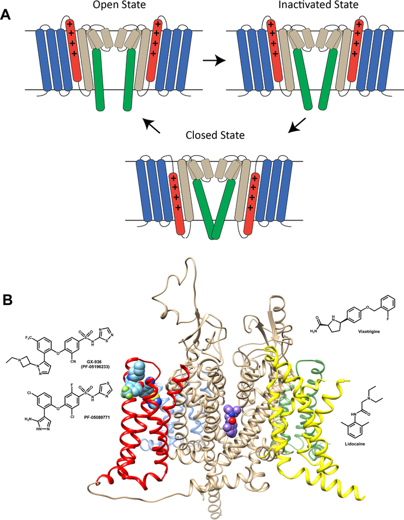
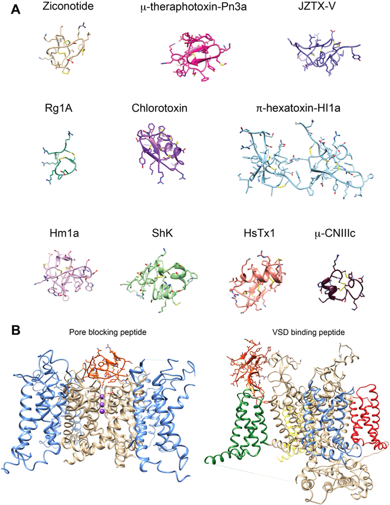
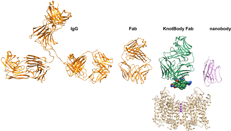
Ion channels play fundamental roles in both excitable and non-excitable tissues and therefore constitute attractive drug targets for myriad neurological, cardiovascular and metabolic diseases as well as for cancer and immunomodulation. However, achieving selectivity for specific ion channel subtypes with small-molecule drugs has been challenging, and there currently is a growing trend to target ion channels with biologics. One approach is to improve the pharmacokinetics of existing or novel venom-derived peptides. In parallel, after initial studies with polyclonal antibodies demonstrated the technical feasibility of inhibiting channel function with antibodies, multiple preclinical programmes are now using the full spectrum of available technologies to generate conventional monoclonal and engineered antibodies or nanobodies against extracellular loops of ion channels. After a summary of the current state of ion channel drug discovery, this Review discusses recent developments using the purinergic receptor channel P2X purinoceptor 7 (P2X7), the voltage-gated potassium channel KV1.3 and the voltage-gated sodium channel NaV1.7 as examples of targeting ion channels with biologics.


Conflict of interest statement

Competing interests statement

The authors declare competing financial interests: see web version for details.

Palle Christophersen is a full-time employee of Saniona A/S. Paul Colussi, is a full-time employee of TetraGenetics Inc.

Figures

Figure 1.
Figure 1.
A) Cartoon representation of the Nav channel gating cycle in closed, open and inactivated states. The voltage sensing domain (VSD) segments are colored blue (S1-S3) and red (S4). The pore-forming domain segments are colored sand (S5, P1-helix, P2-helix, and N-terminal part of S6) and green (C-terminal part of S6). The gating charge carrying arginines in the S4 segment are represented by “+” signs. Key conformational changes in the Nav channel during gating between closed, open, and inactivated states are highlighted by transmembrane movement of the S4 segment (colored in red) and lateral movement of the C-terminal part of S6 segment (colored in green). Inactivation in Nav channels can be either fast and involve one of the cytoplasmic inter-repeat loops “plugging” the inner vestibule or slow and involve a rearrangement of the selectivity filter. B) Small molecule receptor sites within Nav channels. Transmembrane view of Nav channel interaction with pore bound (lidocaine) and voltage-sensor bound (GX-936) small molecule drugs. The Nav channel structure is based on the electric eel Nav1.4 channel structure (pdb: 5XSV) and is shown in ribbon representation. The pore domain is shown in beige and the voltage-sensors are shown in blue (domain I), green (domain II), yellow (domain III), and red (domain IV). Lidocaine is shown in space-filling representation and colored purple. GX-936 is shown in space-filling representation and colored light blue. The chemical structures of vixotrigine, lidocaine, GX-936, and PF-05089771 are shown in 2D representation next to the channel.
Figure 2.
Figure 2.
A) Structures of peptide toxins targeting ion channels. Ribbon representation of peptide toxin structures colored individually and labeled. Disulfide bonds are colored in yellow. Ziconotide (pdb: 1TTK), μ-theraphotoxin-Pn3a (pdb: 5T4R_A), JZTX-v (pdb: 6CGW), Rg1A (pdb: 2JUT), chlorotoxin (pdb: 5L1C), π-hexatoxin-HI1a (pdb: 2N8F), Hm1a (pdb: 2N6O), ShK (pdb:2K9E_A), HsTx1 (pdb: 1QUZ_A) μ-CNIIIc (pdb: 2YEN). B) Peptide toxin receptor sites on ion channels. Left, Pore blocking peptide receptor site in a Kv channel illustrated by the transmembrane view of charybdotoxin in the pore of the Kv1.2-Kv2.1 chimera structure (pdb: 2R9R) and shown in ribbon representation. The pore is shown in beige and the voltage-sensors are shown in blue. Charybdotoxin is shown in orange. Potassium ions within the selectivity filter region are shown in sphere representation and colored purple. Right, voltage sensor binding peptide receptor site in a Nav channel illustrated by the transmembrane view of the voltagesensor bound peptide toxin Dc1a. The Nav structure is the American cockroach NavPaS structure (pdb: 6A90) and shown in ribbon representation. The pore is shown in beige and voltage-sensors are shown in blue (domain I), green (domain II), yellow (domain III), and red (domain Iv). The Dc1a toxin is shown in orange.
Figure 3.
Figure 3.
Figure illustrating the size relation of a full-length immunoglobulin G (IgG2a, pdb: 11GT, ribbon presentation) colored in orange, an antigen-binding fragment (Fab, pdb: 1K4C) colored in orange, a KnotBody Fab colored in green, and a nanobody (pdb: 6C5W) colored in pink. The Ecballium elaterium Trypsin Inhibitor (EETI-II) structure within the KnotBody is shown in space-filling representation. The Kv channel structure is based on the Kv1.2-Kv2.1 chimera (pdb: 2R9R) and shown in ribbon representation and colored in beige. Potassium ions within the selectivity filter region are shown in sphere representation and colored in purple.

References

    1. Alexander SP et al. THE CONCISE GUIDE TO PHARMACOLOGY 2017/18: Overview. Br. J. Pharmacol. 174 Suppl 1, S1–S16 (2017). - PMC - PubMed
    1. Alexander SP et al. THE CONCISE GUIDE TO PHARMACOLOGY 2017/18: voltage-gated ion channels. Br. J. Pharmacol. 174 Suppl 1, S160–S194 (2017). - PMC - PubMed
    1. Alexander SP et al. THE CONCISE GUIDE TO PHARMACOLOGY 2017/18: Ligand- gated ion channels. Br. J. Pharmacol. 174 Suppl 1, S130–S159 (2017). - PMC - PubMed
    1. Alexander SP et al. THE CONCISE GUIDE TO PHARMACOLOGY 2017/18: Other ion channels. Br. J. Pharmacol. 174 Suppl 1, S195–S207 (2017). - PMC - PubMed
    1. Santos R et al. A comprehensive map of molecular drug targets. Nat. Rev. Drug Discov. 16, 19–34 (2017). - PMC - PubMed

Publication types