Review
. 2018 Dec;35(5):406-426.
doi: 10.1055/s-0038-1676342. Epub 2019 Feb 5.

On the Cutting Edge: Wound Care for the Endovascular Specialist

Affiliations
Review

On the Cutting Edge: Wound Care for the Endovascular Specialist

Brandon Olivieri et al. Semin Intervent Radiol. 2018 Dec.

Abstract

Clinical outcomes in patients with critical limb ischemia (CLI) depend not only on endovascular restoration of macrovascular blood flow but also on aggressive periprocedural wound care. Education about this area of CLI therapy is essential not only to maximize the benefits of endovascular therapy but also to facilitate participation in the multidisciplinary care crucial to attaining limb salvage. In this article, we review the advances in wound care products and therapies that have granted the wound care specialist the ability to heal previously nonhealing wounds. We provide a primer on the basic science behind wound healing and the pathogenesis of ischemic wounds, familiarize the reader with methods of tissue viability assessment, and provide an overview of wound debridement techniques, dressings, hyperbaric therapy, and tissue offloading devices. Lastly, we explore emerging technology on the horizons of wound care.

Keywords: critical limb ischemia; debridement; interventional radiology; peripheral arterial disease; ulcer; wound care.


Figures

Fig. 1
Fig. 1
Diagram depicting ideal conceptual framework for addressing each critical limb ischemia patient.
Fig. 2
Fig. 2
Visual tools, such as the Society for Vascular Surgery Wound, Ischemia, and foot Infection (WIfI) classification smartphone application, shown here in use with a critical limb ischemia patient in clinic (a) and as a screenshot (b) , are very useful for improving a patient's understanding of their condition, the associated risk of amputation, and the potential benefits of revascularization.
Fig. 3
Fig. 3
The stages of normal wound healing over time.
Fig. 4
Fig. 4
Selective versus nonselective methods of debridement.
Fig. 5
Fig. 5
Wet-to-dry dressings are a commonly used form of mechanical debridement. In this patient with an infected heel wound, a povidone-iodine antiseptic solution is used to saturate sterile gauze ( a ). This is then applied to the wound ( b, c ), covered with ample dry gauze, and secured in place ( d ). After approximately 4 to 6 hours, the gauze has dried and become adherent at which point it is removed (not shown), performing nonselective mechanical wound debridement.
Fig. 6 (a, b)
Fig. 6 (a, b)
A patient in clinic undergoes sharp debridement of periulcer callous to promote continued proper healing of the wound.
Fig. 7
Fig. 7
A patient with extensive heel gangrene undergoes surgical debridement in the operating room.
Fig. 8
Fig. 8
HTOM with the HyperView device (HyperMed Imaging) uses a handheld spectrometer (a) to quantify tissue perfusion and viability via the absorption of visible light in hemoglobin molecules over a region of interest. In a region of ulceration (b), the levels of superficial subcutaneous tissue oxyhemoglobin (c) , deoxyhemoglobin (d) , and oxygen saturation (e) can be determined by differential light absorption and are color coded. Images reproduced with permission from HyperMed Imaging, Inc., Memphis, TN.
Fig. 9
Fig. 9
NIFA imaging using ICG in use for intraoperative assessment of tissue viability prior to possible amputation in a patient who underwent successful revascularization after initially presenting with right great hallux (a) and left second digit (b) gangrenous changes. Sterile water and ICG solution (c) are injected intravenously. The NIFA camera and display console are then used to assess viability via the detection of ICG (d). Fluorescence imaging of the gangrenous right hallux demonstrates a concordant paucity of flow ( white arrows ) in the gangrenous region in both anteroposterior (e) and lateral projections (f). Given this lack of viability even after revascularization, decision was made to perform amputation of the right hallux (g, h). Postamputation NIFA imaging of the right foot demonstrates flow to the surgical resection margin in both anteroposterior and lateral projections (i, j). Fluorescence imaging of the gangrenous tip of the left second digit (k) demonstrates focal area of diminished flow ( white arrow ) that is discordant with the amount of gangrene on physical exam, indicating a penumbra of viable tissue. Therefore, surgical debridement of the left second toe was performed rather than amputation.
Fig. 10
Fig. 10
An 80-year-old male with history of smoking, hypertension, hyperlipidemia, and critical limb ischemia gangrenous changes to the right second and third toes (a) with underlying metatarsophalangeal joint abscess. AP angiogram of the right infrageniculate circulation (b) demonstrated ostial occlusion of the right anterior tibial artery * (TASC II type D), subtotal occlusion of the peroneal artery (TASC II type C), and 50% proximal posterior tibial artery stenosis (TASC II type A) with no in-line flow to the foot (not shown). Tibial and pedal plantar loop reconstruction was then performed with complete recanalization of the anterior tibial (AT), tibioperoneal trunk (TP), peroneal (P), dorsalis pedis, and lateral plantar (LP) arteries (c, d) . Second and third digits and metatarsophalangeal joint resection with evacuation of abscess was then performed with apparent adequate bleeding post-revascularization (e) . Fluobeam angiography of the resection site was performed demonstrating unexpected areas of hypoperfusion to the wound site and periphery ( white arrows ; f) . Transmetatarsal amputation was performed with adequate macro/microvasculature for stump healing now demonstrated by NIFA (g). Outpatient follow-up showed complete stump healing and ambulation ( h ).
Fig. 11
Fig. 11
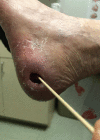
Medial heel ulcer in a critical limb ischemia patient that probes to bone on examination.
Fig. 12
Fig. 12
Pyoderma gangrenosum mimicking a nonhealing wound. (Reprinted with permission from Snyder et al. 91 )
Fig. 13
Fig. 13
Aggressive digital adenocarcinoma mimicking a nonhealed wound. (Reprinted with permission from Vazales et al. 92 )
Fig. 14
Fig. 14
Components of a total contact case.
Fig. 15
Fig. 15
Removable cast walker ( a ) and removable offloading shoe ( b ).
Fig. 16
Fig. 16
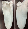
Interior ( a ) and exterior ( b ) of a custom-fitting orthosis.
Fig. 17
Fig. 17
Various offloading shoe inserts.

References

    1. Fowkes F GR, Rudan D, Rudan Iet al.Comparison of global estimates of prevalence and risk factors for peripheral artery disease in 2000 and 2010: a systematic review and analysis Lancet 2013382(9901):1329–1340. - PubMed
    1. Henry J C, Peterson L A, Schlanger R E. Wound healing in peripheral arterial disease: current and future therapy. J Vasc Med Surg. 2014;02(04):157.
    1. Yost M.PAD Costs Economics Amputation Costs Economics, Critical Limb Ischemia, Chronic Venous Disease, Venous Ulcers, Chronic Venus Insufficiency - CLI US Supplement 2016The Sage Group. Available at:http://thesagegroup.us/pages/reports/cli-us-supplement-2016.php. Accessed August 18, 2018
    1. Noone A, Howlander N, Krapcho M, Miller D.SEER Cancer Statistics Review, 1975–2015National Cancer Institute;2018. Available at:https://seer.cancer.gov/csr/1975_2015/. Accessed August 27, 2018
    1. Inter-Society Consensus for the Management of Peripheral Arterial Disease (TASC II) - Journal of Vascular Surgery. Available at:https://www.jvascsurg.org/article/S0741-5214(06)02296-8/fulltext. Accessed August 18, 2018 - PubMed