Review
. 2019 Jan 23:12:1035.
doi: 10.3389/fnins.2018.01035. eCollection 2018.

Glycolysis-Derived Compounds From Astrocytes That Modulate Synaptic Communication

Affiliations
Review

Glycolysis-Derived Compounds From Astrocytes That Modulate Synaptic Communication

Carlos-Alberto Gonçalves et al. Front Neurosci. .

Abstract

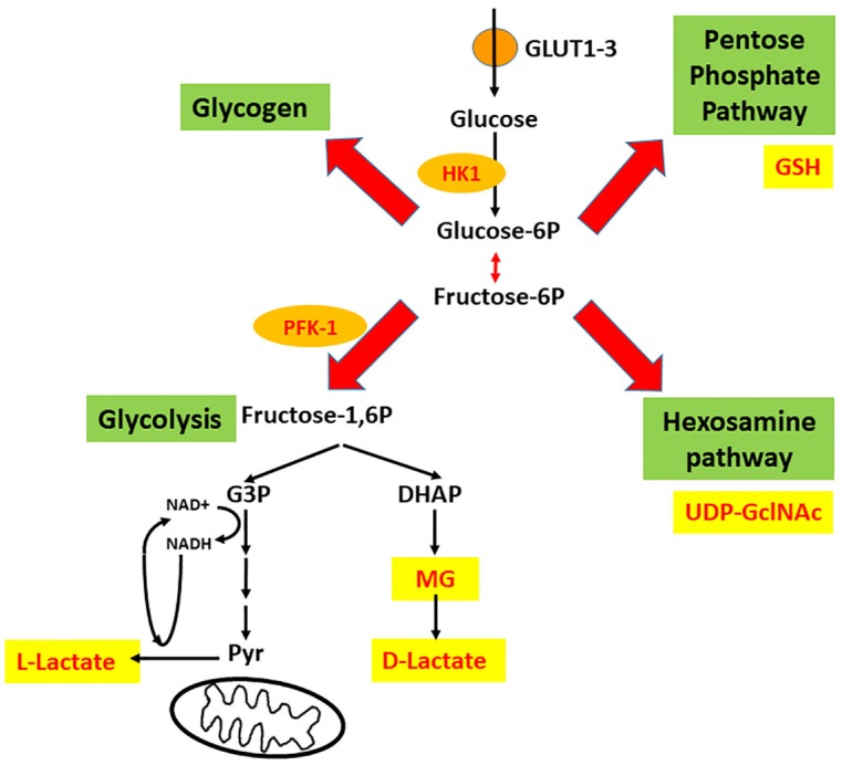
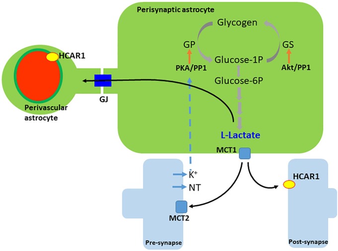
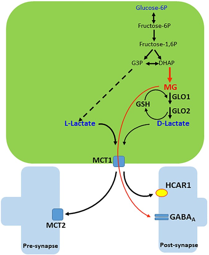
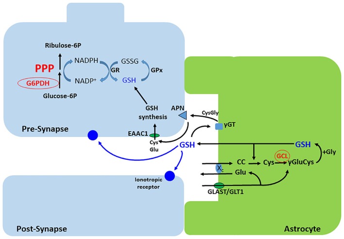
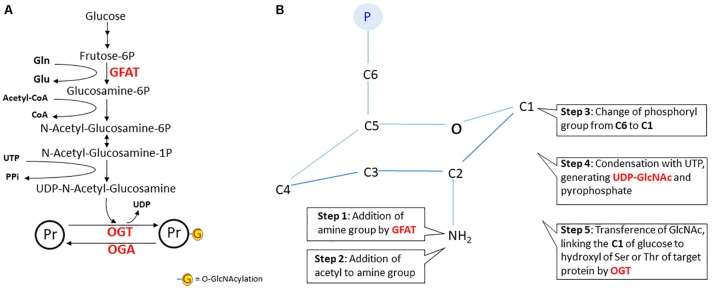
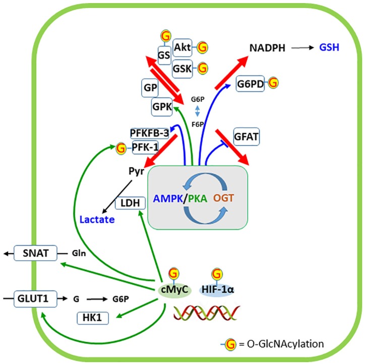
Based on the concept of the tripartite synapse, we have reviewed the role of glucose-derived compounds in glycolytic pathways in astroglial cells. Glucose provides energy and substrate replenishment for brain activity, such as glutamate and lipid synthesis. In addition, glucose metabolism in the astroglial cytoplasm results in products such as lactate, methylglyoxal, and glutathione, which modulate receptors and channels in neurons. Glucose has four potential destinations in neural cells, and it is possible to propose a crossroads in "X" that can be used to describe these four destinations. Glucose-6P can be used either for glycogen synthesis or the pentose phosphate pathway on the left and right arms of the X, respectively. Fructose-6P continues through the glycolysis pathway until pyruvate is formed but can also act as the initial compound in the hexosamine pathway, representing the left and right legs of the X, respectively. We describe each glucose destination and its regulation, indicating the products of these pathways and how they can affect synaptic communication. Extracellular L-lactate, either generated from glucose or from glycogen, binds to HCAR1, a specific receptor that is abundantly localized in perivascular and post-synaptic membranes and regulates synaptic plasticity. Methylglyoxal, a product of a deviation of glycolysis, and its derivative D-lactate are also released by astrocytes and bind to GABAA receptors and HCAR1, respectively. Glutathione, in addition to its antioxidant role, also binds to ionotropic glutamate receptors in the synaptic cleft. Finally, we examined the hexosamine pathway and evaluated the effect of GlcNAc-modification on key proteins that regulate the other glucose destinations.

Keywords: GSH; astrocyte; glycolysis; lactate; methylglyoxal; neurotransmission.


Figures

FIGURE 1
FIGURE 1
Four intracellular destinations of glucose that suggest an intersection in “X.” Glucose enters astrocytes mainly via GLUT1, and neurons mainly via GLUT3 and is trapped by phosphorylation (catalyzed by hexokinase 1, HK1). Afterward, four destinations are possible; these form a crossroads in the shape of an X, where glycogen synthesis and the pentose phosphate pathway (PPP) are the left and right arms of the X, and glycolysis (until pyruvate) and the hexosamine pathway (HP) are the left and right legs of the X. The deviation of glycolysis that generates methylglyoxal (MG) is also indicated. PFK-1, phosphofructokinase-1; G-3P, glyceraldehyde 3-phosphate; DHAP, dihydroxyacetone phosphate.
FIGURE 2
FIGURE 2
Generation and release of lactate in astrocytes. L-lactate is generated either from recently uptaken glucose or from glycogen. Neurotransmitters (NT), and/or extracellular K+, trigger glycogen breakdown until lactate, via cAMP/PKA signaling. Lactate leaves the cell by the monocarboxylate transporter 1 (MCT1) and enters neurons via the monocarboxylate transporter 2 (MCT2). Extracellular lactate also binds to hydroxy-carboxylic acid receptor 1 (HCAR1), which is found more abundantly in perivascular and post-synaptic membranes. Lactate travels among astrocytes through gap junctions (GJ). PKA, protein kinase A; GP, glycogen phosphorylase; GS, glycogen synthase; PP1, protein phosphatase 1.
FIGURE 3
FIGURE 3
Generation and release of MG and D-lactate in astrocytes. Methylglyoxal (MG) is produced from dihydroxyacetone phosphate (DHAP) by a deviation of the glycolytic pathway. MG is condensed with glutathione (GSH) and then, by sequential action of glyoxalases 1 and 2 (GLO1 and 2), generates D-lactate and recycles GSH. D-lactate leaves the cell via monocarboxylate transporter 1 (MCT1) but disturbs lactate and pyruvate flows to mitochondria (not illustrated). MG and D-lactate leave the cell and act on the GABAA receptor and hydroxy-carboxylic acid receptor 1 (HCAR1), respectively. Extrasynaptic GABAA receptors of MG are not illustrated.
FIGURE 4
FIGURE 4
Synthesis and release of GSH in astrocytes. Cystin (cisteinyl-cystein, CC) is uptaken via the xc- exchanger, which releases glutamate. This exchanger is functionally coupled to the Na+- dependent glutamate transporters, GLAST or GLT-1. Cystin is reduced by GSH to two cysteines. The gamma acid group of glutamate then condenses with the amine group of cysteine, forming gamma-glutamyl-cysteine (γ Glu-Cys), by the action of the glutamate cysteine ligase (GCL). Addition of glycine completes the synthesis of GSH that, in part, is exported to modulate ionotropic receptors and as a source of cysteine for GSH synthesis in neurons. The extracellular cysteine from astroglial GSH is generated after sequential action of the extracellular peptidases, γGT and APN. Neurons synthesize low levels of GSH compared with astrocytes, but exhibit a high capacity of GSH regeneration, which depends on NADPH synthesis in the PPP. γGT, gamma peptidyl transpeptidase; APN, aminopeptidase neuronal; GPx, glutathione peroxidase; GR, glutathione reductase; EAAC, excitatory amino acid carrier.
FIGURE 5
FIGURE 5
The hexosamine pathway. In panel A, the steps of the hexosamine pathway (HP) from frutose-6P to UDP-N-acetyl-glucosamine. Notice that glutamine (Gln), Acetyl-CoA and UTP are key substrates in this pathway. The rate-limiting step is catalyzed by glutamine:fructose-6P aminotransferase (GFAT). In panel B, a schematic representation of the structural changes during N-acetyl-glucosamine synthesis. The first reaction is the amination of fructose-6P (fructofuranose) to glucosamine-6P (glucopyranose). It is worth noting that, in step six, carbon 1 of glucopyranose binds to the hydroxyl of serine or threonine of the protein target. In fact, this is a reaction of O-linked β-N-acetylglucosaminylation, but for simplification, the nomenclature widely used is O-GlcNAcylation. However, this gives the wrong idea that linking occurs at the acyl group. It would be better to use NAGylation, since NAG is another (and the simplest) abbreviation of N-AcetylGlucosamine, found in some polymers. Herein, we will maintain the use of the term “O-GlcNAcylation.” Abbreviations: OGT, O-GlcNAc transferase; OGA, O-GlcNAcase; Pr, protein.
FIGURE 6
FIGURE 6
Regulation of cellular destinations of glucose by O-GlcNAc modification of proteins. AMP-activated protein kinase (AMPK) and protein kinase A (PKA) have a narrow interaction in the regulation of glucose metabolism as well as phosphorylate and are targets of O-GlcNAc transferase (OGT). The phosphorylation of specific targets of AMPK or PKA in the four cellular destinations of glucose are indicated by the respective colored arrows. Targets of OGT are indicated by G. Two transcription factors closely related to glucose metabolism (cMyc and HIF-1α) are represented at the double-strand DNA and five proteins whose expressions are regulated by these transcription factors are indicated. G6PD, glucose-6-phosphate dehydrogenase; GFAT, glutamine:fructose-6P aminotransferase; GLUT1, glucose transporter 1; GP, glycogen phosphorylase; GPK, glycogen phosphorylase kinase; GS, glycogen synthase; GSK, glycogen synthase kinase; HIF-1α, hypoxia-inducible factor-1α; HK1, hexokinase 1; LDH, lactate dehydrogenase; PFK-1, phosphofructokinase 1; PFKFB-3, 6-phosphofructo-2-kinase/fructose-2,6-bisphosphatase-3; SNAT, sodium-neutral amino acid transporter (type 3 or 5, in astrocytes).

References

    1. Ahmed K., Tunaru S., Offermanns S. (2009). GPR109A, GPR109B and GPR81, a family of hydroxy-carboxylic acid receptors. Trends Pharmacol. Sci. 30 557–562. 10.1016/j.tips.2009.09.001 - DOI - PubMed
    1. Ahmed K., Tunaru S., Tang C., Müller M., Gille A., Sassmann A., et al. (2010). An autocrine lactate loop mediates insulin-dependent inhibition of lipolysis through GPR81. Cell Metab. 11 311–319. 10.1016/j.cmet.2010.02.012 - DOI - PubMed
    1. Allaman I., Bélanger M., Magistretti P. J. (2015). Methylglyoxal, the dark side of glycolysis. Front. Neurosci. 9:23. 10.3389/fnins.2015.00023 - DOI - PMC - PubMed
    1. Almeida A., Moncada S., Bolaños J. P. (2004). Nitric oxide switches on glycolysis through the AMP protein kinase and 6-phosphofructo-2-kinase pathway. Nat. Cell Biol. 6 45–51. 10.1038/ncb1080 - DOI - PubMed
    1. Anderson C. M., Swanson R. A. (2000). Astrocyte glutamate transport: review of properties, regulation, and physiological functions. Glia 32 1–14. 10.1002/1098-1136(200010)32:1<1::AID-GLIA10>3.0.CO;2-W - DOI - PubMed

LinkOut - more resources