Review
. 2019 Jan 21:12:117.
doi: 10.3389/fncir.2018.00117. eCollection 2018.

General Principles of Neuronal Co-transmission: Insights From Multiple Model Systems

Affiliations
Review

General Principles of Neuronal Co-transmission: Insights From Multiple Model Systems

Erik Svensson et al. Front Neural Circuits. .

Abstract

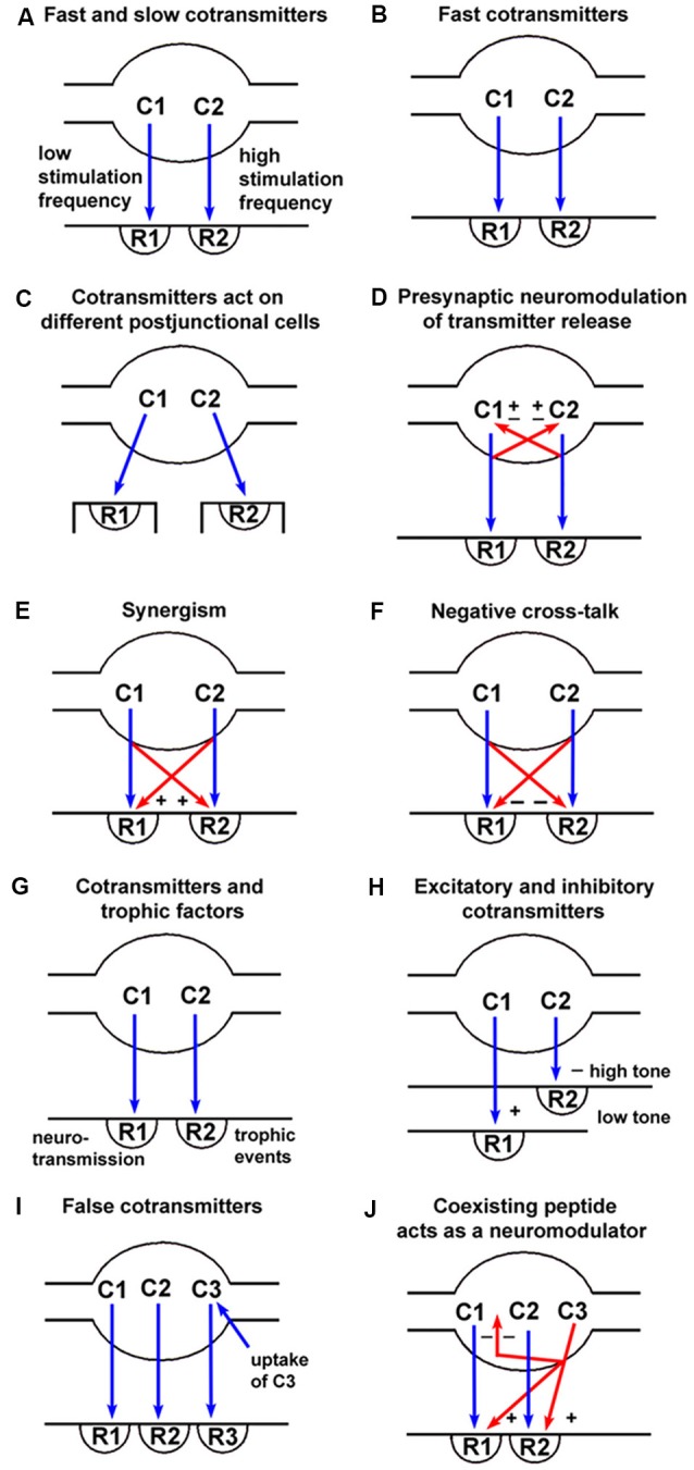
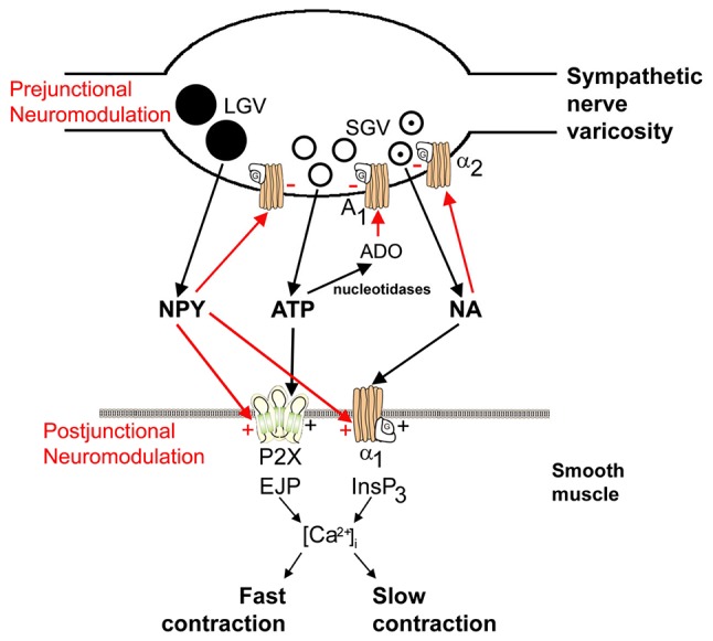
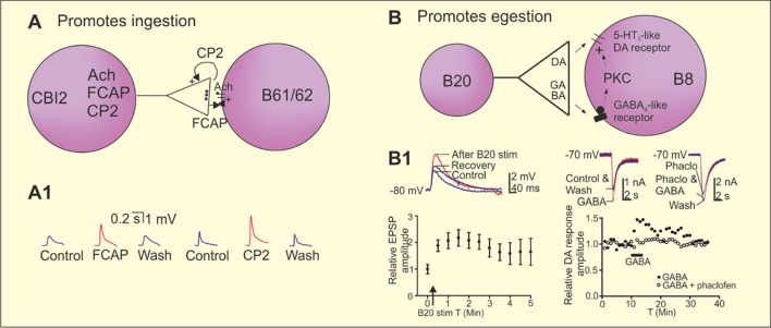
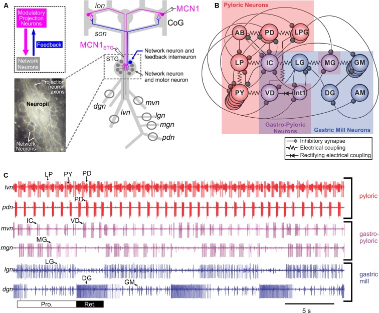
It is now accepted that neurons contain and release multiple transmitter substances. However, we still have only limited insight into the regulation and functional effects of this co-transmission. Given that there are 200 or more neurotransmitters, the chemical complexity of the nervous system is daunting. This is made more-so by the fact that their interacting effects can generate diverse non-linear and novel consequences. The relatively poor history of pharmacological approaches likely reflects the fact that manipulating a transmitter system will not necessarily mimic its roles within the normal chemical environment of the nervous system (e.g., when it acts in parallel with co-transmitters). In this article, co-transmission is discussed in a range of systems [from invertebrate and lower vertebrate models, up to the mammalian peripheral and central nervous system (CNS)] to highlight approaches used, degree of understanding, and open questions and future directions. Finally, we offer some outlines of what we consider to be the general principles of co-transmission, as well as what we think are the most pressing general aspects that need to be addressed to move forward in our understanding of co-transmission.

Keywords: colocalization; corelease; neuromodulation; neuropeptides; neurotransmitter complexity.


Figures

Figure 1
Figure 1
Spectrum of signaling variations offered by co-transmission (blue arrows = neurotransmission; red arrows = pre- or post-junctional neuromodulation). (A) Fast transmission is usually produced by small molecules (C1) released at low frequency nerve stimulation acting on ionotropic receptors (R1), whereas slow transmission is usually produced by release of peptides (C2) or other molecules at high frequency stimulation acting on G-protein-coupled receptors (R2). (B) Co-transmitters C1 and C2 can both be fast messengers acting via ionotropic receptors (R1 and R2). (C) Co-transmitters C1 and C2 act on receptors (R1 and R2) localized on different postjunctional cells. (D) Co-transmitters C1 and C2 not only act postjunctionally via R1 and R2 receptors but can also act as prejunctional modulators to either inhibit (−) or enhance (+) the release of C1 and/or C2. (E) Co-transmitters C1 and C2 act synergistically to enhance the combined responses produced via R1 and R2 receptors. (F) Co-transmitters C1 and C2 act to inhibit the responses evoked via R1 and/or R2 receptors. (G) Co-transmitter C1 evokes neurotransmission via R1 receptors, while C2 evokes long-term (trophic) responses of postjunctional cells via R2 receptors. (H) Co-transmitter C1 produces excitation via R1 receptors when the postjunctional smooth muscle target has low tone, with C2 having little influence; however, when the smooth muscle tone is high, the dominant response might be relaxation produced by C2 via R2 receptors. (I) Substance C3 is taken up by nerve terminals, rather than being synthesized and stored as is true for the co-transmitters C1 and C2. C3 can then be released on nerve stimulation to act on postjunctional R3 receptors. In these circumstances, C3 would be known as a “false transmitter.” (J) A coexisting substance C3 (often a peptide) can be synthesized and stored in a nerve, but not act directly via a postjunctional receptor to produce changes in postjunctional cell activity. It could, however, act as a prejunctional inhibitor (−) of the release of the co-transmitters C1 and C2, or as a postjunctional enhancer (+) of the responses mediated by R1 and R2. (Reproduced from Burnstock (2004), with permission from Elsevier).
Figure 2
Figure 2
Schematic of sympathetic co-transmission. Adenosine 5’-triphosphate (ATP) and noradrenaline (NA) released from small granular vesicles (SGVs) act on P2X and α1 receptors on smooth muscle, respectively. ATP acting on inotropic P2X receptors evokes excitatory junction potentials (EJPs), an increase in intracellular calcium [(Ca2+)]i and fast contraction; while metabotropic α1 adrenoceptors leads to production of inositol triphosphate (InsP3), an increase in (Ca2+)i and slow contraction. Neuropeptide Y (NPY) stored in large granular vesicles (LGVs) acts after release both as a prejunctional inhibitory modulator of release of ATP and NA and as a postjunctional modulatory potentiator of the actions of ATP and NA. Soluble nucleotidases are released from nerve varicosities, and are also present as ectonucleotidases [reproduced from Burnstock and Verkhratsky (2010) with permission from Elsevier].
Figure 3
Figure 3
Co-transmission in the regulation of ingestion and egestion in the Aplysia feeding circuit. (A) Repeated stimulation of the cerebral buccal interneuron 2 (CBI-2) progressively induces the ingestive motor programs in Aplysia. The command neuron CBI-2 uses acetylcholine (Ach) as its fast-excitatory neurotransmitter onto the motor neurons B61/62. CBI-2 also co-localize the two neuropeptides feeding circuitry activating peptide (FCAP) and cerebral peptide 2 (CP-2). Both peptides are released by high frequency stimulation of CBI-2 and contributes to post-tetanic potentiation (PTP) of the fast-cholinergic transmission at the CBI-2 to B61/62 synapses, however by different mechanisms. CP-2 is acting presynaptically and FCAP postsynaptically. (A1) Recording traces showing the potentiation of the cholinergic EPSP by FCAP and CP-2 in B8. (B) Repeated stimulation of the esophageal nerve and high frequency stimulation of the interneuron B20 that co-localizes dopamine and γ-aminobutyric acid (GABA) induces egestion and a short-term potentiation of the fast dopaminergic EPSPs between B20 and the motor neuron B8. Dopamine acts on a 5-HT3-like receptor and GABA contributes to the potentiation of the B20 to B8 EPSP by a postsynaptic mechanism that involves activation a GABAB receptor of protein kinase C (PKC). (B1) High frequency stimulation of B20 potentiates the dopaminergic EPSPs in B8. GABA potentiated fast dopaminergic responses in B8 and the effect is blocked by the GABAB receptor antagonist phaclofen. Adapted from Koh et al. (2003), Koh and Weiss (2005) and Svensson et al. (2014).
Figure 4
Figure 4
The crab Cancer borealis stomatogastric nervous system (STNS). (A) Schematic of the isolated STNS of the crab Cancer borealis. The inset shows a whole-mount image of the desheathed stomatogastric ganglion (STG) under dark-field illumination (anterior, top; posterior, bottom). As it is evident in both the schematic and the inset, the 26 neuronal somata form a single layer surrounding the neuropil. Circles on nerves indicate recording sites for traces shown in part (C). (B) Schematic of the gastric mill and pyloric circuit. The arrangement of neurons in the schematic represents the relative timing of activity for each neuron during the gastric mill and pyloric rhythms. Specifically, the neurons that exhibit pyloric rhythm-timed activity (“pyloric neurons” and “gastropyloric neurons”) are displayed such that the top-row neurons are co-active, followed by the middle-row neurons and then the bottom-row neurons, after which the top-row neurons are again active. The neurons that exhibit gastric mill rhythm (GMR)-timed activity (“gastric mill neurons” and “gastropyloric neurons”) are displayed such that the top-row neurons are co-active and burst in alternation with the bottom-row neurons. As shown, there are eight gastric mill circuit neuron types, one of which is present as four apparently equivalent copies (GM neurons). All eight neuron types contribute to gastric mill pattern generation, whereas only two (LG and Int1) are also rhythm generator neurons (Coleman et al., ; Bartos et al., ; Saideman et al., 2007). There are seven pyloric circuit neuron types, including the rhythm generator (“pacemaker”) group AB/PD/LPG (Marder and Bucher, 2007). Three of these neuron types are present as multiple, apparently equivalent copies (PD: 2; LPG: 2; PY: 5). (C) Simultaneous extracellular nerve recordings of the gastric mill and pyloric rhythms during tonic stimulation of the modulatory projection neuron modulatory commissural neuron 1 (MCN1) are shown. The pyloric rhythm exhibits a rhythmically repeating triphasic pattern (for example, lateral ventricular nerve (lvn): PD, LP, PY) that is continuously active, in vivo and in vitro, with a cycle period of ~1 s. The GMR (cycle period ~10–20 s) is silent except when driven by modulatory neurons (for example, MCN1), which themselves require activation in vivo and in vitro. It is a rhythmically repeating biphasic pattern, consisting of teeth protraction (Pro.) and teeth retraction (Ret.), which drives the motor response (chewing). Note that some neurons exhibit activity patterns time-locked to both rhythms (gastropyloric neurons). CoG, commissural ganglion; dgn, dorsal gastric nerve; ion, inferior oesophageal nerve; lgn, lateral gastric nerve; mgn, medial gastric nerve; mvn, medial ventricular nerve; pdn, pyloric dilator nerve; son, superior oesophageal nerve; stn, stomatogastric nerve.Panels (A) and (C) are adapted with permission Nusbaum et al. (2017), Nature Reviews Neuroscience. Panel (B) is adapted with permission from Nusbaum and Beenhakker (2002), Nature. STG photo courtesy of Marie Suver, New York University, USA, and Wolfgang Stein, Illinois State University, USA.
Figure 5
Figure 5
The microcircuit response to peptidergic neuron activity is not necessarily mimicked by bath application of that neuropeptide. (A) Extracellular recordings of identified neurons in the crab Cancer borealis STG, which are active during the GMR (LG and DG neurons), pyloric rhythm (PD neuron) or both rhythms (IC and VD neurons). In the isolated crab STG, bath-applied proctolin (far left set of responses) selectively excites the pyloric rhythm (Marder et al., ; Nusbaum and Marder, 1989a). This action mimics the response to activation of only one [modulatory proctolin neuron (MPN)] of the three proctolinergic projection neurons that innervate the STG (MPN, MCN1 and MCN7), even though MPN also contains a small-molecule co-transmitter (GABA; Blitz et al., 1999). As indicated, MPN also inhibits two projection neurons [MCN1 and commissural projection neuron 2 (CPN2)] by releasing GABA from a separate axon projecting to a separate location (CoG; Blitz and Nusbaum, 1997, 1999). The other two proctolinergic projection neurons (MCN1 and MCN7) also influence STG microcircuit activity but elicit activity patterns from the circuit neurons that are distinct from proctolin bath application (Coleman and Nusbaum, ; Blitz et al., 1999). MCN1-released C. borealis tachykinin-related peptide Ia (CabTRP Ia) and GABA are pivotal for MCN1 activation of the GMR, whereas its release of CabTRP Ia and proctolin dominates its excitation of the pyloric rhythm (see part B). The MCN7 actions on these rhythms result partly from proctolin and probably also from one or more yet-to-be-identified co-transmitters (indicated by “?”). In the figure, pyloric rhythm activity is shown in red; GMR activity is shown in blue; gastropyloric activity is shown in purple. (B) In the crab STG, MCN1 influences all pyloric, gastropyloric and gastric mill neurons. The figure shows a representation of responsiveness of each STG circuit neuron to the MCN1-released co-transmitters proctolin (white w/black border), CabTRP Ia (green) and GABA (black; Swensen and Marder, ; Stein et al., 2007). Examples of convergent peptide co-transmitter action (proctolin and CabTRP Ia), selective peptide co-transmitter action (CabTRP Ia) and selective GABA action are shown. In some cases, the STG neuron only responds to the indicated co-transmitter (or co-transmitters; for example, Int1). In other cases, the STG neuron does respond to an additional co-transmitter but not when it is released from MCN1 (for example, LG responds to applied GABA but not GABA released from MCN1). No information is available regarding whether these co-transmitters are colocalized to all MCN1 terminals or are localized to separate terminals for their release. Panel (A) is adapted with permission from Nusbaum et al. (2001), Elsevier. Panel (B) is adapted with permission from Nusbaum et al. (2017), Nature Reviews Neuroscience.
Figure 6
Figure 6
Activation of hypothalamic orexin neurons that impact wakefulness-promoting histamine neurons generates distinct signature excitatory responses resulting from the co-release of glutamate and the neuropeptide hypocretin/orexin (HO). (Top panel) A cre-recombinase approach was used to express the light-activating excitatory protein channelrhodopsin (ChR2) in orexin neurons in orexin-cre transgenic mice. Brief pulses of light were sufficient to evoke transmitter release from orexin terminals that then impacted postsynaptic histamine neurons, which were identified by intrinsic signature currents and post hoc immunoprocessing for histamine decarboxylase reactivity. (Bottom panel) Low frequency stimulation led to the synaptic release of glutamate only while under high firing regimes both glutamate and orexin were co-released. In response to this co-transmission, the evoked responses measured on histamine neurons were sequential, where glutamate release and its corresponding postsynaptic excitation was fast and transient, lasting only while orexin neurons were active while orexin independently excited histamine neurons in a delayed fashion and whose response outlasted the stimulation duration. These results indicate that glutamate/orexin cotransmission may translate distinct features of orexin activity into parallel, nonredundant signals for regulating distinct circuits important for generating appropriate levels of arousal for maintaining wakefulness. Figure adapted with permission from Schöne et al. (2014) and the publisher.
Figure 7
Figure 7
Co-transmission in higher brain circuits (neocortex and basal ganglia). (A) In the cortical column GABAergic interneurons co-localize a range of different neuropeptides that can contribute to the tuning of activity of the cortical column. The neocortex also receives innervation from subcortical nuclei that can influence the tuning of cortical network activity. The projection from ventral tegmental area (VTA) co-localize glutamate, dopamine and/or the neuropeptides neurotensin and CCK. The cholinergic projections from forebrain neurons co-release GABA and thus activates nAchR and GABAA receptors in cortical neurons. (A1). Traces showing nicotinergic receptor and GABAA receptor antagonism of optogenetically generated synaptic potentials. (B) In the basal ganglia, GABAergic medium spiny neurons (MSN) in the direct pathway co-localize substance P and dynorphins, while MSN in the indirect pathway co-localized enkephalins. Cholinergic interneurons in striatum co-localize glutamate and have excitatory action onto MSN. Dopaminergic neurons from substantia nigra pars compacta also co-localize and co-release GABA and glutamate. This input to striatum will thus activate GABAA, AMPA, NMDA, and D1 (dMSN) or D2 (iMSN) receptors on MSN in the dorsal striatum. (B1) Traces showing that optogenetic activation of dopaminergic neurons activates GABAA receptors and glutamatergic AMPA and NMDA receptors. Adapted from Lobo (2009), El Mestikawy et al. (2011), Higley et al. (2011), Tritsch et al. (2012, 2016), Kabanova et al. (2015), Saunders et al. (2015a,b), Morales and Margolis (2017).

References

    1. Abbracchio M. P., Burnstock G., Verkhratsky A., Zimmermann H. (2009). Purinergic signalling in the nervous system: an overview. Trends Neurosci. 32, 19–29. 10.1016/j.tins.2008.10.001 - DOI - PubMed
    1. Abraham W. C. (2008). Metaplasticity: tuning synapses and networks for plasticity. Nat. Rev. Neurosci. 9:387. 10.1038/nrn2356 - DOI - PubMed
    1. Abrahams V. C., Koelle G. B., Smart P. (1957). Histochemical demonstration of cholinesterases in the hypothalamus of the dog. J. Physiol. 139, 137–144. 10.1113/jphysiol.1957.sp005881 - DOI - PMC - PubMed
    1. Adamantidis A. R., Zhang F., Aravanis A. M., Deisseroth K., de Lecea L. (2007). Neural substrates of awakening probed with optogenetic control of hypocretin neurons. Nature 450, 420–424. 10.1038/nature06310 - DOI - PMC - PubMed
    1. Adams M. E., O’Shea M. (1983). Peptide cotransmitter at a neuromuscular junction. Science 221, 286–289. 10.1126/science.6134339 - DOI - PubMed

Publication types

Substances

LinkOut - more resources