. 2019 Jan 8:2019:7670854.
doi: 10.1155/2019/7670854. eCollection 2019.

Naringenin Attenuates Myocardial Ischemia-Reperfusion Injury via cGMP-PKGI α Signaling and In Vivo and In Vitro Studies

Affiliations

Naringenin Attenuates Myocardial Ischemia-Reperfusion Injury via cGMP-PKGI α Signaling and In Vivo and In Vitro Studies

Li-Ming Yu et al. Oxid Med Cell Longev. .

Abstract

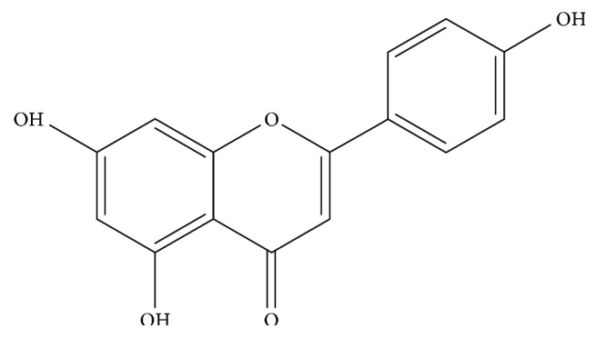
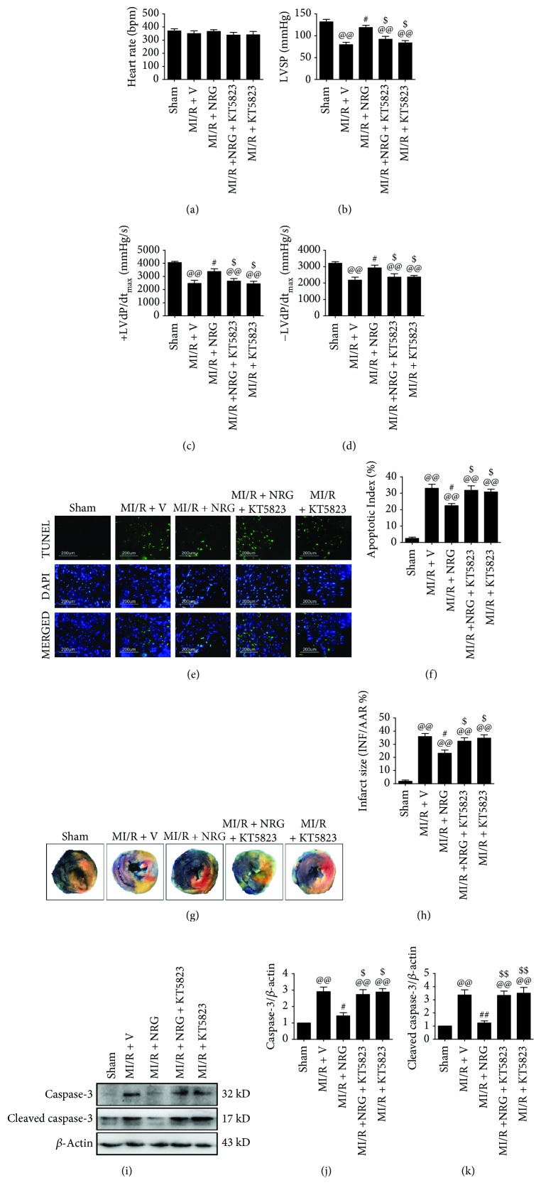
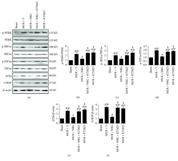
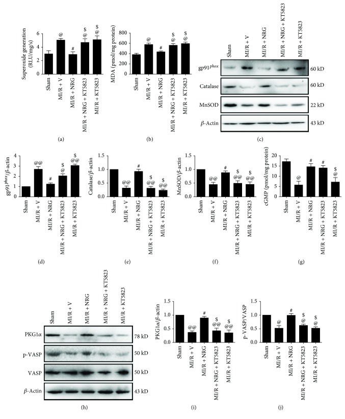
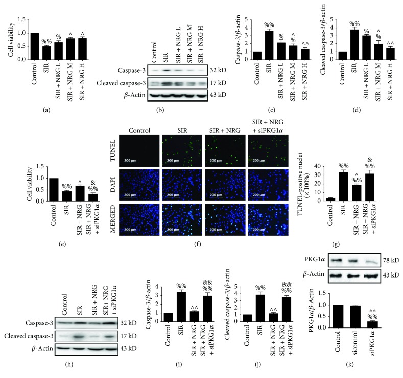
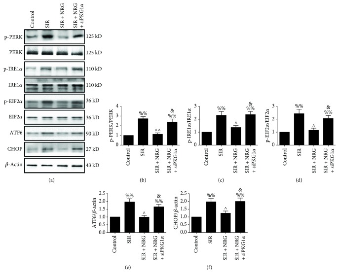
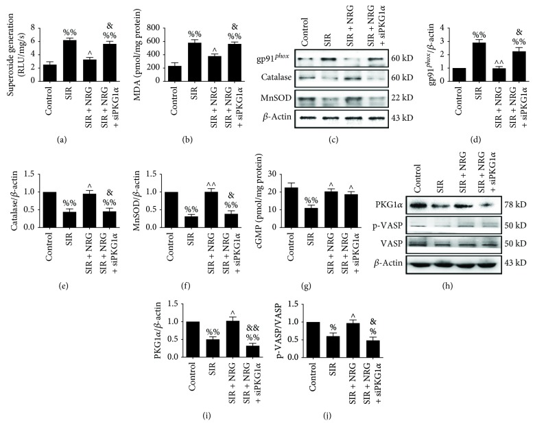
Endoplasmic reticulum (ER) stress and oxidative stress contribute greatly to myocardial ischemia-reperfusion (MI/R) injury. Naringenin, a flavonoid derived from the citrus genus, exerts cardioprotective effects. However, the effects of naringenin on ER stress as well as oxidative stress under MI/R condition and the detailed mechanisms remain poorly defined. This study investigated the protective effect of naringenin on MI/R-injured heart with a focus on cyclic guanosine monophosphate- (cGMP-) dependent protein kinase (PKG) signaling. Sprague-Dawley rats were treated with naringenin (50 mg/kg/d) and subjected to MI/R surgery with or without KT5823 (2 mg/kg, a selective inhibitor of PKG) cotreatment. Cellular experiment was conducted on H9c2 cardiomyoblasts subjected to simulated ischemia-reperfusion treatment. Before the treatment, the cells were incubated with naringenin (80 μmol/L). PKGIα siRNA was employed to inhibit PKG signaling. Our in vivo and in vitro data showed that naringenin effectively improved heart function while it attenuated myocardial apoptosis and infarction. Furthermore, pretreatment with naringenin suppressed MI/R-induced oxidative stress as well as ER stress as evidenced by decreased superoxide generation, myocardial MDA level, gp91 phox expression, and phosphorylation of PERK, IRE1α, and EIF2α as well as reduced ATF6 and CHOP. Importantly, naringenin significantly activated myocardial cGMP-PKGIα signaling while inhibition of PKG signaling with KT5823 (in vivo) or siRNA (in vitro) not only abolished these actions but also blunted naringenin's inhibitory effects against oxidative stress and ER stress. In summary, our study demonstrates that naringenin treatment protects against MI/R injury by reducing oxidative stress and ER stress via cGMP-PKGIα signaling. Its cardioprotective effect deserves further clinical study.


Figures

Figure 1
Figure 1
Molecular structure of naringenin (NRG).
Figure 2
Figure 2
Evaluation of cardiac function, myocardial apoptosis, and infarction. The animals were given naringenin treatment by oral gavage at a dose of 50 mg/kg/d for 5 days and then subjected to MI/R surgery. KT5823 (2 mg/kg) was intravenously administered to the animals at the beginning of myocardial ischemia. Cardiac function and myocardial apoptosis were measured after 4 hours of reperfusion. Myocardial infarct size was measured after 6 hours of reperfusion. (a) Heart rate (HR). (b) Left ventricular systolic pressure (LVSP). (c, d) The instantaneous first derivative of left ventricular pressure (+dP/dtmax and −dP/dtmax). (e) Representative photomicrographs of TUNEL staining (200x). Green fluorescence shows TUNEL-positive nuclei; blue fluorescence shows nuclei of total cardiomyocytes. (f) Percentage of TUNEL-positive nuclei. (g) Representative photographs of Evan's Blue-TTC staining. (h) Myocardial infarct size expressed as percentage of area-at-risk (AAR). (i) Representative blots. (j) Caspase-3 expression. (k) Cleaved caspase-3 expression. Data are expressed as mean ± SEM. n = 6 per group. @@P < 0.01vs. sham group, #P < 0.05/##P < 0.01vs. MI/R + V group, and $P < 0.05/$$P < 0.01vs. MI/R + NRG group. MI/R: myocardial ischemia-reperfusion; V: vehicle; NRG: naringenin.
Figure 3
Figure 3
Evaluation of myocardial endoplasmic reticulum stress level. Western blot analysis was conducted after 4 hours of reperfusion. (a) Representative blots, (b) PERK (Thr980) phosphorylation level, (c) IRE1α (Ser724) phosphorylation level, (d) EIF2α (Ser51) phosphorylation level, (e) ATF6 expression, and (f) CHOP expression. Data are expressed as mean ± SEM. n = 6 per group. @@P < 0.01vs. sham group, #P < 0.05/##P < 0.01vs. MI/R + V group, and$P < 0.05/$$P < 0.01vs. MI/R + NRG group. MI/R: myocardial ischemia-reperfusion; V: vehicle; NRG: naringenin.
Figure 4
Figure 4
Evaluation of myocardial oxidative stress markers and cGMP-PKGIα signaling. All the measurements were carried out after 4 hours of reperfusion. (a) Myocardial superoxide generation, (b) myocardial malondialdehyde (MDA) contents, (c) representative blots, (d) gp91phox expression, (e) catalase expression, (f) MnSOD expression, (g) myocardial cGMP level, (h) representative blots, (i) PKGIα expression, and (j) VASP (Ser239) phosphorylation. Data are expressed as mean ± SEMn = 6 per group. @P < 0.05/@@P < 0.01vs. sham group, #P < 0.05vs. MI/R + V group, and $P < 0.05/$$P < 0.01vs. MI/R + NRG group. MI/R: myocardial ischemia-reperfusion; V: vehicle; NRG: naringenin.
Figure 5
Figure 5
Evaluation of cell viability and apoptosis. Initially, H9c2 cardiomyoblasts received low (L), medium (M), or high (H) concentrations of naringenin treatment (40, 80, or 160 μmol/L) for 6 hours and subjected to simulated ischemia-reperfusion injury. Cell viability and apoptosis were assessed (a-d). Next, H9c2 cells were transfected with PKGIα siRNA and administered with or without naringenin (80 μmol/L, 6 hours). Then, they emulated ischemia-reperfusion SIR treatment. Cell viability and apoptosis were measured after 4 hours of simulated reperfusion (e-j). (a) Cell viability, (b) representative blots, (c) caspase-3 expression, (d) cleaved caspase-3 expression, (e) cell viability, and (f) representative photomicrographs of TUNEL staining (200x). Green fluorescence shows TUNEL-positive nuclei; blue fluorescence shows nuclei of total cardiomyocytes. (g) Percentage of TUNEL-positive nuclei, (h) representative blots, (i) caspase-3 expression, (j) cleaved caspase-3 expression, (k) the knockdown capacity of PKGIα siRNA were evaluated by Western blotting. Data are expressed as mean ± SEM. n = 6 per group. %P < 0.05/%%P < 0.01vs. control group, P < 0.05/∧∧P < 0.01vs. SIR group, &P < 0.05/&&P < 0.01vs. SIR + NRG group, and ∗∗P < 0.01vs. sicontrol group. SIR: simulated myocardial ischemia-reperfusion; NRG: naringenin.
Figure 6
Figure 6
Evaluation of cellular endoplasmic reticulum stress level. All the measurements were carried out after 4 hours of simulated reperfusion. (a) Representative blots, (b) PERK (Thr980) phosphorylation level, (c) IRE1α (Ser724) phosphorylation level, (d) EIF2α (Ser51) phosphorylation level, (e) ATF6 expression, and (f) CHOP expression. Data are expressed as mean ± SEM. n = 6 per group. %%P < 0.01vs. control group, P < 0.05/∧∧P < 0.01vs. SIR group, and &P < 0.05vs. SIR + NRG group. SIR: simulated myocardial ischemia-reperfusion; NRG: naringenin.
Figure 7
Figure 7
Evaluation of myocardial oxidative stress markers and cGMP-PKGIα signaling. All the measurements were carried out after 4 hours of simulated reperfusion. (a) Cellular superoxide generation, (b) cellular malondialdehyde (MDA) contents, (c) representative blots, (d) gp91phox expression, (e) catalase expression, (f) MnSOD expression, (g) cellular cGMP level, (h) representative blots, (i) PKGIα expression, and (j) VASP (Ser239) phosphorylation. Data are expressed as mean ± SEM. n = 6 per group. %P < 0.05/%%P < 0.01 vs. control group, P < 0.05/∧∧P < 0.01vs. SIR group, and &P < 0.05/&&P < 0.01vs. SIR + NRG group. SIR: simulated myocardial ischemia-reperfusion; NRG: naringenin.

References

    1. Hausenloy D. J., Barrabes J. A., Botker H. E., et al. Ischaemic conditioning and targeting reperfusion injury: a 30 year voyage of discovery. Basic Research in Cardiology. 2016;111(6):p. 70. doi: 10.1007/s00395-016-0588-8. - DOI - PMC - PubMed
    1. Hausenloy D. J., Garcia-Dorado D., Botker H. E., et al. Novel targets and future strategies for acute cardioprotection: Position Paper of the European Society of Cardiology Working Group on Cellular Biology of the Heart. Cardiovascular Research. 2017;113(6):564–585. doi: 10.1093/cvr/cvx049. - DOI - PubMed
    1. Perrino C., Barabasi A. L., Condorelli G., et al. Epigenomic and transcriptomic approaches in the post-genomic era: path to novel targets for diagnosis and therapy of the ischaemic heart? Position Paper of the European Society of Cardiology Working Group on Cellular Biology of the Heart. Cardiovascular Research. 2017;113(7):725–736. doi: 10.1093/cvr/cvx070. - DOI - PMC - PubMed
    1. Van de Werf F. The history of coronary reperfusion. European Heart Journal. 2014;35(37):2510–2515. doi: 10.1093/eurheartj/ehu268. - DOI - PubMed
    1. Shi Z., Fu F., Yu L., et al. Vasonatrin peptide attenuates myocardial ischemia-reperfusion injury in diabetic rats and underlying mechanisms. American Journal of Physiology. Heart and Circulatory Physiology. 2015;308(4):H281–H290. doi: 10.1152/ajpheart.00666.2014. - DOI - PubMed