Review
. 2019 Jan 7:2:7.
doi: 10.1038/s42003-018-0237-x. eCollection 2019.

Z-DNA and Z-RNA in human disease

Affiliations
Review

Z-DNA and Z-RNA in human disease

Alan Herbert. Commun Biol. .

Abstract

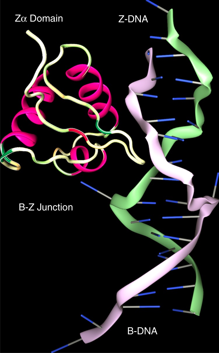
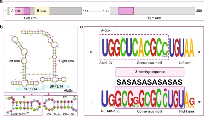
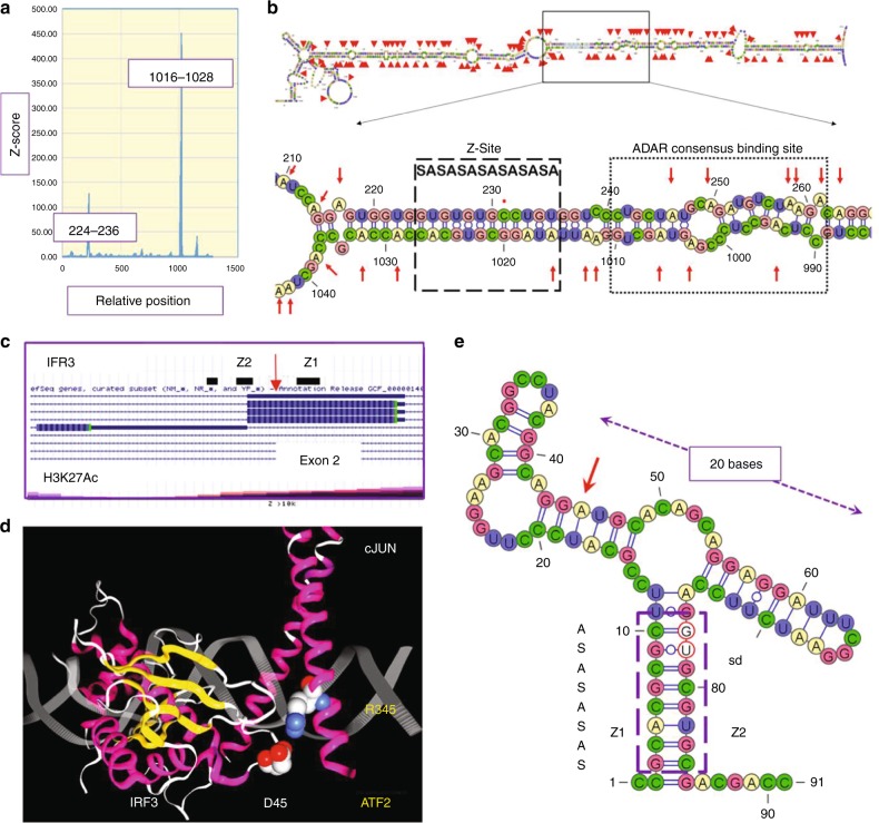
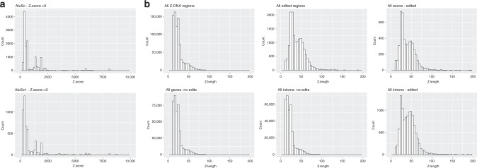
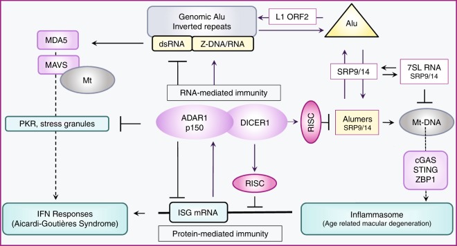
Left-handed Z-DNA/Z-RNA is bound with high affinity by the Zα domain protein family that includes ADAR (a double-stranded RNA editing enzyme), ZBP1 and viral orthologs regulating innate immunity. Loss-of-function mutations in ADAR p150 allow persistent activation of the interferon system by Alu dsRNAs and are causal for Aicardi-Goutières Syndrome. Heterodimers of ADAR and DICER1 regulate the switch from RNA- to protein-centric immunity. Loss of DICER1 function produces age-related macular degeneration, a different type of Alu-mediated disease. The overlap of Z-forming sites with those for the signal recognition particle likely limits invasion of primate genomes by Alu retrotransposons.


Conflict of interest statement

The author declares no competing interests.

Figures

Fig. 1
Fig. 1
The B−Z junction. Z-DNA is a conformer of B-DNA stabilized both by negative superhelical stress and by binding of the Zα domain of ADAR1. Zα is conformation specific, contacting the DNA backbone through the α3 helix and its carboxy-terminal β Hairpin but not making any sequence-specific contacts with bases. Formation of the B−Z junction is driven entropically by the eversion of two bases from the helix, a process further favored when non-Watson-Crick basepairs, such as mismatches, are present at this position (PDB structure 2ACJ)
Fig. 2
Fig. 2
The properties of Alu repeats. a Alu repeats consist of a right and left arm derived originally from the 7SL RNA present in the signal recognition protein (SRP). Transcription is driven by the A and B-boxes of the left arm that are promoters for RNA polymerase III. Alu retrotransposition requires binding of the SRP9/14 heterodimer, using sites on both arms (purple box in upper panel). The site on the left arm overlaps the A-Box. b Each Alu arm folds independently with SRP9/14 binding sites as visualized with VARNA. c The left-hand SRP9/14 sequence is constrained by the interaction of the A-Box with Pol III, with very little variation apparent in the WebLogo motif for the Alu family RepeatMasker consensus sequences. The right-hand site shows more variation and has a consensus logo favoring a 6 bairpair Z-forming alternating C−G motif that can be extended to a full turn of the Z-helix by flipping the out-of-alternation cytosine residue highlighted with a purple dot. The alternating syn-anti (SA) of the Z-conformation is annotated
Fig. 3
Fig. 3
Mapping of Z-formation to editing sites. a The CTSSgene region chr1:150703475-150704757 (hg19) was analyzed for Z-DNA formation using the ZHUNT3 algorithm that generates the probability of Z-formation based on statistical mechanical calculations. Z-sites map to a 5′ inverted AluSx repeat and a 3′ AluJo. b The dsRNA foldback structure with editing sites indicated by red arrows contains an alternating syn-anti (SA) Z-forming segment (box with dashed lines). A dot marks the one cytosine out of alternation. The Z-site lies adjacent to the consensus ADAR binding site found by CLIP-seq (box with dotted lines). Editing sites extend 150 basepairs on either side of it. c Map of IRF3 with editing site and H3K12Ac status. d Nonsynonymous edit of exon2 of IRF3 changes readout of codon 45 from Glutamate (D45) to Glycine, the residue in loop 1 that hydrogen bonds to R345 of ATF2(PDB structure 1T2K). e A proposed minimal editing substrate. The Z-stem (box with dashes), formed by Z1 and Z2 sequences, incorporates the splice donor site (sd, bases circled in red) from exon 2. The edited A (red arrow) lies in the left arm of a 20 base long helix of similar structure to other ncRNA substrates
Fig. 4
Fig. 4
Edited genes are enriched for higher Z-scores. a Histogram of Z-scores less than 10,000 for AluSc annotated sequences from hg19 is right-shifted compared to AluSx1 sequences. b Histogram of the length of Z-elements with scores >10,000, showing enrichment of longer segments in edited genes in both exons (5′ UTR, Coding Sequences and 3′ UTR) and introns compared to nonedited genes
Fig. 5
Fig. 5
The Alu cycle and disease. The Alu cycle of retrotransposition involves the LINE1 retrotransposase (L1 ORF2), the SRP9/14 dimer and Alu dimers. Some genomic insertions result in formation of Alu inverted repeats. Transcripts from these regions fold to form dsRNA. ADAR in partnership with DICER1 regulates protein-mediated immune responses to Alu transcripts through a series of RNA-based switches. In the resting state, repression of interferon-stimulated genes (ISG) by the RISC complex is enhanced through protein−protein interactions between ADAR and DICER1. ADAR also reduces PKR (protein kinase, dsRNA activated (EIF2AK2)) stress-related responses by editing dsRNA. ADAR p150 and PKR expression is stimulated by interferon. Decreased ADAR activity or increased production of dsRNA promotes translation of other ISG, leading to amplification of interferon responses through the dsRNA sensor MDA5 (encoded by IFIH1) that promotes the assembly of MAVS filaments on the mitochondrial (mt) surface. Loss of ADAR function is causal for Aicardi-Goutières Syndrome, a disease where the persistent activation of the interferon system is driven by endogenous dsRNA formed in part from Alu inverted repeats. Loss of DICER1 function in age-related macular degeneration leads to accumulation of Alumers, loss of mt integrity, release of mt nucleic acids, inflammasome activation and cell death

References

    1. Herbert A, et al. A Z-DNA binding domain present in the human editing enzyme, double-stranded RNA adenosine deaminase. Proc. Natl. Acad. Sci. USA. 1997;94:8421–8426. - PMC - PubMed
    1. Herbert AG, Spitzner JR, Lowenhaupt K, Rich A. Z-DNA binding protein from chicken blood nuclei. Proc. Natl Acad. Sci. USA. 1993;90:3339–3342. - PMC - PubMed
    1. Kim U, Wang Y, Sanford T, Zeng Y, Nishikura K. Molecular cloning of cDNA for double-stranded RNA adenosine deaminase, a candidate enzyme for nuclear RNA editing. Proc. Natl Acad. Sci. USA. 1994;91:11457–11461. - PMC - PubMed
    1. Patterson JB, Thomis DC, Hans SL, Samuel CE. Mechanism of interferon action: double-stranded RNA-specific adenosine deaminase from human cells is inducible by alpha and gamma interferons. Virology. 1995;210:508–511. - PubMed
    1. Schade M, et al. A 6 bp Z-DNA hairpin binds two Z alpha domains from the human RNA editing enzyme ADAR1. FEBS Lett. 1999;458:27–31. - PubMed

MeSH terms

Supplementary concepts