. 2019 Jan 30:2:42.
doi: 10.1038/s42003-019-0286-9. eCollection 2019.

A high-throughput integrated microfluidics method enables tyrosine autophosphorylation discovery

Affiliations

A high-throughput integrated microfluidics method enables tyrosine autophosphorylation discovery

Hadas Nevenzal et al. Commun Biol. .

Abstract

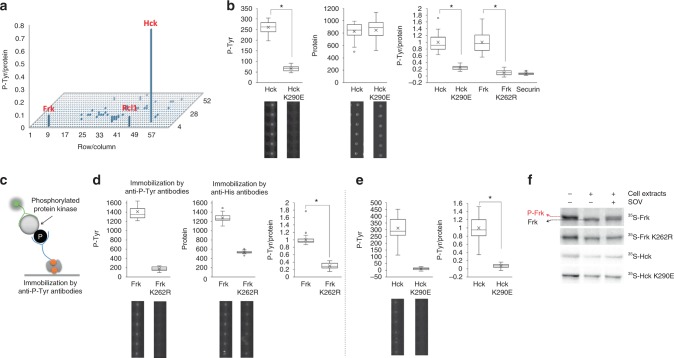
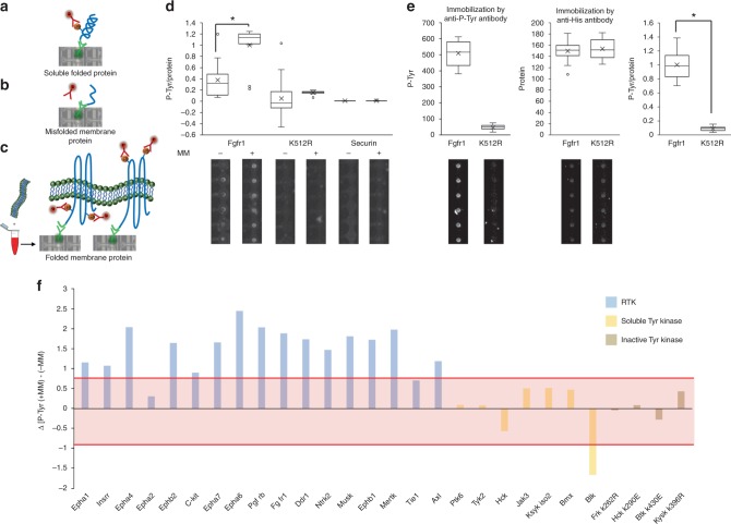
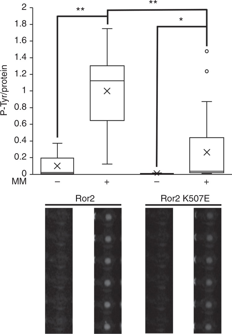
Autophosphorylation of receptor and non-receptor tyrosine kinases is a common molecular switch with broad implications for pathogeneses and therapy of cancer and other human diseases. Technologies for large-scale discovery and analysis of autophosphorylation are limited by the inherent difficulty to distinguish between phosphorylation and autophosphorylation in vivo and by the complexity associated with functional assays of receptors kinases in vitro. Here, we report a method for the direct detection and analysis of tyrosine autophosphorylation using integrated microfluidics and freshly synthesized protein arrays. We demonstrate the efficacy of our platform in detecting autophosphorylation activity of soluble and transmembrane tyrosine kinases, and the dependency of in vitro autophosphorylation assays on membranes. Our method, Integrated Microfluidics for Autophosphorylation Discovery (IMAD), is high-throughput, requires low reaction volumes and can be applied in basic and translational research settings. To our knowledge, it is the first demonstration of posttranslational modification analysis of membrane protein arrays.


Conflict of interest statement

The authors declare no competing interests.

Figures

Fig. 1
Fig. 1
Device and strategy. Integrated microfluidic device combined with His/Myc-double-tagged ORF library spotted on glass (the observed DNA in the picture encodes for FRK protein), allow parallel expressions of thousands of proteins ready for biochemical assays. Each unit cell comprises DNA and protein chambers isolated by valves. a The entire microfluidics device. b Target proteins are expressed in DNA chambers following incubation with reticulocyte lysate, diffused, and immobilized in protein chambers via His tag. Overall, 10 μl reagents are sufficient to cover a chip of thousands unit cells. c Proteins are expressed in mammalian cell lysates. Thus, enzymes with inherent autophosphorylation activity are expected to be functional and undergo autophosphorylation during expression. d, e Arrayed proteins and phosphorylated Tyr (P-Tyr) are both quantified in situ using Cy3-coupled anti-Myc antibodies and Cy5-coupled anti-phosphorylated-Tyr antibodies, respectively. Finally, a net autophosphorylation signal is determined
Fig. 2
Fig. 2
Large-scale Tyr autophosphorylation analysis of freshly expressed protein arrays. a An array of 882 human proteins was expressed on chip in quadruplicates. Protein and P-Tyr signals were quantified as described in Fig. 1. Each bar represents an average P-Tyr-to-Protein ratio of a single protein (n = 4). Top 3 hits are indicated. Source data is provided in Supplementary Figure 2. b Assay validation. Hck and Frk kinases, their inactive variants (K290E; K262R) and the non-kinase protein Securin, were expressed in tube, deposited on chip, and assayed for autophosphorylation activity as described in Fig. 1. Average P-Tyr normalized to protein levels are shown (n = 17–36; *P < 0.05). Data are normalized to maximal activity for each kinase. Absolute P-Tyr and protein levels, and representative raw data are shown for wild type and inactive Hck to clarify the methodology. Source data is provided in Supplementary Figures 4 and 5. c Schematic presentation of anti-P-Tyr immunoprecipitation. d, e Immunoprecipitation of autophosphorylated proteins on chip. d Frk wt or e Hck wt and their kinase dead derivates (K262R, K290E respectively) were immobilized on chip using biotinylated anti-P-Tyr antibody. Proteins expression value was evaluated using anti-His antibody for immobilization and anti C-myc. Representative raw data is shown below (n = 34 for (d) and n = 63 (e); *P < 0.01). Results were normalized to each protein expression level (P-Tyr/Protein) as well as to maximum activity (Frk or Hck phosphorylation level). Source data is provided in Supplementary Figures 6 and 7. f 35S-labeled Frk wt, Hck wt and their kinase dead derivates were in vitro transcribed and expressed using rabbit reticulocyte lysate and incubated for 30 min, 37 °C in parallel with HEK293 cell extracts supplemented with sodium orthovanadate (1 mM), as depicted in the plot. Protein’s mobility shift was assayed by SDS–PAGE and autoradiography. Source data is provided in Supplementary Figures 8 and 9
Fig. 3
Fig. 3
Autophosphorylation analysis of Tyr kinase array. An ORF library comprising 17 soluble Tyr kinases and 11 negative controls (5 inactive Tyr kinases and 6 non-Tyr kinases) was generated by assembly PCR. ORFs were spotted in quadruplicates. A freshly expressed protein array was generated and assayed for autophosphorylation as described in Fig. 1. Bars represent an average and standard deviation values of P-Tyr signals (n = 4) after background P-Tyr signal subtraction and normalization to protein level. A cutoff value of 0.05 was calculated by receiver operating characteristic analysis
Fig. 4
Fig. 4
A functional array of receptor Tyr kinases (RTKs). Challenges and mitigation. Soluble protein kinases properly folded in cell lysates exhibiting specific autophosphorylation activity that can be detected by anti-P-Tyr antibodies (DNA spot in the picture encodes for FRK protein) (a). Conversely, membrane proteins are misfolded with limited and/or ectopic kinase activity, if at all (b). A proper folding of membrane proteins is achieved in reticulocyte lysate supplemented with microsomal membranes. Following immobilization and detection, bona fide autophosphorylation activity can be assayed on chip (c). The observed DNA spots in b and c encode for the expression of wild-type FGFR1. d Assay validation. Wild type and inactive mutant (K512R) Fgfr1, and Securin (negative control), were expressed in reticulocyte lysate supplemented with microsomal membranes (+MM) or mock (−MM), deposited on chip and assayed for autophosphorylation. Average P-Tyr to protein levels are shown (n = 11–25; *P < 0.01). Data are normalized to maximal activity. Representative raw data showing P-Tyr detection are shown. Source data is provided in Supplementary Figures 10–12. e Immunoprecipitation of autophosphorylated membrane protein on chip. Fgfr1 and kinase dead (K512R) were immobilized on chip using biotinylated anti-P-Tyr antibody. Proteins expression value was evaluated using anti-His antibody for immobilization and labeling with Green Lysine. Representative raw data is shown below (n = 20; *P < 0.01). Results were normalized to each protein expression level (P-Tyr/Protein) as well as to maximum activity (Fgfr1 phosphorylation level). Source data is provided in Supplementary Data Figure 12. f A global dependency of RTK activity assays on membranes revealed on chip. An ORF library comprising 17 RTK, 7 soluble Tyr kinases, and 4 inactive Tyr kinases was spotted on chip in quadruplicates. Proteins were expressed in reticulocyte lysate supplemented with microsomal membranes (MM) or mock (−MM). An average P-Tyr signal from three experiments was determined for the arrayed proteins, and the difference: [P-Tyr(+MM)] - [P-Tyr(−MM)] was plotted. A cutoff (±0.84) marking significant impact of microsomal membranes on autophosphorylation activity was determined by receiver operating characteristic analysis as 3× standard deviation from P-Tyr values calculated for inactive kinases. Membrane effect on autophosphorylation is considered significant if P-Tyr values are either above (positive cutoff) or below (negative cutoff) the red region
Fig. 5
Fig. 5
A membrane-dependent autophosphorylation activity for Ror2. Myc/His-tagged Ror2 and its inactive derivate (Ror2 K507E) were expressed in reticulocyte lysate supplemented with microsomal membranes (MM) or mock (−MM), and immobilized to protein chambers for an on-chip autophosphorylation activity assay. An average P-Tyr/Protein level ratio is shown (n = 13; *P < 0.05, **P < 0.001). Data are normalized to maximal activity. Representative raw data are shown. Error bars = 1 SE. Source data is provided in Supplementary Figure 14

References

    1. Jona G, Snyder M. Recent developments in analytical and functional protein microarrays. Curr. Opin. Mol. Ther. 2003;5:271–277. - PubMed
    1. Zhu H, Snyder M. Protein arrays and microarrays. Curr. Opin. Chem. Biol. 2001;5:40–45. doi: 10.1016/S1367-5931(00)00170-8. - DOI - PubMed
    1. Gerber D, Maerkl SJ, Quake SR. An in vitro microfluidic approach to generating protein-interaction networks. Nat. Methods. 2009;6:71–74. doi: 10.1038/nmeth.1289. - DOI - PMC - PubMed
    1. He M, Taussig MJ. Single step generation of protein arrays from DNA by cell-free expression and in situ immobilisation (PISA method) Nucleic Acids Res. 2001;29:E73–73. doi: 10.1093/nar/29.15.e73. - DOI - PMC - PubMed
    1. Yu X, Petritis B, LaBaer J. Advancing translational research with next-generation protein microarrays. Proteomics. 2016;16:1238–1250. doi: 10.1002/pmic.201500374. - DOI - PMC - PubMed

Publication types

MeSH terms

Substances