. 2019 May;137(5):801-823.
doi: 10.1007/s00401-019-01967-4. Epub 2019 Feb 7.

Beta-amyloid pathology in human brain microvessel extracts from the parietal cortex: relation with cerebral amyloid angiopathy and Alzheimer's disease

Affiliations

Beta-amyloid pathology in human brain microvessel extracts from the parietal cortex: relation with cerebral amyloid angiopathy and Alzheimer's disease

Philippe Bourassa et al. Acta Neuropathol. 2019 May.

Abstract

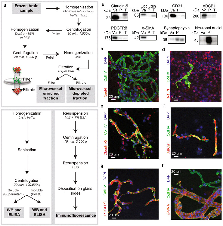
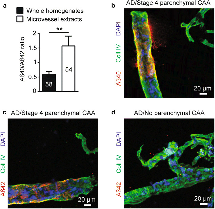
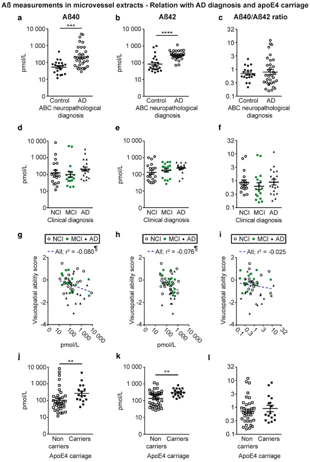
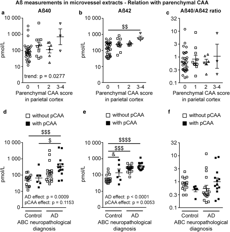
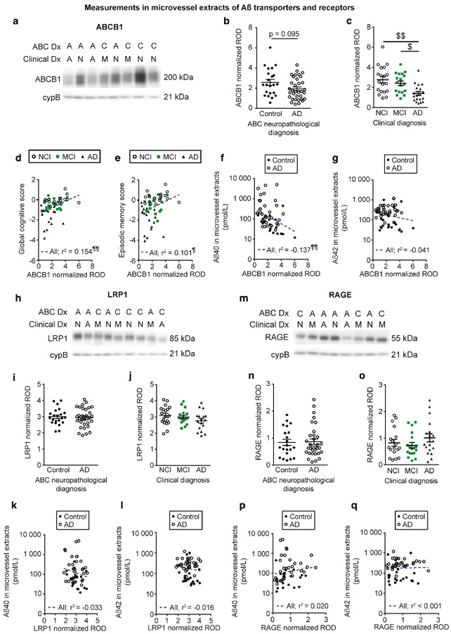
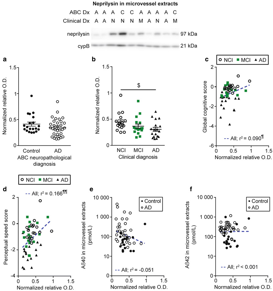
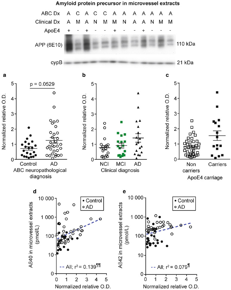
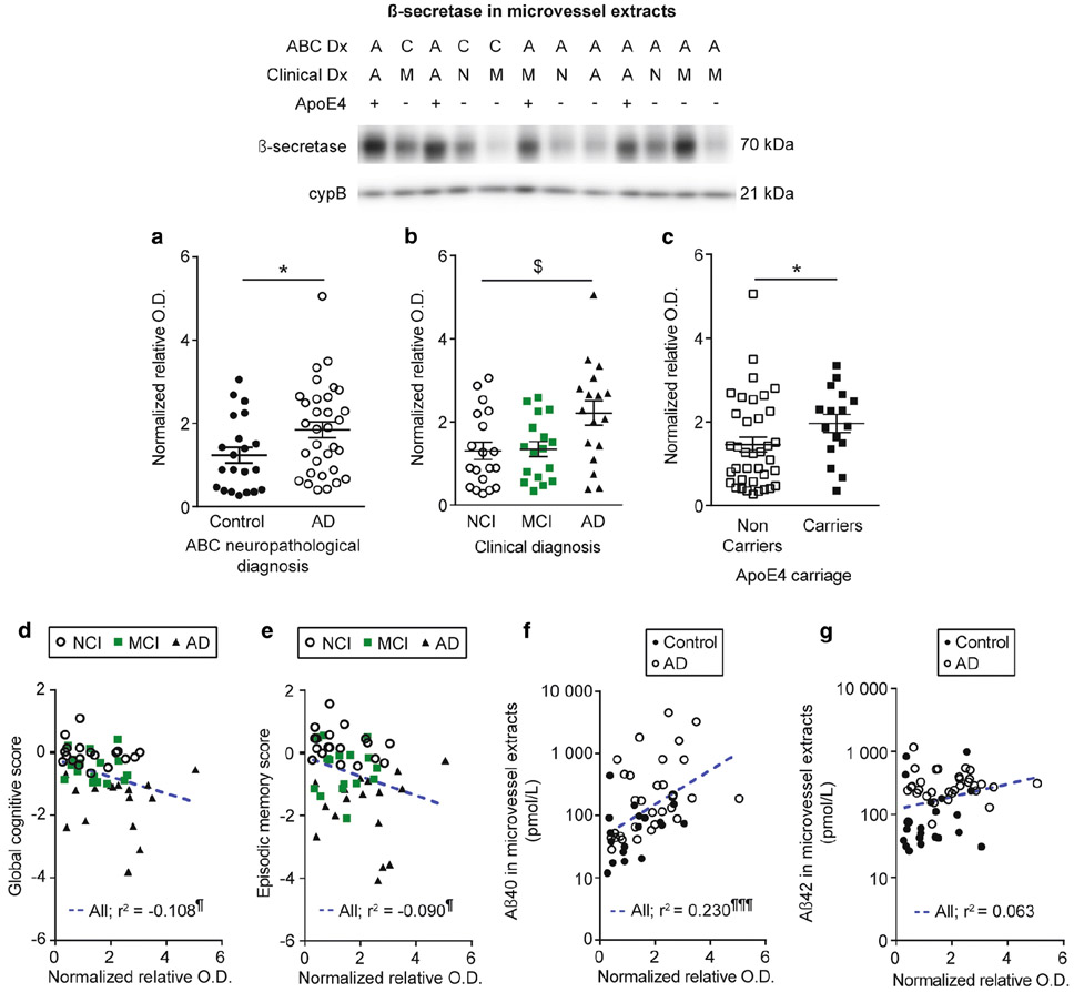
Several pieces of evidence suggest that blood-brain barrier (BBB) dysfunction is implicated in the pathophysiology of Alzheimer's disease (AD), exemplified by the frequent occurrence of cerebral amyloid angiopathy (CAA) and the defective clearance of Aβ peptides. However, the specific role of brain microvascular cells in these anomalies remains elusive. In this study, we validated by Western, ELISA and immunofluorescence analyses a procedure to generate microvasculature-enriched fractions from frozen samples of human cerebral cortex. We then investigated Aβ and proteins involved in its clearance or production in microvessel extracts generated from the parietal cortex of 60 volunteers in the Religious Orders Study. Volunteers were categorized as AD (n = 38) or controls (n = 22) based on the ABC scoring method presented in the revised guidelines for the neuropathological diagnosis of AD. Higher ELISA-determined concentrations of vascular Aβ40 and Aβ42 were found in persons with a neuropathological diagnosis of AD, in apoE4 carriers and in participants with advanced parenchymal CAA, compared to respective age-matched controls. Vascular levels of two proteins involved in Aβ clearance, ABCB1 and neprilysin, were lower in persons with AD and positively correlated with cognitive function, while being inversely correlated to vascular Aβ40. In contrast, BACE1, a protein necessary for Aβ production, was increased in individuals with AD and in apoE4 carriers, negatively correlated to cognitive function and positively correlated to Aβ40 in microvessel extracts. The present report indicates that concentrating microvessels from frozen human brain samples facilitates the quantitative biochemical analysis of cerebrovascular dysfunction in CNS disorders. Data generated overall show that microvessels extracted from individuals with parenchymal CAA-AD contained more Aβ and BACE1 and less ABCB1 and neprilysin, evidencing a pattern of dysfunction in brain microvascular cells contributing to CAA and AD pathology and symptoms.

Keywords: Alzheimer’s disease; Beta-amyloid; Blood–brain barrier; Brain microvascular cells; Cerebral amyloid angiopathy.


Conflict of interest statement

Conflict of interest: The authors declare that they have no conflict of interest.

Figures

Fig.1
Fig.1
Validation of our microvessel enrichment protocol. a) Workflow leading to the separation of the microvessel-enriched and the microvessel-depleted parenchymal fractions, respectively illustrated in green and red in the filtration scheme, from whole homogenates of cerebral cortex, and subsequent steps allowing for Western blot, ELISA and immunofluorescence experiments. b) Microvessel extracts show enrichment in endothelial markers like claudin-5, occludin, CD31/PECAM and ABCB1/P-glycoprotein, and mural cell markers PDGFRβ and α-SMA, while synaptophysin and NeuN, both neuronal markers, are rather enriched in the microvessel-depleted parenchymal fraction. All samples were generated from frozen blocks of parietal cortices. Consecutive bands were taken for all representative photo examples. The same amount (8 μg) of proteins per sample was loaded. c-f) Validation by immunofluorescence showed that microvessels, stained with type IV collagen, a marker of basal membrane (green), are concentrated in the vascular fraction (panel c) compared to the microvessel-depleted parenchymal fraction (panel d). On the contrary, immunostaining of neuronal nuclei (NeuN, red) in both fractions revealed that neurons are predominantly found in the microvessel-depleted parenchymal fraction (panel d). Type IV collagen-positive microvessels were also stained by claudin-5 (panel e), ABCB1 (panel f), PDGFRβ (panel g) and α-SMA (panel h) antibodies. Magnification used: 20X. Scale bar: 20 μm. Abbreviations: P, microvessel-depleted parenchymal fraction; T, total homogenate; Va, vascular fraction, enriched in microvessels; WB, Western blot
Fig. 2
Fig. 2
Localization of Aβ peptides in microvessel extracts from the parietal cortex. a) Concentrations of Aβ40, Aβ42 and Aβ40/Aβ42 ratios were determined in brain microvascular extracts by ELISA. A 3-fold higher Aβ40/Aβ42 ratio was observed in brain microvessel extracts compared to whole homogenates from the same parietal cortex samples. Data are represented as mean ± S.E.M. Sample size is indicated in the graph bars. Statistical analysis: unpaired Student’s t-test. ** p < 0.01. b-d) Immunolabeling of Aβ40 and Aβ42 following formic acid pretreatment revealed that both peptides accumulated on larger vessels at stage 4 parenchymal CAA in parietal cortex while no immunoreactivity was observed at stage 0 parenchymal CAA. Markedly, for both stages, no signal was found in smaller capillary-like vessels. Magnification used: 20X. Scale bar: 20 μm
Fig. 3
Fig. 3
Aβ40 and Aβ42 concentrations in brain microvessels are increased in persons with AD and in apoE4 carriers. Concentrations of Aβ40, Aβ42 and Aβ40/Aβ42 ratios were determined in brain microvascular extracts by ELISA. a-c) Participants were divided according to their neuropathological diagnosis based on the ABC criteria; d-f) their clinical diagnosis and j-l) their apoE4 allele carriage. We observed increased concentrations of Aβ in individuals with a neuropathological diagnosis of AD (a, b) and in apoE4 carriers (j, k). For all groups, no significant variation was found for the Aβ40/Aβ42 ratios (c, f and l). Data were log transformed for statistical analysis and are represented as scatterplots with a logarithmic scale. Horizontal bars indicate mean ± S.E.M. Statistical analysis: Mann-Whitney test. * p < 0.05, ** p < 0.01, **** p < 0.0001. Correlative analysis revealed that Aβ40 and Aβ42 levels in microvessel extracts were both negatively associated with visuospatial ability scores (g, h). Statistical analysis: Pearson correlation coefficient. ¶ p < 0.05. Abbreviations: AD, Alzheimer’s disease; MCI, mild cognitive impairment; NCI, healthy controls with no cognitive impairment
Fig. 4
Fig. 4
Aβ40 and Aβ42 concentrations in brain microvessels are increased in participants with higher stages of parenchymal CAA in the parietal cortex. Concentrations of Aβ40, Aβ42 and Aβ40/Aβ42 ratios were determined in brain microvascular extracts by ELISA. a-c) Participants were divided according to their parenchymal CAA staging in the parietal cortex as following: 0, no deposition; 1, scattered segmental but not circumferential deposition; 2, circumferential deposition up to 10 vessels; 3, circumferential deposition up to 75% of the region; 4, circumferential deposition over 75% of the total region. Stages 3 and 4 were pooled together for statistical analysis. Aβ42 concentrations were elevated in individuals with stage 3 and 4 parenchymal CAA (panel b) while only an upward trend was noted for Aβ40 concentrations in the same group (panel a). Statistical analysis: Kruskal-Wallis non parametric one-way analysis of variance followed by a Dunn’s post hoc test. $$ p < 0.01. d-f) Individuals were grouped based on their ABC neuropathological diagnosis and subdivided based on the presence or not of parenchymal CAA, regardless of the severity. Neuropathological diagnosis of AD was associated with higher Aβ40 and Aβ42 vascular concentrations (panels d and e), while parenchymal CAA was associated with increased levels of Aβ42 only (panel e). Highest concentrations of Aβ40 were found in participants with AD and parenchymal CAA. Statistical analysis: Kruskal-Wallis non parametric one-way analysis of variance followed by a Dunn’s post hoc test. $ p < 0.05, $$$ p < 0.001, $$$$ p < 0.0001 and two-way analysis of variance followed by a Bonferroni’s post hoc test. & p < 0.05. Data were log transformed for statistical analysis and are represented as scatterplots with a logarithmic scale. Horizontal bars indicate mean ± S.E.M. Abbreviations: pCAA, parenchymal CAA
Fig. 5
Fig. 5
Aβ efflux transporter ABCB1 is reduced in vascular fractions from individuals with AD. ABCB1 (p-glycoprotein), LRP1 and RAGE levels in microvessel extracts were determined by Western blot. Data were normalized with cyclophilin B. Representative immunoblotting examples for ABCB1, LRP1 and RAGE (panels a, h and m). All samples, loaded in a random order, were run on the same immunoblot experiment for quantification. Consecutive bands are shown. No difference was found when participants were divided according to their AD neuropathological diagnosis (panels b, i and n). When individuals were grouped based on their clinical diagnosis, a significant reduction was noted for ABCB1 in AD while no difference was observed for LRP1 and RAGE (panels c, j and o). Statistical analysis: Kruskal-Wallis one-way analysis of variance followed by a Dunn’s post hoc test. $ p < 0.05, $$ p < 0.01. ABCB1 levels in microvessel extracts were positively associated to global cognition and episodic memory (panels d and e). In microvessel extracts, among transporters investigated only ABCB1 levels were inversely correlated to Aβ40 content (Panel f). No significant correlation was observed between Aβ42 concentrations in microvessel extracts and any of the transporter investigated (panels g, l and q). Statistical analysis: Pearson correlation coefficient. ¶ p < 0.05, ¶¶ p < 0.01. Abbreviations: A/AD, Alzheimer’s disease; ABC Dx, ABC neuropathological diagnosis; C, control; Clinical Dx, clinical diagnosis; cypB, cyclophilin B; M/MCI, mild cognitive impairment; N/NCI, healthy controls with no cognitive impairment; ROD, relative optical density
Fig. 6
Fig. 6
Neprilysin levels are reduced in brain microvessels from individuals with AD and correlated to cognitive function and Aβ40. Neprilysin levels in microvessel extracts were determined by Western blot. Data were normalized with cyclophilin B. No difference was found when participants were divided according to their AD neuropathological assessment (panel a) while a significant decrease was observed in individuals with AD based on clinical diagnosis (panel b). All samples, loaded in a random order, were run on the same immunoblot experiment. Consecutive bands are shown. Data are represented as scatterplots. Horizontal bars indicate mean ± S.E.M. Statistical analysis: Kruskal-Wallis ANOVA; $ p < 0.05. Neprilysin levels in microvessel extracts were positively associated to global cognition and perceptual speed (panels c and d). A trend towards a negative correlation between vascular neprilysin levels and Aβ40 (panel e) was noted while no significant association was found with Aβ42 (panel f). Statistical analysis: Pearson correlation coefficient. ¶ p < 0.05, ¶¶ p < 0.01. Abbreviations: A/AD, Alzheimer’s disease; ABC Dx, ABC neuropathological diagnosis; C, control; Clinical Dx, clinical diagnosis; cypB, cyclophilin B; M/MCI, mild cognitive impairment; N/NCI, healthy controls with no cognitive impairment; relative O.D., relative optical density
Fig. 7
Fig. 7
Amyloid protein precursor levels are correlated to Aβ peptides concentrations in microvessel extracts. Amyloid protein precursor (APP) levels in microvessel extracts were determined by Western blot. Data were normalized with cyclophilin B. a-c) Participants were divided according to a) their AD neuropathological diagnosis; b) their clinical diagnosis and c) their apoE4 allele carriage. No difference was observed in each of these comparisons. All samples, loaded in a random order, were run on the same immunoblot experiment. Consecutive bands were taken for the representative photo example. Data are represented as scatterplots. Horizontal bars indicate mean ± S.E.M. Statistical analysis: Mann Whitney test and Kruskal-Wallis ANOVA; p > 0.05. Linear regression analyses revealed that APP levels were positively correlated to both Aβ40 (panel e) and Aβ42 (panel f) concentrations in microvessel extracts. Statistical analysis: Pearson correlation coefficient. ¶ p < 0.05, ¶¶ p < 0.01. Abbreviations: −, ApoE4 non-carrier; +, ApoE4 carrier; A/AD, Alzheimer’s disease; ABC Dx, ABC neuropathological diagnosis; APP, amyloid protein precursor; C, control; Clinical Dx, clinical diagnosis; cypB, cyclophilin B; M/MCI, mild cognitive impairment; N/NCI, healthy controls with no cognitive impairment; relative O.D., relative optical density
Fig. 8
Fig. 8
β-secretase levels are increased in individuals with AD and in apoE4 carriers, and are correlated to cognition and Aβ40 peptides in microvessel extracts. β-secretase (BACE1) levels in microvessel extracts were determined by Western blot. Data were normalized with cyclophilin B. a-c) Participants were divided according to a) their AD neuropathological diagnosis; b) their clinical diagnosis and c) their apoE4 allele carriage. We observed an increase in BACE1 levels in individuals with a neuropathological diagnosis of AD (panel a) or a clinical diagnosis of AD (panel b). We also observed an increase for apoE4 carriers compared to non-carriers (panel c). All samples, loaded in a random order, were run on the same immunoblot experiment. Consecutive bands were taken for the representative photo example. Data are represented as scatterplots. Horizontal bars indicate mean ± S.E.M. Statistical analysis: Mann Whitney test, * p < 0.05; Kruskal-Wallis one-way analysis of variance followed by a Dunn’s post hoc test, $ p < 0.05. Linear regression analyses showed that BACE1 levels were negatively associated with global cognition (panel d) and episodic memory (panel e). In addition, BACE1 levels were positively correlated to Aβ40 concentrations in microvessel extracts (panel f). In addition, a trend towards a positive correlation was observed for Aβ42 (panel g). Statistical analysis: Pearson correlation coefficient. ¶ p < 0.05, ¶¶¶ p < 0.001. Abbreviations: −, ApoE4 non-carrier; +, ApoE4 carrier; A/AD, Alzheimer’s disease; ABC Dx, ABC neuropathological diagnosis; C, control; Clinical Dx, clinical diagnosis; cypB, cyclophilin B; M/MCI, mild cognitive impairment; N/NCI, healthy controls with no cognitive impairment; relative O.D., relative optical density.

References

    1. Aggarwal NT, Wilson RS, Beck TL, Bienias JL, Bennett DA (2005) Mild cognitive impairment in different functional domains and incident Alzheimer's disease. J Neurol Neurosurg Psychiatry 76: 1479–1484 - PMC - PubMed
    1. Alata W, Paris-Robidas S, Emond V, Bourasset F, Calon F (2014) Brain uptake of a fluorescent vector targeting the transferrin receptor: a novel application of in situ brain perfusion. Mol Pharm 11: 243–253 - PubMed
    1. Alata W, Ye Y, St-Amour I, Vandal M, Calon F (2015) Human apolipoprotein E varepsilon4 expression impairs cerebral vascularization and blood-brain barrier function in mice. J Cereb Blood Flow Metab 35: 86–94 - PMC - PubMed
    1. Alonzo NC, Hyman BT, Rebeck GW, Greenberg SM (1998) Progression of cerebral amyloid angiopathy: accumulation of amyloid-beta40 in affected vessels. J Neuropathol Exp Neurol 57: 353–359 - PubMed
    1. Arvanitakis Z, Grodstein F, Bienias JL, Schneider JA, Wilson RS, Kelly JF, Evans DA, Bennett DA (2008) Relation of NSAIDs to incident AD, change in cognitive function, and AD pathology. Neurology 70: 2219–2225 - PubMed

Publication types

MeSH terms

Substances