. 2019 Jun;139(6):769-777.
doi: 10.1007/s00402-019-03131-9. Epub 2019 Feb 7.

Risk factors for nonunion after intramedullary nailing of subtrochanteric femoral fractures

Affiliations

Risk factors for nonunion after intramedullary nailing of subtrochanteric femoral fractures

Dietmar Krappinger et al. Arch Orthop Trauma Surg. 2019 Jun.

Abstract

Introduction: Nonunion is a common complication after intramedullary nailing of subtrochanteric femoral fractures. A more detailed knowledge, particularly of avoidable risk factors for subtrochanteric fracture nonunion, is thus desired to develop strategies for reducing nonunion rates. The aim of the present study therefore was to analyse a wide range of parameters as potential risk factors for nonunion after intramedullary nailing of subtrochanteric fractures.

Materials and methods: Seventy-four patients who sustained a subtrochanteric fracture and were treated by femoral intramedullary nailing at a single level 1 trauma centre within a 6-year period were included in this study. A total of 15 patient-related, fracture-related, surgery-related, mechanical and biological parameters were analysed as potential risk factors for nonunion. Furthermore, the accuracy of each of these parameters to predict nonunion was calculated.

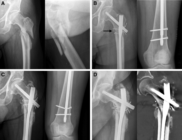
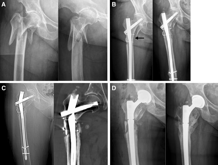
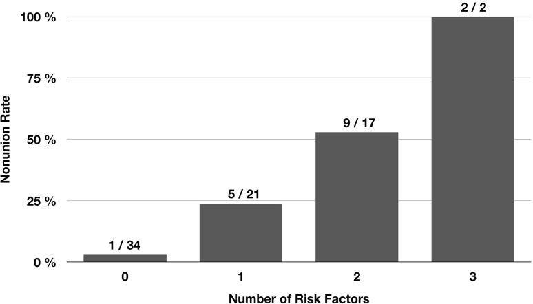
Results: Nonunion occurred in 17 of 74 patients (23.0%). Of the 15 potential risk factors analysed, only 3 were found to have a significant effect on the nonunion rate (p < 0.05): postoperative varus malalignment, postoperative lack of medial cortical support and autodynamisation of the nail within the first 12 weeks post-surgery. Accuracy of each of these 3 parameters to predict nonunion was > 0.70. Furthermore, the nonunion rate significantly increased with the number of risk factors (no risk factor: 2.9%, one risk factor: 23.8%, two risk factors: 52.9%, and three risk factors: 100% [Chi-square test, p = 0.001)].

Conclusions: Our study indicates that intraoperative correction of varus malalignment and restoration of the medial cortical support are the most critical factors to prevent nonunion after intramedullary nailing of subtrochanteric femoral fractures. In addition, autodynamisation of the nail within the first 3 months post-surgery is a strong predictor for failure and should result in revision surgery.

Keywords: Femoral fracture; Femoral nailing; Intramedullary nailing; Nonunion; Pseudarthrosis; Risk factors; Subtrochanteric femoral fracture; Subtrochanteric fracture.


Conflict of interest statement

All authors declare that they have no conflict of interest. No funding was received for the study.

Figures

Fig. 1
Fig. 1
Schematic illustration of varus malalignment, lack of medial cortical support and combination of both. a Varus malalignment, but restored medial cortical support. b Lack of medial cortical support due to nonanatomic reduction (left), a large displaced medial butterfly fragment (middle) or medial comminution (right), but no varus malalignment. c Varus malalignment combined with lack of medial cortical support due to nonanatomic reduction (lack of medial cortical support due to medial comminution or a displaced medial fracture fragment not depicted for reasons of clarity)
Fig. 2
Fig. 2
Seventy two-year-old male after a fall at home. a Radiographs obtained at admission showing a Seinsheimer Type IV fracture. b Postoperative radiographs after open reduction, cerclage wiring and intramedullary nailing: varus malalignment (as indicated by the displacement of the proximal medial cortex into the medullary canal with slight varus angulation relative to the distal medial cortex) and lack of medial cortical support due to nonanatomic reduction (black arrow), distal static and dynamic locking. c Unscheduled radiographs after 9 weeks due to persistent pain: no loss of reduction, timely callus formation, breakage of the static locking bolt and autodynamisation of the nail. There was no breakage of the static locking bolt at the previous routine controls. d Unscheduled radiograph (left) and CT scan (right) after 7 months due to suddenly increasing pain: nail breakage and no fracture healing
Fig. 3
Fig. 3
Eighty three-year-old female after a simple fall at home. a Radiographs obtained at admission showing a Seinsheimer Type V fracture. b Postoperative radiograph (left) after open reduction, cerclage wiring and intramedullary nailing: restoration of the subtrochanteric medial cortical support (black arrow) and no varus malalignment. The lesser trochanter fragment was not reduced. Scheduled radiograph after 12 weeks: no loss of reduction, timely callus formation, but breakage of the static locking bolt and autodynamisation of the nail. c Unscheduled radiograph (left) and CT scan (right) after 10 months due to suddenly increasing pain: nail breakage and no fracture healing. d Postoperative X-ray (left) after conversion to total hip arthroplasty using a modular revision stem anchored in the diaphyseal isthmus. The nonunion was not addressed surgically. Scheduled radiograph (right) 12 months after revision surgery: no component loosening and fracture healing
Fig. 4
Fig. 4
Effect of the number of risk factors on the nonunion rate. Autodynamisation, varus malalignment and lack of medial cortical support were defined as risk factors for nonunion. The nonunion rate significantly increased with the number of risk factors

References

    1. Ekström W, Németh G, Samnegård E, Dalen N, Tidermark J. Quality of life after a subtrochanteric fracture: a prospective cohort study on 87 elderly patients. Injury. 2009;40:371–376. doi: 10.1016/j.injury.2008.09.010. - DOI - PubMed
    1. Codesido P, Mejía A, Riego J, Ojeda-Thies C. Subtrochanteric fractures in elderly people treated with intramedullary fixation: quality of life and complications following open reduction and cerclage wiring versus closed reduction. Arch Orthop Trauma Surg. 2017;137:1077–1085. doi: 10.1007/s00402-017-2722-y. - DOI - PubMed
    1. Egol KA, Park JH, Rosenberg ZS, Peck V, Tejwani NC. Healing delayed but generally reliable after bisphosphonate-associated complete femur fractures treated with IM nails. Clin Orthop Relat Res. 2014;472:2728–2734. doi: 10.1007/s11999-013-2963-1. - DOI - PMC - PubMed
    1. Bogdan Y, Tornetta P, Einhorn TA, Guy P, Leveille L, Robinson J, et al. Healing time and complications in operatively treated atypical femur fractures associated with bisphosphonate use: a multicenter retrospective cohort. J Orthop Trauma. 2016;30:177–181. doi: 10.1097/BOT.0000000000000516. - DOI - PubMed
    1. Kim KK, Won Y, Smith DH, Lee GS, Lee HY. Clinical results of complex subtrochanteric femoral fractures with long cephalomedullary hip nail. Hip Pelvis. 2017;29:113–119. doi: 10.5371/hp.2017.29.2.113. - DOI - PMC - PubMed

MeSH terms