. 2019 Mar 7;4(5):e122475.
doi: 10.1172/jci.insight.122475.

Sialylation of MUC4β N-glycans by ST6GAL1 orchestrates human airway epithelial cell differentiation associated with type-2 inflammation

Affiliations

Sialylation of MUC4β N-glycans by ST6GAL1 orchestrates human airway epithelial cell differentiation associated with type-2 inflammation

Xiuxia Zhou et al. JCI Insight. .

Abstract

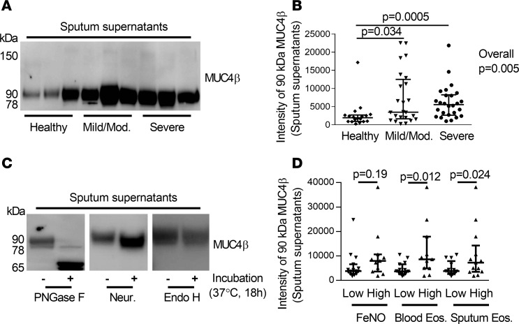
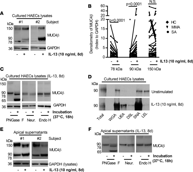
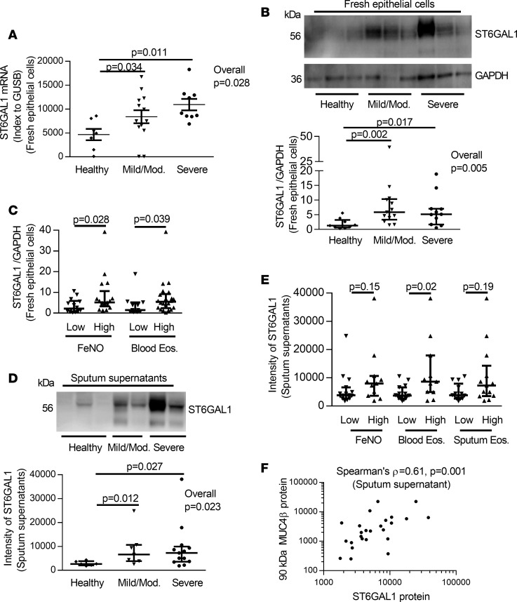
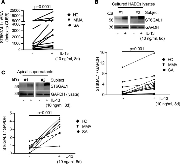
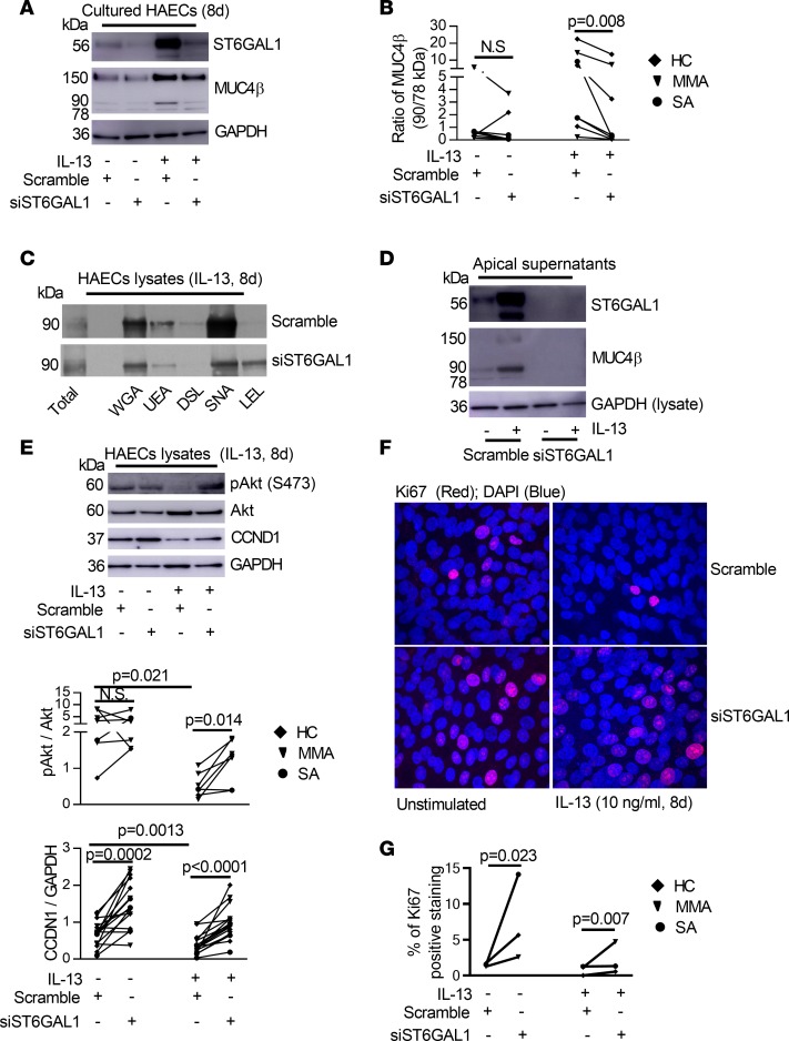
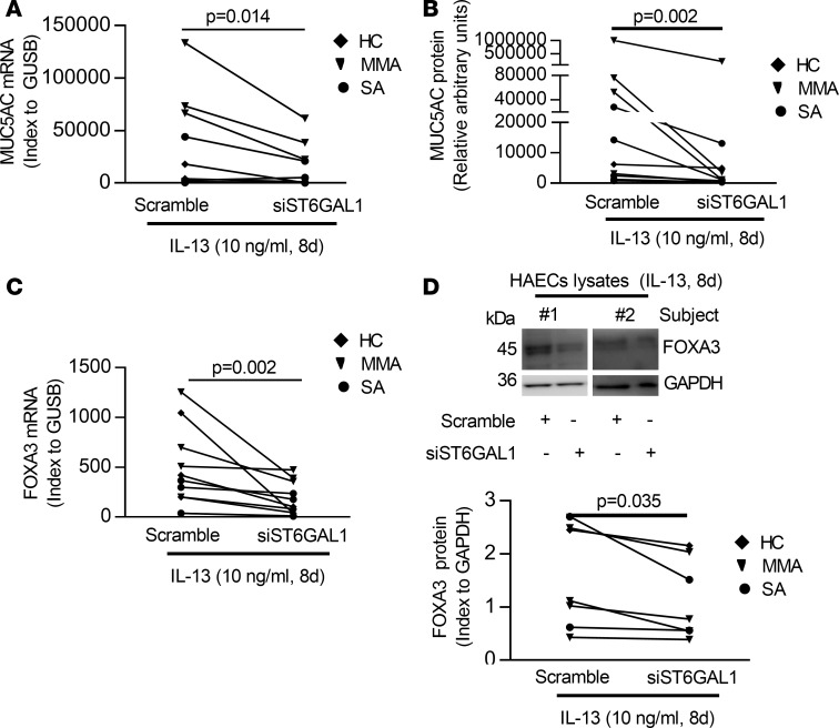
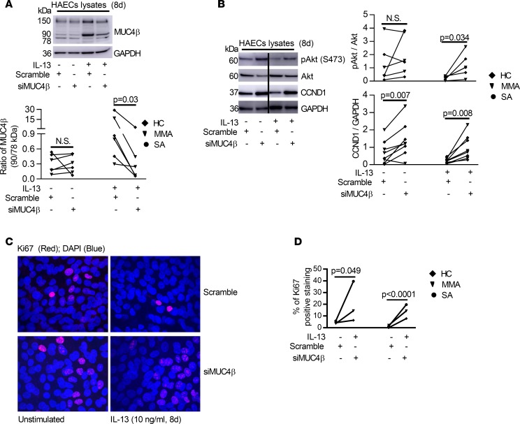
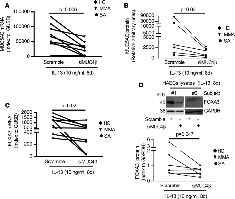
Although type-2-induced (T2-induced) epithelial dysfunction is likely to profoundly alter epithelial differentiation and repair in asthma, the mechanisms for these effects are poorly understood. A role for specific mucins, heavily N-glycosylated epithelial glycoproteins, in orchestrating epithelial cell fate in response to T2 stimuli has not previously been investigated. Levels of a sialylated MUC4β isoform were found to be increased in airway specimens from asthmatic patients in association with T2 inflammation. We hypothesized that IL-13 would increase sialylation of MUC4β, thereby altering its function and that the β-galactoside α-2,6-sialyltransferase 1 (ST6GAL1) would regulate the sialylation. Using human biologic specimens and cultured primary human airway epithelial cells (HAECs),we demonstrated that IL-13 increases ST6GAL1-mediated sialylation of MUC4β and that both were increased in asthma, particularly in sputum supernatant and/or fresh isolated HAECs with elevated T2 biomarkers. ST6GAL1-induced sialylation of MUC4β altered its lectin binding and secretion. Both ST6GAL1 and MUC4β inhibited epithelial cell proliferation while promoting goblet cell differentiation. These in vivo and in vitro data provide strong evidence for a critical role for ST6GAL1-induced sialylation of MUC4β in epithelial dysfunction associated with T2-high asthma, thereby identifying specific sialylation pathways as potential targets in asthma.

Keywords: Asthma; Inflammation; Pulmonology; Th2 response.


Conflict of interest statement

Conflict of interest: ERB has served as a paid consultant for AstraZeneca/MedImmune, GSK, Novartis, Regeneron, and Sanofi Genzyme. SEW has been site and study PI on studies sponsored by AstraZeneca, GSK, and Sanofi-Aventis.

Figures

Figure 1
Figure 1. N-glycosylated 90-kDa MUC4β was higher in asthmatic sputum supernatants and associated with a T2 signature.
(A and B) Western blot and densitometry of MUC4β (18 HC, 24 MMA, 26 SA). Since there were no loading controls for sputum supernatant, the same amount of 40 μg total proteins were loaded for each sample, and 1 internal control sample was loaded for each gel. (C) Deglycosylation with PNGase F, neuraminidase (Neur.), and Endo H (n = 3). Untreated samples were added reaction buffer without above enzymes. (D) The 90-kDa MUC4β isoform was associated with the T2 inflammation markers blood and sputum eosinophils but not FeNO. The data are presented as median with interquantile range. Wilcoxon tests identified the overall differences among the groups. When overall P ≤ 0.05, intergroup comparisons were further assessed using Wilcoxon signed rank. One-way analysis Wilcoxon test was used for 2 independent group comparison.
Figure 2
Figure 2. Effects of IL-13 on MUC4β N-glycosylation/secretion and lectin-binding capacity in 8-day ALI-cultured HAECs.
(A and B) Representative Western blot of 78-, 90-, 150-kDa MUC4β isoforms influenced by IL-13 (indexed to GAPDH) (n = 37). (C) Deglycosylation with PNGase F, neuraminidase (Neur.), and Endo H in IL-13–stimulated HAECs (n = 3). (D) Representative Western blot of lectin (WGA, UEA, DSL, SNA, and LEL) binding of 90-kDa MUC4β in the absence and presence of IL-13 for 8 days (n = 3). (E) Representative Western blot of apical supernatant MUC4β isoforms in ALI-cultured HAECs. (F) Deglycosylation with PNGase F, neuraminidase, and Endo H in IL-13–stimulated apical supernatant (n = 3). Nonparametric paired t test identified the difference between IL-13 and unstimulated condition.
Figure 3
Figure 3. The levels of ST6GAL1 mRNA/protein and association with T2 inflammation in freshly isolated epithelial cells and sputum supernatant among HC, MMA, and SA.
(A) ST6GAL1 mRNA by qRT-PCR in freshly isolated epithelial cells (7 HC, 13 MMA, 9 SA). (B) Representative Western blot and densitometry of ST6GAL1 protein (9 HC, 13 MMA, and 11 SA) in freshly isolated epithelial cells. (C) Presence of T2 inflammation (FeNO and blood eosinophils) associated with increased epithelial cell ST6GAL1 protein. (D) Representative Western blot and densitometry of sputum supernatant ST6GAL1 protein (6 HC, 7 MMA, 13 SA). (E) Sputum supernatant ST6GAL1 protein was associated with blood eosinophils but not sputum eosinophils and FeNO. (F) ST6GAL1 protein positively correlated with 90-kDa MUC4β protein in sputum supernatant. The data are presented as median with interquantile range. Wilcoxon tests identified the overall differences for multiple comparisons, followed by Wilcoxon each pair comparison when overall P ≤ 0.05. One-way analysis Wilcoxon test was used for independent comparison between 2 groups. Spearman’s ρ was applied for nonparametric correlation analysis.
Figure 4
Figure 4. IL-13–induced ST6GAL1 mRNA and protein expression in intracellular and apical supernatants in ALI-cultured HAECs.
(A) IL-13 increased ST6GAL1 mRNA expression. (B) Representative Western blot and densitometry of ST6GAL1 in IL-13–stimulated HAECs (indexed to GAPDH). (C) Representative Western blot and densitometry of apical supernatant ST6GAL1 (indexed to whole cell lysates GAPDH; since there were no loading controls for apical supernatant and the cells directly controlled secretion). Nonparametric paired t test identified the difference between IL-13 and unstimulated condition.
Figure 5
Figure 5. ST6GAL1 knockdown decreased sialylated MUC4β but enhanced proliferative signaling in ALI-cultured HAECs.
(A) Representative Western blot of ST6GAL1 and MUC4β after ST6GAL1 knockdown in HAECs. (B) The ratio of 90-kDa/78-kDa MUC4β was decreased by ST6GAL1 knockdown in IL-13 condition. (C) Representative Western blot of MUC4β lectin pulldown after ST6GAL1 knockdown in IL-13–stimulated HAECs for 8 days (n = 3). (D) Representative Western blot of apical secreted ST6GAL1 and MUC4β in the absence and presence of IL-13 (n = 5). (E) Representative Western blot and densitometry of pAkt/Akt and CCND1/GAPDH by ST6GAL1 knockdown in the absence and presence of IL-13 for 8 days. (F and G) Representative Ki67 immunofluorescent staining and quantification by ST6GAL1 knockdown in the absence and presence of IL-13 for 8 days in ALI-cultured HAECs (n = 3). Original magnification, ×600. Nonparametric paired t test identified the difference between IL-13 and unstimulated condition and siST6GAL1 vs. scramble.
Figure 6
Figure 6. ST6GAL1 knockdown attenuated T2-caused goblet cell differentiation in ALI-cultured HAECs.
(A and B) Knockdown of ST6GAL1 decreased the MUC5AC mRNA by qRT-PCR and protein by ELISA. (C) Knockdown of ST6GAL1 decreased FOXA3 mRNA by qRT-PCR. (D) Representative Western blot of FOXA3 and densitometry (index to GAPDH). Nonparametric paired t test identified the difference between siST6GAL1 and scramble.
Figure 7
Figure 7. Knockdown of MUC4β primarily decreased its N-glycosylated/sialylated isoform but increased proliferative signaling in 8-day ALI-cultured HAECs in the absence or presence of IL-13.
(A) Representative Western blot of MUC4β and densitometry of MUC4β ratios (90 kDa/78 kDa) after MUC4β knockdown. (B) Representative Western blot and densitometry of pAkt (indexed to total Akt) and CCND1 (indexed to GAPDH) by MUC4β knockdown. (C and D) Representative Ki67 immunofluorescent staining and quantification by MUC4β knockdown in the absence and presence of IL-13–stimulated HAECs (n = 3). For transfection experiments, the same scramble siRNA was utilized as a negative control well/condition for both siST6GAL1 and siMUC4β. Therefore, the Ki67 immunofluorescent images for scramble siRNA are identical to the images in Figure 5F. Original magnification, ×600. Nonparametric paired t test identified the difference between siMUC4β and scramble.
Figure 8
Figure 8. Knockdown of MUC4β decreased T2-induced goblet cell differentiation in 8-day IL-13–stimulated ALI-cultured HAECs.
(A and B) Knockdown of MUC4β decreased the MUC5AC mRNA by qRT-PCR and protein by ELISA. (C) Knockdown of MUC4β decreased FOXA3 mRNA by qRT-PCR. (D) Representative Western blot and densitometry of FOXA3 (index to GAPDH) in MUC4β-silenced HAECs under IL-13 conditions. Nonparametric paired t test identified the difference between siMUC4β and scramble.

References

    1. Wawrzyniak P, et al. Regulation of bronchial epithelial barrier integrity by type 2 cytokines and histone deacetylases in asthmatic patients. J Allergy Clin Immunol. 2017;139(1):93–103. doi: 10.1016/j.jaci.2016.03.050. - DOI - PubMed
    1. Morcillo EJ, Cortijo J. Mucus and MUC in asthma. Curr Opin Pulm Med. 2006;12(1):1–6. doi: 10.1097/01.mcp.0000198064.27586.37. - DOI - PubMed
    1. Fahy JV. Goblet cell and mucin gene abnormalities in asthma. Chest. 2002;122(6 Suppl):320S–326S. - PubMed
    1. Dorscheid DR, Wojcik KR, Yule K, White SR. Role of cell surface glycosylation in mediating repair of human airway epithelial cell monolayers. Am J Physiol Lung Cell Mol Physiol. 2001;281(4):L982–L992. doi: 10.1152/ajplung.2001.281.4.L982. - DOI - PubMed
    1. Spiro RG. Protein glycosylation: nature, distribution, enzymatic formation, and disease implications of glycopeptide bonds. Glycobiology. 2002;12(4):43R–56R. doi: 10.1093/glycob/12.4.43R. - DOI - PubMed

Publication types

MeSH terms