Review
. 2019 May;25(3):242-248.
doi: 10.1097/MCP.0000000000000567.

Computed tomography scan contribution to the diagnosis of community-acquired pneumonia

Affiliations
Review

Computed tomography scan contribution to the diagnosis of community-acquired pneumonia

Nicolas Garin et al. Curr Opin Pulm Med. 2019 May.

Abstract

Purpose of review: Pneumonia is a frequent disease mainly affecting older and multimorbid patients. Symptoms and signs lack sensitivity and specificity, and chest X-ray has poor accuracy. Hence, an initial diagnosis of pneumonia has limited predictive value for the presence of pneumonia. Overdiagnosis of pneumonia leads to inappropriate antibiotic use and may delay the appropriate management of mimicking diseases. Alternative imaging strategies including computed tomography (CT)-scan or lung ultrasonography may improve the diagnosis of pneumonia. We review the recent evidence and perspectives regarding their contribution to the diagnosis and management of patients with suspected pneumonia.

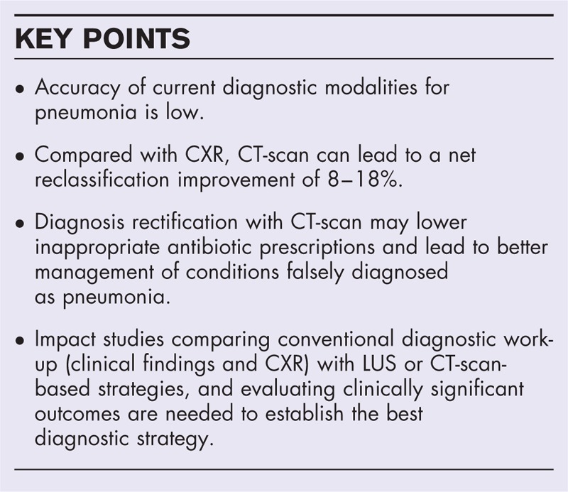
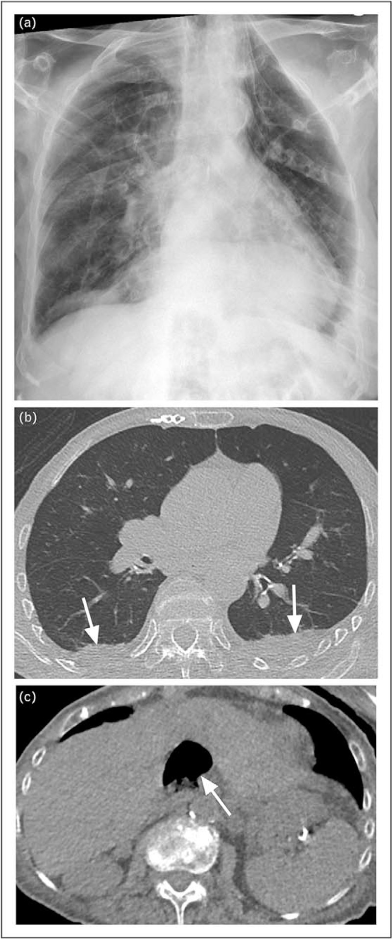
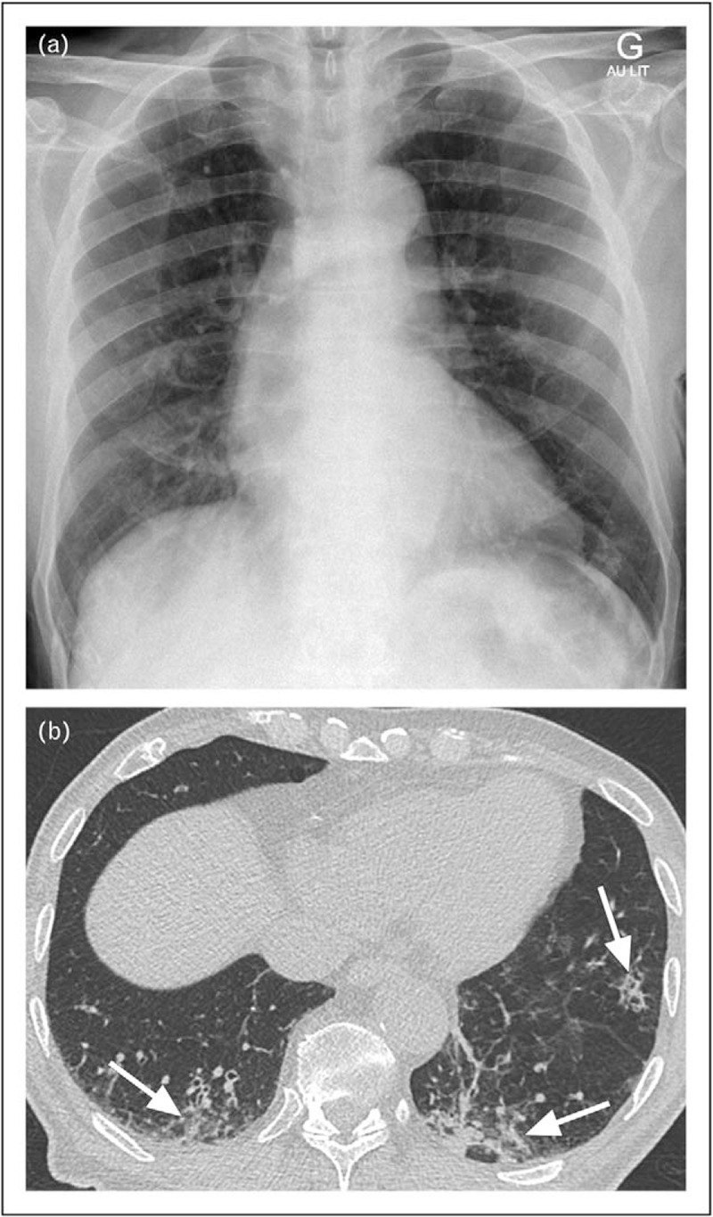
Recent findings: Two studies assessed the diagnostic accuracy of CT-scan in emergency department or hospitalized patients suspected of pneumonia. CT-scan led to a net reclassification improvement of 8 and 18% of patients, and was particularly helpful to rule out the diagnosis, allowing a lowering of the number of inappropriate antibiotic prescriptions.

Summary: CT-scan reduces overdiagnosis of pneumonia and allows a better identification of alternative diagnoses. The impact on clinical outcomes of a strategy incorporating CT-scan for patients suspected of pneumonia should be evaluated, along with its cost-effectiveness.


Figures

Box 1
Box 1
no caption available
FIGURE 1
FIGURE 1
Images of 96-year-old woman with suspicion of community-acquired pneumonia. a–c, frontal chest radiograph acquired in sitting position (a), unenhanced low-dose CT of lung bases in lung window setting (b) and CT of upper abdomen in soft tissue window setting (c). The radiograph (a) is difficult to interpret, with an enlarged heart because of the antero-posterior incidence of X-rays and the patient's left hand superimposed on the left lung base. A left basal consolidation was initially suspected. Consolidation could not be confirmed on subsequent CT (b). Bilateral pleural effusions (arrows, b) were considered to be related to congestive heart failure. The lowermost CT slices covering the upper abdomen showed a liver cavity containing debris and air (arrow, c), held responsible for pain referred to the lung bases. CT, computed tomography.
FIGURE 2
FIGURE 2
Images of 77-year-old man with suspicion of community-acquired pneumonia. a and b, frontal bedside chest radiograph (a) and unenhanced low-dose CT image of lung bases in lung window setting (b). No consolidation is obvious on the chest radiograph (a). The CT, however, shows faint infiltrates (arrows, b) affecting also nondependent lung regions. CT, computed tomography.
FIGURE 3
FIGURE 3
Images of 82-year-old woman with suspicion of community-acquired pneumonia. a and b, frontal upright chest radiograph (a) and axial image of unenhanced low-dose CT covering the lung bases, lung window setting (b). The radiograph (a) shows signs of congestive heart failure in the form of cardiomegaly, perivascular haze and right-sided pleural effusion with adjacent atelectases. On CT, some of the atelectacic bands (arrows, b) are not directly adjacent to the pleural effusion, and an infectious component cannot be excluded. CT, computed tomography.
FIGURE 4
FIGURE 4
Images of 83-year-old woman with suspicion of community-acquired pneumonia. a and b, frontal chest radiograph acquired in sitting position (a) and axial image of unenhanced low-dose CT in lung window setting (b). The radiograph (a) shows a diffusely irregular pulmonary parenchyma with superimposed areas of confluence (arrow, a). The corresponding CT image confirms small areas of airspace disease anterior to the right oblique fissure (arrows, b). Note reticular infiltrates predominating in subpleural regions (arrowheads, b), consistent with underlying chronic interstitial lung disease. CT, computed tomography.

References

    1. GBD 2015 LRI Collaborators.. Estimates of the global, regional, and national morbidity, mortality, and aetiologies of lower respiratory tract infections in 195 countries: a systematic analysis for the Global Burden of Disease Study 2015. Lancet Infect Dis 2017; 17:1133–1161. - PMC - PubMed
    1. Jain S, Self WH, Wunderink RG, et al. Community-acquired pneumonia requiring hospitalization among U.S. adults. N Engl J Med 2015; 373:415–427. - PMC - PubMed
    1. Ewig S, Birkner N, Strauss R, et al. New perspectives on community-acquired pneumonia in 388 406 patients. Results from a nationwide mandatory performance measurement programme in healthcare quality. Thorax 2009; 64:1062–1069. - PMC - PubMed
    1. Shapiro DJ, Hicks LA, Pavia AT, Hersh AL. Antibiotic prescribing for adults in ambulatory care in the USA, 2007-09. J Antimicrob Chemother 2014; 69:234–240. - PubMed
    1. Fridkin S, Baggs J, Fagan R, et al. Vital signs: improving antibiotic use among hospitalized patients. MMWR Morb Mortal Wkly Rep 2014; 63:194–200. - PMC - PubMed

MeSH terms

Substances