

Interplay between spinal cord and cerebral cortex metabolism in amyotrophic lateral sclerosis
- PMID: 30730551
- PMCID: PMC6061793
- DOI: 10.1093/brain/awy152
Interplay between spinal cord and cerebral cortex metabolism in amyotrophic lateral sclerosis
Abstract
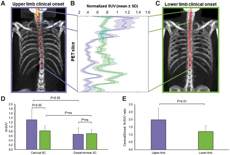
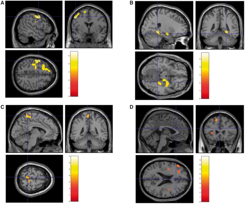
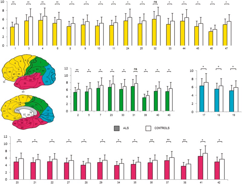
We recently reported the potential of Hough transform in delineating spinal cord metabolism by 18F-fluorodeoxyglucose PET/CT scanning in amyotrophic lateral sclerosis. The present study aimed to verify the relationship between spinal cord and brain metabolism in 44 prospectively recruited patients affected by amyotrophic lateral sclerosis submitted to 18F-fluorodeoxyglucose brain and whole-body PET/CT. Patients were studied to highlight the presence of brain hypo- or hypermetabolism with respect to healthy controls, and multiple regression analysis was performed to evaluate the correlation between spinal cord and brain metabolism. Our results confirmed higher 18F-fluorodeoxyglucose uptake in both cervical and dorsal spinal cord in patients with amyotrophic lateral sclerosis with respect to controls. This finding was paralleled by the opposite pattern in the brain cortex that showed a generalized reduction in tracer uptake. This hypometabolism was particularly evident in wide regions of the frontal-dorsolateral cortex while it did not involve the midbrain. Bulbar and spinal disease onset was associated with similar degree of metabolic activation in the spinal cord. However, among spinal onset patients, upper limb presentation was associated with a more pronounced metabolic activation of cervical segment. Obtained data suggest a differential neuro-pathological state or temporal sequence in disease progression.
Figures
References
-
- Abrahams S, Goldstein LH, Kew JJ, Brooks DJ, Lloyd CM, Frith CD et al. . Frontal lobe dysfunction in amyotrophic lateral sclerosis: a PET study. Brain 1996; 119: 2105–20. - PubMed
-
- Abrahams S, Goldstein LH, Suckling J, Ng V, Simmons A, Chitnis X et al. . Frontotemporal white matter changes in amyotrophic lateral sclerosis. J Neurol 2005; 252: 321–31. - PubMed
-
- Beltrametti MC, Massone AM, Piana M. Hough transform of special classes of curves. SIAM J Imaging Sci 2013; 6: 391–412.
Publication types
MeSH terms
Substances
LinkOut - more resources
Full Text Sources
Medical
Molecular Biology Databases
