

Highly invasive and poorly differentiated corneal squamous cell carcinoma in a dog
- PMID: 30732595
- PMCID: PMC6367762
- DOI: 10.1186/s12917-019-1790-3
Highly invasive and poorly differentiated corneal squamous cell carcinoma in a dog
Abstract
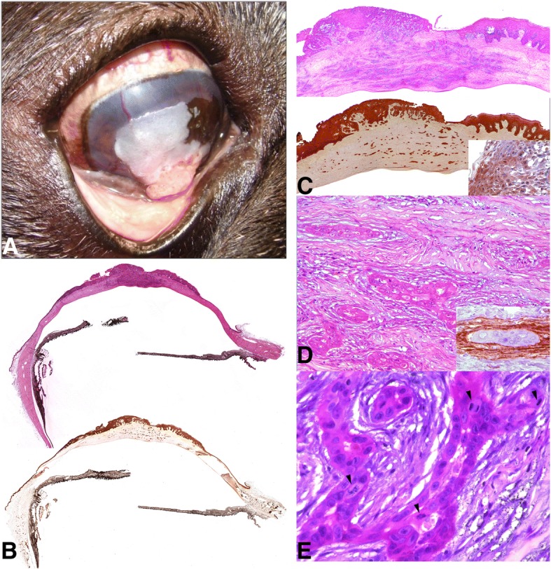
Background: Primary corneal tumors are unusual in dogs although there has been a rise in the prevalence of canine corneal squamous cell carcinoma in the past decades due to different factors. Exposure to ultraviolet radiation, presence of chronic keratitis or history of superficial trauma are some of them. We report for the first time a highly infiltrative corneal neoplasia with both exophytic and deep stromal growth, which presented atypical histologic features of a squamous cell carcinoma.
Case presentation: An adult male French bulldog was referred with an exophytic, pink to white gelatinous mass occupying approximately 70% of the central cornea on the right eye. Histological findings from the excisional biopsy were consistent with corneal carcinoma and transconjunctival enucleation was performed at the request of the owner. A final diagnosis of primary corneal squamous cell carcinoma was done based on the squamous differentiation observed in the neoplastic cells of the superficial layers. However, cells in deeper layers were less differentiated, showed pseudoacinar formations and did not expressed marker for stratified squamous epithelium (i.e., cytokeratin 5/6). The dramatic thickening of the cornea and the fact of observing neoplastic cells almost at the level of the Descemet's membrane make this case very unusual as the squamous cell carcinoma in dogs tends to involve the superficial stroma or colonize the corneal surface as an exophytic lesion.
Conclusions: Based on the histological findings, a high infiltrative and poorly differentiated corneal squamous cell carcinoma was diagnosed. In terms of clinical relevance, our results suggest that corneal lesions compatible with neoplasia need an early diagnosis in order to prevent the aggressive growth of the tumor and the enucleation of the eye.
Keywords: Corneal neoplasia; Dog; Primary corneal squamous cell carcinoma.
Conflict of interest statement
Ethics approval
The dog’s owner gave his permission for the publication of this clinical case.
Consent for publication
Not applicable
Competing interests
The authors declare that they have no competing interests.
Publisher’s Note
Springer Nature remains neutral with regard to jurisdictional claims in published maps and institutional affiliations.
Figures
References
Publication types
MeSH terms
Grants and funding
LinkOut - more resources
Full Text Sources
Medical
Research Materials
Miscellaneous
