. 2019 Feb 7;7(1):16.
doi: 10.1186/s40478-019-0666-x.

White matter capillaries in vascular and neurodegenerative dementias

Affiliations

White matter capillaries in vascular and neurodegenerative dementias

Yoshiki Hase et al. Acta Neuropathol Commun. .

Abstract

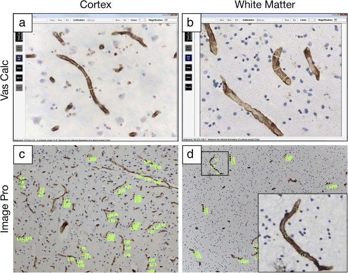
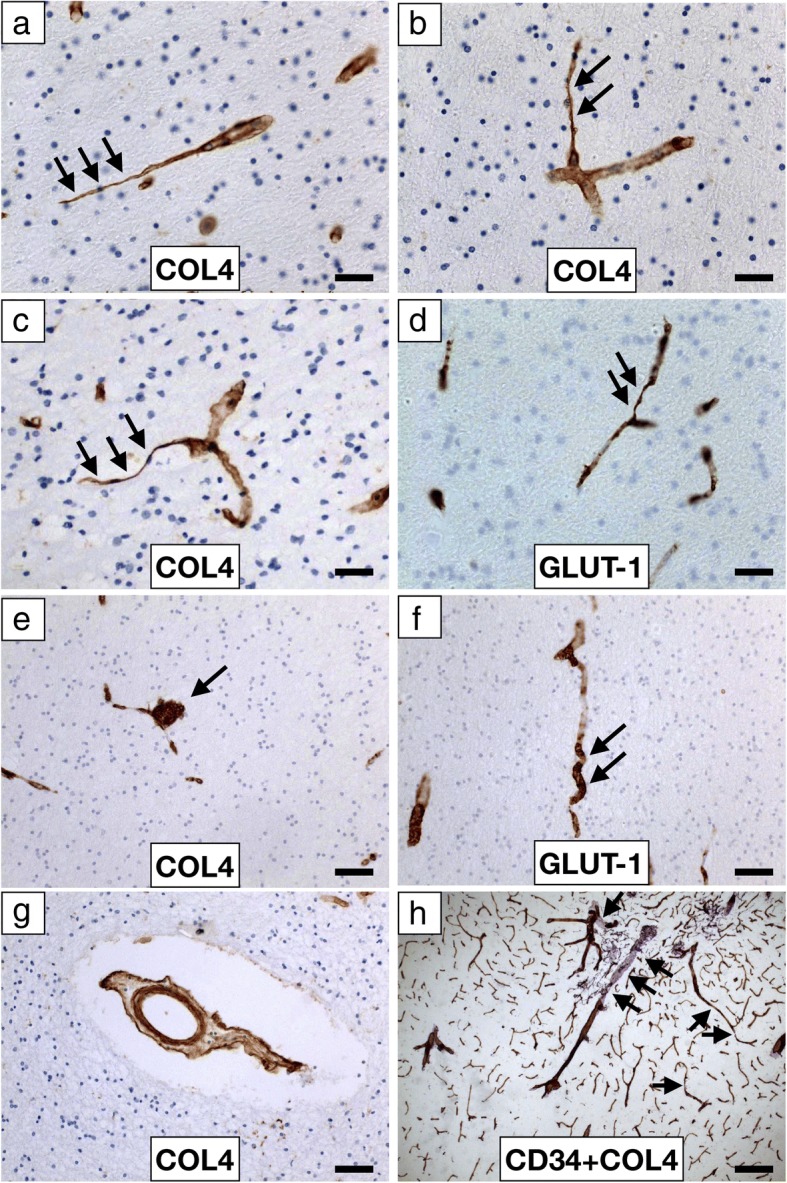
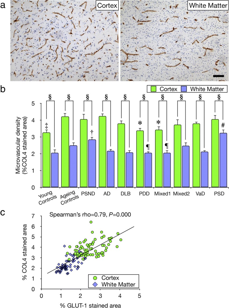
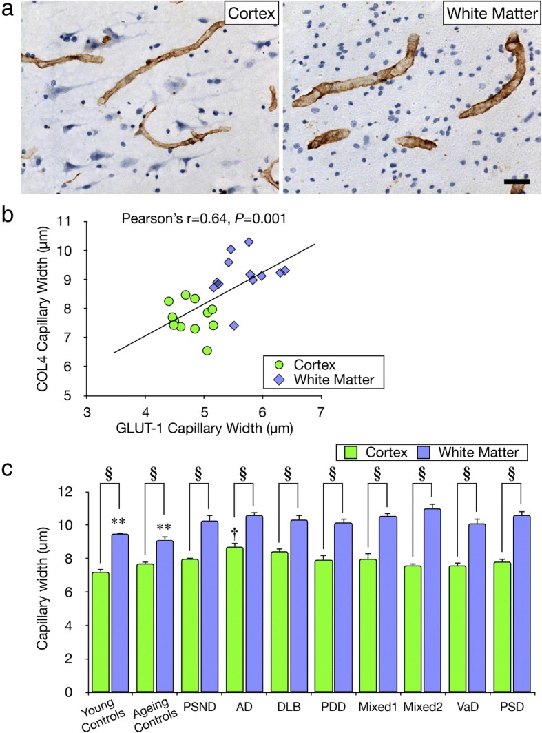
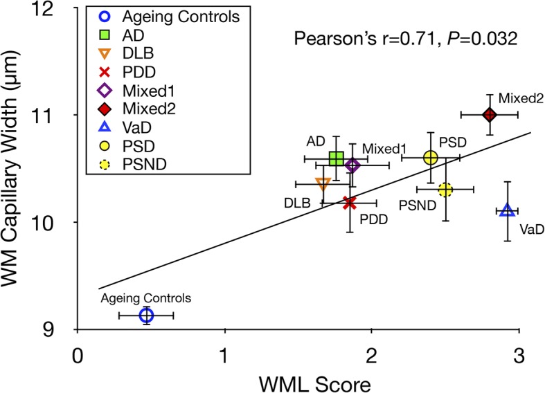
Previous studies suggest white matter (WM) integrity is vulnerable to chronic hypoperfusion during brain ageing. We assessed ~ 0.7 million capillary profiles in the frontal lobe WM across several dementias comprising Alzheimer's disease, dementia with Lewy bodies, Parkinson's disease with dementia, vascular dementia, mixed dementias, post-stroke dementia as well as post-stroke no dementia and similar age ageing and young controls without significant brain pathology. Standard histopathological methods were used to determine microvascular pathology and capillary width and densities in 153 subjects using markers of the basement membrane (collagen IV; COL4) and endothelium (glucose transporter-1; GLUT-1). Variable microvascular pathology including coiled, tortuous, collapsed and degenerated capillaries as well as occasional microaneurysms was present in all dementias. As expected, WM microvascular densities were 20-49% lower than in the overlying cortex. This differential in density between WM and cortex was clearly demonstrated by COL4, which was highly correlated with GLUT-1 densities (Spearman's rho = 0.79, P = 0.000). WM COL4 immunopositive microvascular densities were decreased by ~ 18% across the neurodegenerative dementias. However, we found WM COL4 densities were increased by ~ 57% in post-stroke dementia versus ageing and young controls and other dementias. Using three different methods to measure capillary diameters, we found WM capillaries to be significantly wider by 19-45% compared to those in overlying neocortex apparent with both COL4 and GLUT-1. Remarkably, WM capillary widths were increased by ~ 20% across all dementias compared to ageing and young controls (P < 0.01). We also noted mean WM pathology scores incorporating myelin loss, arteriolosclerosis and perivascular spacing were correlated with COL4 immunopositive capillary widths (Pearson's r = 0.71, P = 0.032). Our key finding indicates that WM capillaries are wider compared to those in the overlying neocortex in controls but they dilate further during dementia pathogenesis. We suggest capillaries undergo restructuring in the deep WM in different dementias. This reflects compensatory changes to retain WM perfusion and integrity during hypoperfusive states in ageing-related dementias.

Keywords: Alzheimer’s disease; Dementia; Dementia with Lewy bodies; Microvascular pathology; Mixed dementia; Parkinson’s disease with dementia; Post-stroke dementia; Small vessel disease; Vascular dementia.


Conflict of interest statement

Ethics approval and consent to participate

Ethical approvals were granted by local research ethics committees of the Newcastle upon Tyne Foundation Hospitals Trust. Permission for use of brains for post-mortem research was also granted by consent from next-of-kin or family. All the brain tissues were retained in and obtained from the Newcastle Brain Tissue Resource.

Consent for publication

All the authors have approved publication and see various versions of the manuscript.

Competing interests

The authors declare that they have no competing interests.

Publisher’s Note

Springer Nature remains neutral with regard to jurisdictional claims in published maps and institutional affiliations.

Figures

Fig. 1
Fig. 1
Methods used to quantify microvascular morphology a-d, Representative images of collagen-IV (COL4) stained microvessels in the cortex (a, c) and WM (b, d). a-b, Screen shots of profiles of capillaries indicating how widths (diameters) were measured longitudinally using the VasCalc method using 40x objective lens. c-d, Images of capillaries with indications (in green markers) where widths along the vessel were measured using the Image-Pro Analyser method using 10x objective lens
Fig. 2
Fig. 2
Microvascular pathology in the frontal WM in dementia a-h, Low and High power representative images of COL4 (a, b, c, e, g), GLUT-1 (d, f) and CD34 + COL4 (h) immunostained capillaries and microvessel in the WM. Collapsed and string vessels (arrows) were observed using both markers in VaD and PSD with similar profiles in all dementias. e, a microaneurysm-like structure (arrow) in a PSD case detected using COL4. f, a GLUT-1 immunmopostivie tortuous capillary (arrows) in AD. g, COL4 immunopositive ‘bagged’ vessel with increased perivascular space in a PSD case. h, CD34 and COL4 positive profiles of arterioles and capillaries at the juxtaposition of the grey and WM showing several collapsed and string capillaries (arrows). Scale bar represents 25 μm (a, b, c and d); 50 μm (e, f, and g); 100 μm (h)
Fig. 3
Fig. 3
Quantification of microvascular density a, Typical images of COL4 immunostained capillaries in the cortex and WM used to quantify densities. Scale bar represents 50 μm. b, Histogram showing microvascular densities in the WM and cortex in controls and different dementias. In the WM, mean microvascular density was consistently lower by ~ 49% compared to cortex in all controls and dementia groups (§P < 0.01). In different dementias, microvascular density in the WM was decreased by ~ 18% compared to ageing controls, particularly in PDD and Mixed dementia 1 group (P < 0.05), whereas PSD and PSND showed ~ 57% higher microvascular density (#P < 0.012 vs all disease and control groups other than PSND; P < 0.025 vs all disease groups other than ageing controls, Mixed 2 and PSD). In the cortex, dementia subjects showed ~ 20% lower vascular density compared with ageing controls, particularly in the PDD and Mixed dementia 1 group (*P < 0.05). Young control group showed less vascular density by ~ 23% compared with ageing controls (P < 0.01). c, Correlation of microvascular densities between COL4 and GLUT-1 immunostained areas in dementia subjects. Spearman’s correlation analysis revealed a strong positive correlation (rho = 0.79, P = 0.000)
Fig. 4
Fig. 4
Quantification of capillary width a, Representative images of COL4 immunostained capillaries in the cortex and WM used to determine capillary width. Scale bar represents 25 μm. b, Correlation of mean capillary widths assessed by COL4 and GLUT-1 immunostaining in ageing controls. Pearson’s analysis revealed that mean capillary width in both the WM and the cortex was positively correlated (r = 0.64, P = 0.001). c, Histogram showing mean capillary width in the WM and cortex in controls and dementia groups. In the WM, mean capillary width was consistently larger by 19–45% compared to cortex in all control and dementia groups (§P < 0.01). In all the dementias, capillary width in the WM was consistently greater by < 20% compared to ageing and young controls (**P < 0.01). In the cortex, mean capillary widths in dementia subjects were not significantly wider compared with ageing and young controls, but only AD subjects showed wider capillaries compared with ageing and young controls (P < 0.01)
Fig. 5
Fig. 5
Relationship between WM capillary width and WM pathology Plot shows correlation between mean capillary widths assessed by COL4 immunostaining in all the dementias and ageing controls versus WML scores. Pearson’s analysis revealed that mean capillary width in the WM in dementia was positively correlated with WM damage (r = 0.71, P = 0.032). Although there were some age-related changes in WM in the controls, it was clear that all dementias were a disparate group as a whole exhibiting high WML scores and wider capillaries

References

    1. Allan LM, Rowan EN, Firbank MJ, Thomas AJ, Parry SW, Polvikoski TM, O'Brien JT, Kalaria RN. Long term incidence of dementia, predictors of mortality and pathological diagnosis in older stroke survivors. Brain. 2011;134:3716–3727. doi: 10.1093/brain/awr273. - DOI - PMC - PubMed
    1. Arvanitakis Z, Leurgans SE, Barnes LL, Bennett DA, Schneider JA. Microinfarct pathology, dementia, and cognitive systems. Stroke. 2011;42:722–727. doi: 10.1161/STROKEAHA.110.595082. - DOI - PMC - PubMed
    1. Ballard C, McKeith I, O'Brien J, Kalaria R, Jaros E, Ince P, Perry R. Neuropathological substrates of dementia and depression in vascular dementia, with a particular focus on cases with small infarct volumes. Dement Geriatr Cogn Disord. 2000;11:59–65. doi: 10.1159/000017215. - DOI - PubMed
    1. Baradaran H, Mtui EE, Richardson JE, Delgado D, Dunning A, Marshall RS, Sanelli PC, Gupta A. White matter diffusion abnormalities in carotid artery disease: a systematic review and meta-analysis. J Neuroimaging. 2016;26:481–488. doi: 10.1111/jon.12347.. - DOI - PubMed
    1. Barker R, Ashby EL, Wellington D, Barrow VM, Palmer JC, Kehoe PG, Esiri MM, Love S. Pathophysiology of white matter perfusion in Alzheimer's disease and vascular dementia. Brain. 2014;137:1524–1532. doi: 10.1093/brain/awu040.. - DOI - PMC - PubMed

Publication types