Review
. 2019 Feb;12(2):283-296.
doi: 10.1016/j.jcmg.2018.11.026.

Imaging and Impact of Myocardial Fibrosis in Aortic Stenosis

Affiliations
Review

Imaging and Impact of Myocardial Fibrosis in Aortic Stenosis

Rong Bing et al. JACC Cardiovasc Imaging. 2019 Feb.

Abstract

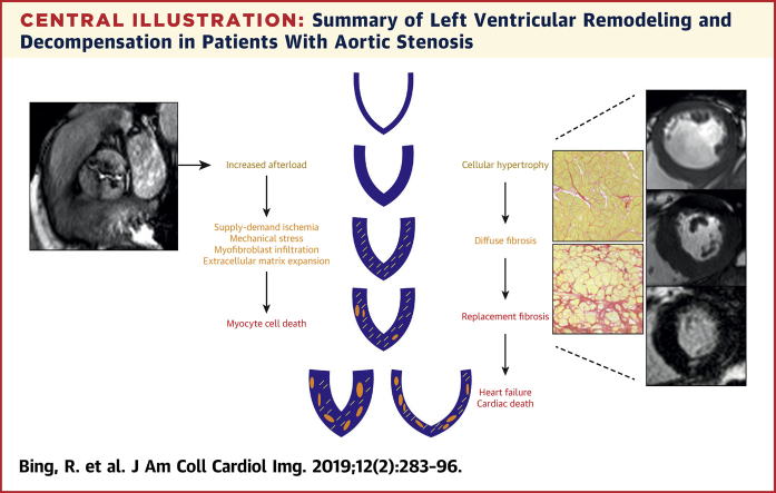
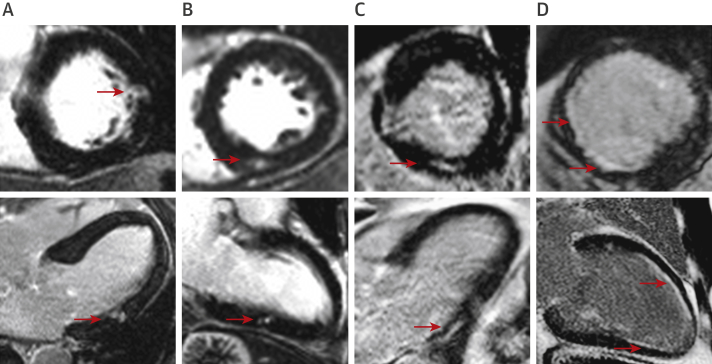
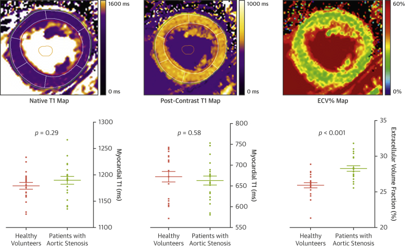
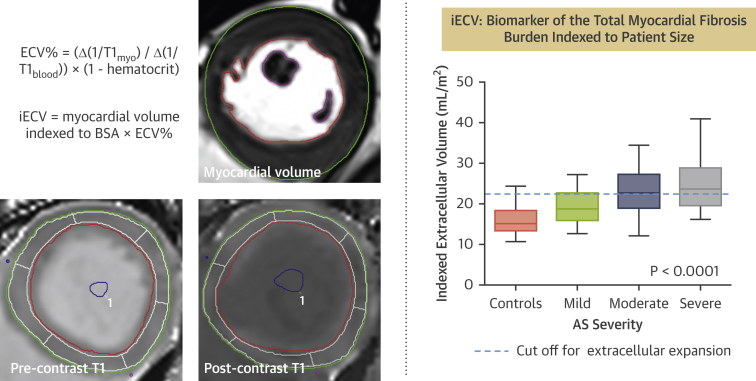
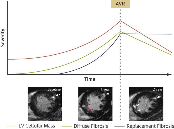
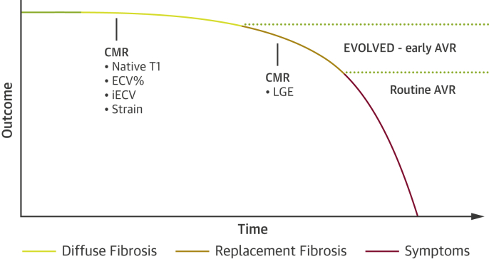
Aortic stenosis is characterized both by progressive valve narrowing and the left ventricular remodeling response that ensues. The only effective treatment is aortic valve replacement, which is usually recommended in patients with severe stenosis and evidence of left ventricular decompensation. At present, left ventricular decompensation is most frequently identified by the development of typical symptoms or a marked reduction in left ventricular ejection fraction <50%. However, there is growing interest in using the assessment of myocardial fibrosis as an earlier and more objective marker of left ventricular decompensation, particularly in asymptomatic patients, where guidelines currently rely on nonrandomized data and expert consensus. Myocardial fibrosis has major functional consequences, is the key pathological process driving left ventricular decompensation, and can be divided into 2 categories. Replacement fibrosis is irreversible and identified using late gadolinium enhancement on cardiac magnetic resonance, while diffuse fibrosis occurs earlier, is potentially reversible, and can be quantified with cardiac magnetic resonance T1 mapping techniques. There is a substantial body of observational data in this field, but there is now a need for randomized clinical trials of myocardial imaging in aortic stenosis to optimize patient management. This review will discuss the role that myocardial fibrosis plays in aortic stenosis, how it can be imaged, and how these approaches might be used to track myocardial health and improve the timing of aortic valve replacement.

Keywords: T(1) mapping; aortic stenosis; cardiac magnetic resonance; late gadolinium enhancement; myocardial fibrosis.


Figures

None
Graphical abstract
Central Illustration
Summary of Left Ventricular Remodeling and Decompensation in Patients With Aortic Stenosis Schematic of the left ventricular remodeling response in aortic stenosis, describing the transition from hypertrophy to fibrosis, heart failure, and cardiac death.
Figure 1
Figure 1
Late Gadolinium Enhancement Patterns in Aortic Stenosis Each panel shows short-axis (top) and corresponding long-axis (bottom) late gadolinium images from cardiac magnetic resonance scans. (A to C) Focal noninfarct late gadolinium enhancement typical of the replacement fibrosis seen in aortic stenosis. (D) Subendocardial late gadolinium enhancement in coronary artery territories, consistent with scar due to infarction rather than focal noninfarct fibrosis. Areas of infarction such as these should be excluded when calculating extracellular volume fraction. Red arrows indicate areas of late gadolinium enhancement.
Figure 2
Figure 2
T1 Mapping Three different cardiac magnetic resonance T1 maps are demonstrated. Native T1 and post-contrast T1 maps are generated by the signal intensity encoded within each voxel, depending on the T1 relaxation time; color coding according to T1 times is applied for visual reference. ECV% maps are generated using the formula ECV% = (Δ[1/T1myo]/Δ[1/T1blood]) × (1 − hematocrit), where Δ(1/T1) is the difference in myocardial or blood T1 pre-contrast and post-contrast. ECV% can be used to assess the proportion of the myocardium comprised by extracellular space. Note that there is significant overlap between health and disease with native and post-contrast T1, in contrast to ECV%. Graphs adapted from Chin et al. by permission of Oxford University Press. ECV% = extracellular volume fraction; iECV = indexed extracellular volume.
Figure 3
Figure 3
iECV calculation The cardiac magnetic resonance short-axis images provide examples of the pre-contrast and post-contrast contours required to calculate iECV. Systolic and diastolic contours are drawn using the short-axis stack to calculate myocardial volume, which is necessary to derive iECV. Color look-up tables have not been applied to the T1 images. iECV provides a surrogate of the total myocardial fibrosis burden according to the formula demonstrated in the figure. iECV demonstrates good correlation with histological fibrosis burden and severity of aortic stenosis. Graph adapted from Chin et al. , Creative Commons Attribution License: https://creativecommons.org/licenses/by/4.0/. BSA = body surface area; other abbreviations as in Figure 2.
Figure 4
Figure 4
Schematic for the Development of Myocardial Fibrosis in Aortic Stenosis and Response to AVR As aortic stenosis progresses, left ventricular (LV) mass gradually increases, followed by the development of diffuse fibrosis. Replacement fibrosis occurs later but accelerates rapidly once established. Following relief of pressure-loading conditions after aortic valve replacement (AVR), LV cellular mass and extracellular matrix both regress at different rates. The burden of replacement fibrosis, however, persists. The insets show short-axis cardiac magnetic resonance late gadolinium enhancement imaging slices of a patient with aortic stenosis. At baseline, there is focal late gadolinium enhancement representing discrete focal replacement fibrosis (white arrow). After 1 year, the burden of this replacement fibrosis has increased with the development of several new discrete deposits (red arrows). The patient subsequently underwent AVR. One year later, despite regression of LV mass, there is no regression of replacement fibrosis (white arrows).
Figure 5
Figure 5
Proposed Integration of Myocardial Fibrosis Into the Classical Description of the Natural History of Aortic Stenosis Adaption of the outcome curve originally proposed by Braunwald in 1968 . Prior to the onset of symptoms, there is a long latent period in aortic stenosis where subclinical myocardial changes take place, including the development of reversible diffuse fibrosis followed by irreversible replacement fibrosis. These changes may be assessed with the imaging modalities denoted in the figure. Exploratory data suggest that diffuse fibrosis is associated with an adverse long-term outcome in aortic stenosis. The prognostic data related to the noninfarct pattern of late gadolinium enhancement (LGE) as a marker of replacement fibrosis is comparatively robust, establishing LGE as a powerful independent predictor of long-term clinical outcomes. According to current guidelines and routine clinical practice, AVR is performed after the onset of symptoms. Future and ongoing trials, including the EVOLVED trial, are required to determine whether targeted early intervention utilizing cardiac magnetic resonance (CMR) to detect fibrosis will lead to improved clinical outcomes. Abbreviations as in Figures 2 and 4.

References

    1. Nkomo V.T., Gardin J.M., Skelton T.N., Gottdiener J.S., Scott C.G., Enriquez-Sarano M. Burden of valvular heart diseases: a population-based study. Lancet. 2006;368:1005–1011. - PubMed
    1. Iung B., Vahanian A. Epidemiology of valvular heart disease in the adult. Nat Rev Cardiol. 2011;8:162–172. - PubMed
    1. Osnabrugge R.L., Mylotte D., Head S.J. Aortic stenosis in the elderly: disease prevalence and number of candidates for transcatheter aortic valve replacement: a meta-analysis and modeling study. J Am Coll Cardiol. 2013;62:1002–1012. - PubMed
    1. Dweck M.R., Boon N.A., Newby D.E. Calcific aortic stenosis: a disease of the valve and the myocardium. J Am Coll Cardiol. 2012;60:1854–1863. - PubMed
    1. Rosenhek R., Zilberszac R., Schemper M. Natural history of very severe aortic stenosis. Circulation. 2010;121:151–156. - PubMed

Publication types

MeSH terms