. 2019 Mar;60(3):299-303.
doi: 10.2967/jnumed.119.226498. Epub 2019 Feb 7.

First Human Imaging Studies with the EXPLORER Total-Body PET Scanner

Affiliations

First Human Imaging Studies with the EXPLORER Total-Body PET Scanner

Ramsey D Badawi et al. J Nucl Med. 2019 Mar.

Abstract

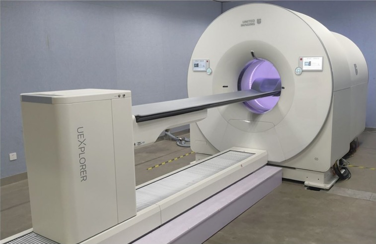
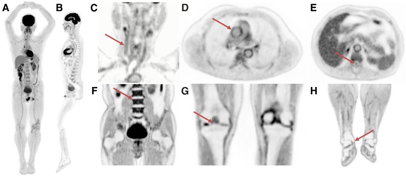
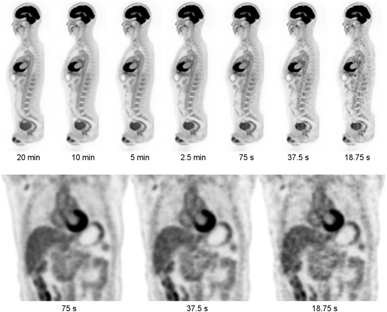
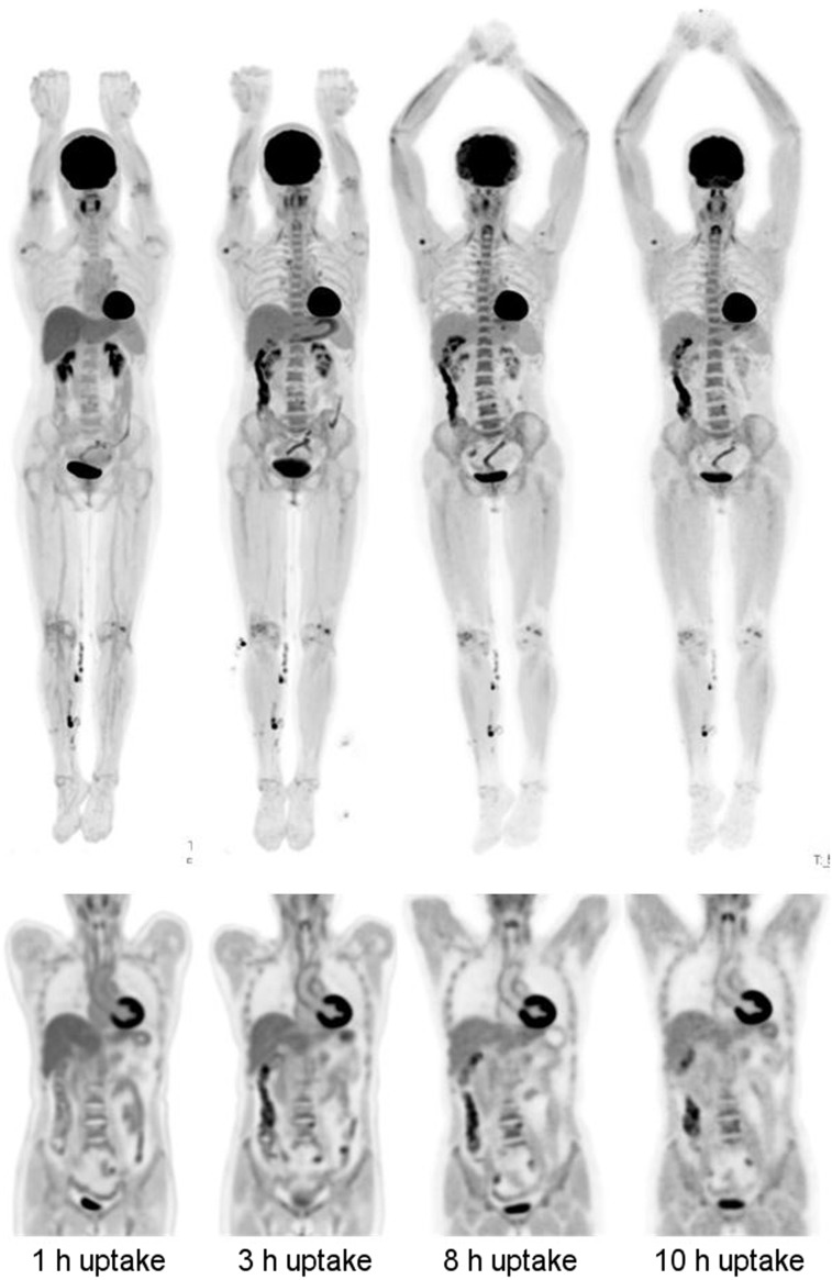
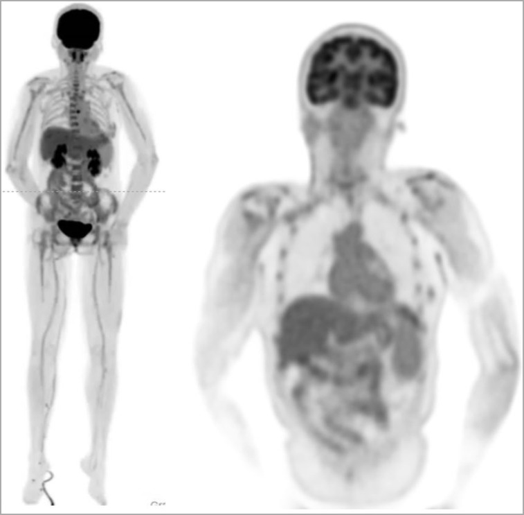
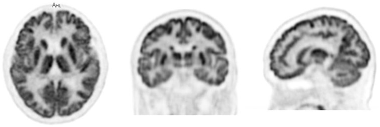
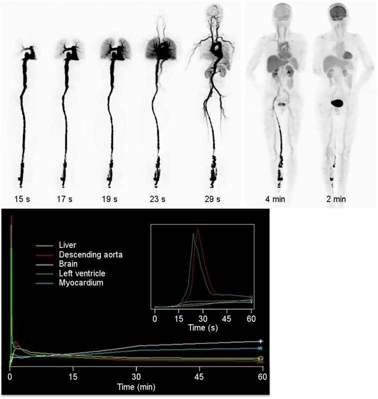
Within the EXPLORER Consortium, the construction of the world's first total-body PET/CT scanner has recently been completed. The 194-cm axial field of view of the EXPLORER PET/CT scanner is sufficient to cover, for the first time, the entire human adult body in a single acquisition in more than 99% of the population and allows total-body pharmacokinetic studies with frame durations as short as 1 s. The large increase in sensitivity arising from total-body coverage as well as increased solid angle for detection at any point within the body allows whole-body 18F-FDG PET studies to be acquired with unprecedented count density, improving the signal-to-noise ratio of the resulting images. Alternatively, the sensitivity gain can be used to acquire diagnostic PET images with very small amounts of activity in the field of view (25 MBq, 0.7 mCi or less), with very short acquisition times (∼1 min or less) or at later time points after the tracer's administration. We report here on the first human imaging studies on the EXPLORER scanner using a range of different protocols that provide initial evidence in support of these claims. These case studies provide the foundation for future carefully controlled trials to quantitatively evaluate the improvements possible through total-body PET imaging.

Keywords: EXPLORER; FDG; PET; PET/CT; instrumentation; total-body PET.


Figures

FIGURE 1.
FIGURE 1.
Photograph of completed EXPLORER total-body PET/CT scanner.
FIGURE 2.
FIGURE 2.
Selected views from total-body scan of subject 1. (A) Total-body MIP. (B) Total-body sagittal view. (C) Head/neck view, with arrow indicating walls of right carotid artery. (D) Chest view, showing walls of major blood vessels, with ascending aorta indicated by arrow. (E) Midthoracic view with spinal canal indicated by arrow. (F) Abdomen and pelvis, showing clear delineation of endplates of vertebral bodies (arrow points to superior endplate of L3). (G) Knees, with bone spur indicated by arrow. (H) Lower extremities, with arrow showing delineation of medial tibial malleolus.
FIGURE 3.
FIGURE 3.
Views from subject 1 as function of scan duration (290 MBq injected, 82 min uptake period). (Top) Sagittal views from 20 min to 18.75 s. (Bottom) Coronal views at 75, 37.5, and 18.75 s.
FIGURE 4.
FIGURE 4.
Delayed imaging for subject 2 (256 MBq injected, 14 min scan duration). (Left-to-right) Images from scans performed at 1, 3, 8, and 10 h after injection. (Top row) MIP images. (Bottom row) Coronal views of thorax and abdomen. Head motion artifacts are visible in 8-h scan.
FIGURE 5.
FIGURE 5.
Low-dose images from subject 4. Subject was injected with 25 MBq and scanned for 10 min after 52.5 min of uptake time. Total-body MIPs (left) and coronal view of upper body (right). Tracer was injected into a vein in right lower leg.
FIGURE 6.
FIGURE 6.
Dedicated brain scan from subject 3. Motion correction has not been implemented at this time.
FIGURE 7.
FIGURE 7.
Total-body dynamic imaging. (Top) Selections of rotating MIPs from dynamic scan of subject 2. Frame duration is 1 s, except for the 2 left-most images, which have frame durations of 1 min. (Bottom) Time–activity curves for selected anatomic regions. (Bottom inset) Time–activity curves for first minute of acquisition.

References

    1. Cherry SR, Badawi RD, Karp JS, Moses WW, Price P, Jones T. Total-body imaging: transforming the role of positron emission tomography. Sci Transl Med. 2017;9:eaaf6169. - PMC - PubMed
    1. Cherry SR, Jones T, Karp JS, Qi J, Moses WW, Badawi RD. Total-body PET: maximizing sensitivity to create new opportunities for clinical research and patient care. J Nucl Med. 2018;59:3–12. - PMC - PubMed
    1. Poon JK, Dahlbom ML, Moses WW, et al. Optimal whole-body PET scanner configurations for different volumes of LSO scintillator: a simulation study. Phys Med Biol. 2012;57:4077–4094. - PMC - PubMed
    1. Badawi RD, Poon JK, Surti S, et al. EXPLORER, an ultrasensitive total-body PET scanner: application feasibility simulations. Paper presented at: the World Molecular Imaging Congress, Savannah, Georgia, September 2013.
    1. Viswanath V, Daube-Witherspoon ME, Schmall JP, et al. Development of PET for total-body imaging. Acta Phys Pol B. 2017;48:1555–1566.

Publication types