Review
. 2019 Feb 28;47(1):399-410.
doi: 10.1042/BST20180608. Epub 2019 Feb 7.

Twenty years of Mediator complex structural studies

Affiliations
Review

Twenty years of Mediator complex structural studies

Alexis Verger et al. Biochem Soc Trans. .

Abstract

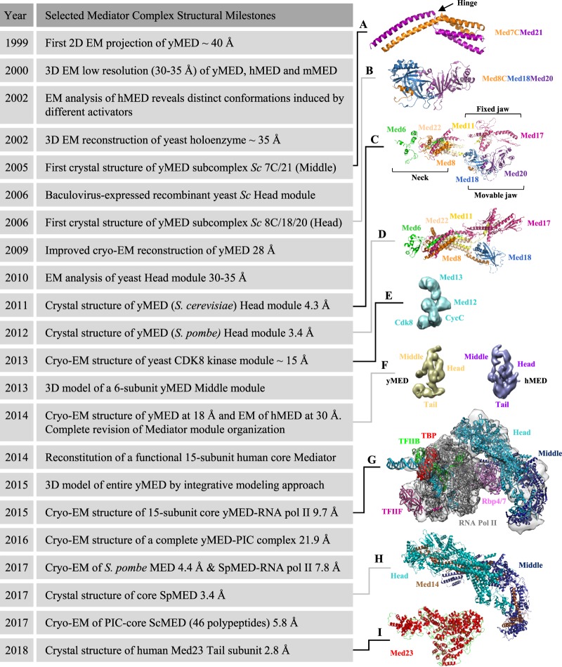
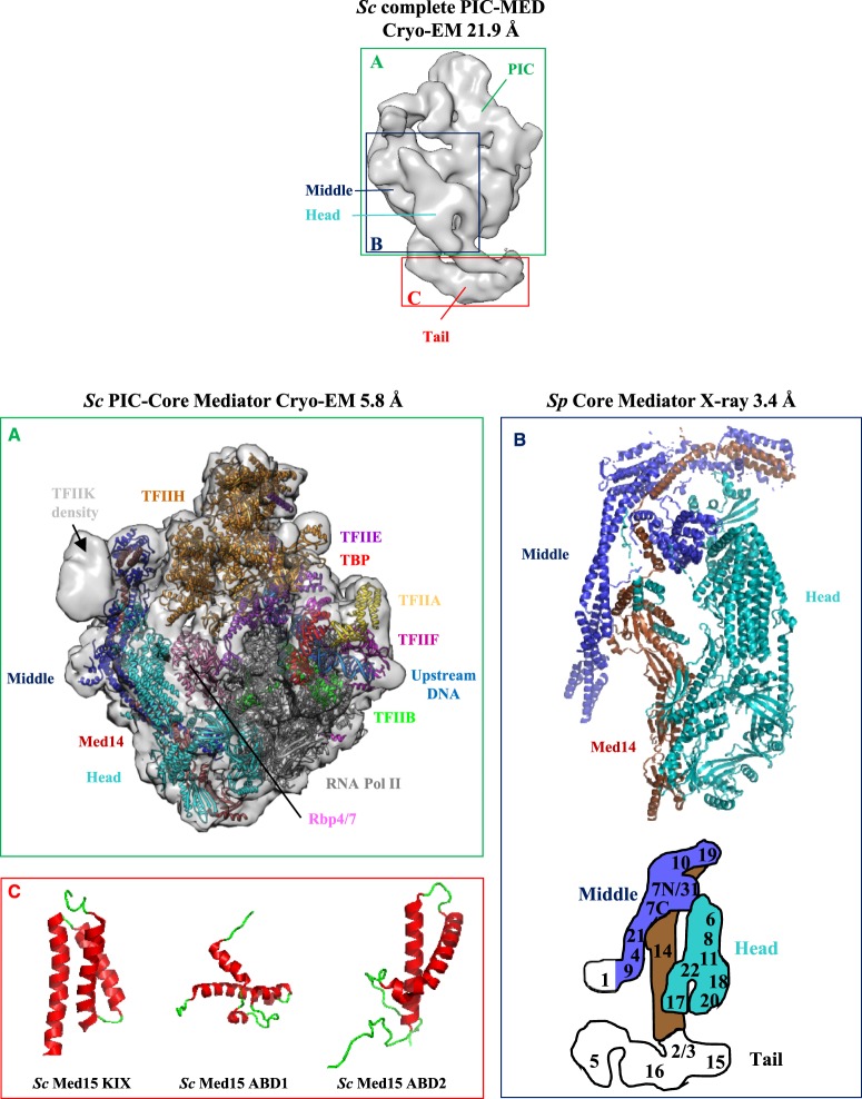
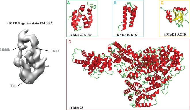
Mediator is a large multiprotein complex conserved in all eukaryotes that plays an essential role in transcriptional regulation. Mediator comprises 25 subunits in yeast and 30 subunits in humans that form three main modules and a separable four-subunit kinase module. For nearly 20 years, because of its size and complexity, Mediator has posed a formidable challenge to structural biologists. The first two-dimensional electron microscopy (EM) projection map of Mediator leading to the canonical view of its division in three topological modules named Head, Middle and Tail, was published in 1999. Within the last few years, optimization of Mediator purification combined with technical and methodological advances in cryo-electron microscopy (cryo-EM) have revealed unprecedented details of Mediator subunit organization, interactions with RNA polymerase II and parts of its core structure at high resolution. To celebrate the twentieth anniversary of the first Mediator EM reconstruction, we look back on the structural studies of Mediator complex from a historical perspective and discuss them in the light of our current understanding of its role in transcriptional regulation.

Keywords: Cryo-EM; Mediator complex; structural biology; transcription.


Conflict of interest statement

The Authors declare that there are no competing interests associated with the manuscript.

Figures

Figure 1.
Figure 1.. Timeline of selected milestones in Mediator complex structural studies.
From the top down, the depicted structures are (A) S. cerevisiae Med7C/Med21 complex (protein data bank (PDB) 1YKH [42]). The conserved flexible hinge is indicated. (B) S. cerevisiae Med8C/18/20 submodule (PDB 2HZS [43]), (C) S. cerevisiae Head module (PDB 3RJ1 [53]). The three major domains described initially in 2011 (fixed jaw, movable jaw and neck) are indicated. (D) S. pombe Head module (PDB 4H63 [55]), (E) S. cerevisiae CDK8 kinase module (electron microscopy data bank (EMD)-5588 [99]), (F) S. cerevisiae (EMD-2634) and Homo sapiens (EMD-2635) [57] Mediator complex, (G) S. cerevisiae RNA Pol II–core Mediator transcription initiation complex (EMD-2786 [48]), (H) S. pombe core Mediator (PDB 5N9J [47]) and (I) Homo sapiens MED23 subunit (PDB 6H02 [72]). For details, refer to the text. For a complete collection of structural data on Mediator, see Supplementary Table S1 adapted from [28] with permission from Elsevier. Figures were prepared with PyMol [116] or UCSF Chimera [117]. y, yeast; h, human; m, murine; Sc, Saccharomyces cerevisiae, Sp, Schizosaccharomyces pombe.
Figure 2.
Figure 2.. Structural studies of yeast Mediator complex.
Cryo-EM three-dimensional reconstruction of a complete S. cerevisiae Mediator–RNA Pol II preinitiation complex (Sc PIC-MED) at 21.9 Å resolution (EMD-8308 [69]). PIC, Mediator Head, Middle and Tail modules are indicated. Individual panels display high-resolution structures of ((A), in green) S. cerevisiae PIC-core Mediator at 5.8 Å resolution (EMD-3850 [49]) in semi-transparent rendering with the docked atomic model (PDB 5OQM) in ribbon representation. The cryo-EM density for the mobile Kin28–Ccl1 kinase complex of TFIIH (TFIIK) is indicated. ((B), in dark blue) S. pombe core Mediator structure at 3.4 Å resolution (PDB 5N9J [47]). For the sake of simplicity, the Mediator Head module is depicted in cyan and the Middle module in blue, with their respective subunits displayed in ribbon representation. The Med14 subunit is in brown. For a more detailed description of core Mediator structure and its 13 defined submodules, see Supplementary Figure S1. The localization of Mediator subunits is schematized below in the Mediator diagram adapted from [50,57] with the same color code (Head module in cyan, Middle module in blue and Tail module in white). The Middle module Med1 subunit was included in recombinant core Mediator but is lacking in the crystal structure [47]. Med1 is thus colored in white. ((C), in red) NMR structures of S. cerevisiae Med15 KIX domain (PDB 2K0N [77]), Med15 ABD1 domain (PDB 2LPB [74]) and Med15 ABD2 domain (PDB 6ALY [78]).
Figure 3.
Figure 3.. Structural studies of human Mediator complex.
EM map of Human Mediator at 30 Å resolution (EMD-2635 [57]). Mediator Head, Middle and Tail modules are indicated. Individual panels display high-resolution structures of ((A), in green) the N-terminal domain of Human Med26 (PDB 5ODD [64]), ((B), in cyan) KIX domain of human Med15 (PDB 2GUT [80]), ((C), in yellow) ACID/PTOV domain of human Med25 (PDB 2L23, 2XNF, 2L6U and 2KY6 [73,75,76,79]) and ((D), in red) human Med23 (PDB 6H02 [72]). Med26 associates with the Middle domain via interactions with Med4, Med7 and Med19 [57]. Note that Med26 N- and C-terminal domains support interactions with TFIID and transcription elongation factors and with Mediator complex, respectively [65]. Med15 and Med23 have been assigned to the Tail module and Med25, which engages in an extensive network of interactions with other Tail subunits is believed to be a component of the Tail [57]. For details, refer to the text.

References

    1. Kelleher, R.J. III, Flanagan P.M. and Kornberg R.D. (1990) A novel mediator between activator proteins and the RNA polymerase II transcription apparatus. Cell 61, 1209–1215 10.1016/0092-8674(90)90685-8 - DOI - PubMed
    1. Flanagan P.M., Kelleher, R.J. III, Sayre M.H., Tschochner H. and Kornberg R.D. (1991) A mediator required for activation of RNA polymerase II transcription in vitro. Nature 350, 436–438 10.1038/350436a0 - DOI - PubMed
    1. Nonet M.L. and Young R.A. (1989) Intragenic and extragenic suppressors of mutations in the heptapeptide repeat domain of Saccharomyces cerevisiae RNA polymerase II. Genetics 123, 715–724 PMID: - PMC - PubMed
    1. Thompson C.M., Koleske A.J., Chao D.M. and Young R.A. (1993) A multisubunit complex associated with the RNA polymerase II CTD and TATA-binding protein in yeast. Cell 73, 1361–1375 10.1016/0092-8674(93)90362-T - DOI - PubMed
    1. Kim Y.J., Björklund S., Li Y., Sayre M.H. and Kornberg R.D. (1994) A multiprotein mediator of transcriptional activation and its interaction with the C-terminal repeat domain of RNA polymerase II. Cell 77, 599–608 10.1016/0092-8674(94)90221-6 - DOI - PubMed

Publication types

MeSH terms

Substances