. 2019 Feb 7;10(1):638.
doi: 10.1038/s41467-019-08568-5.

Human substance P receptor binding mode of the antagonist drug aprepitant by NMR and crystallography

Affiliations

Human substance P receptor binding mode of the antagonist drug aprepitant by NMR and crystallography

Shuanghong Chen et al. Nat Commun. .

Abstract

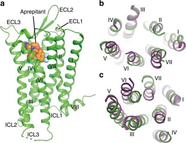
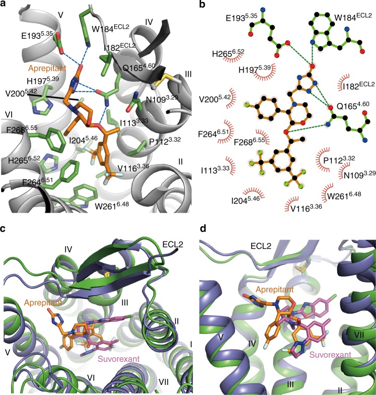
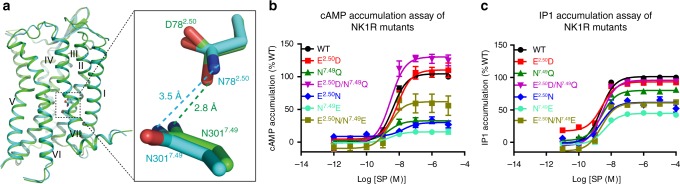
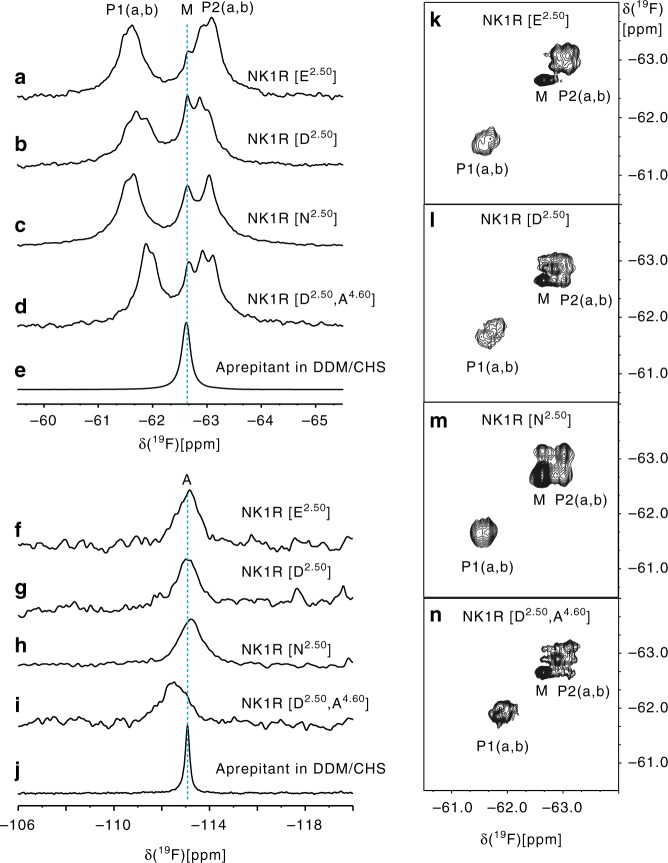
Neurokinin 1 receptor (NK1R) has key regulating functions in the central and peripheral nervous systems, and NK1R antagonists such as aprepitant have been approved for treating chemotherapy-induced nausea and vomiting. However, the lack of data on NK1R structure and biochemistry has limited further drug development targeting this receptor. Here, we combine NMR spectroscopy and X-ray crystallography to provide dynamic and static characterisation of the binding mode of aprepitant in complexes with human NK1R variants. 19F-NMR showed a slow off-rate in the binding site, where aprepitant occupies multiple substates that exchange with frequencies in the millisecond range. The environment of the bound ligand is affected by the amino acid in position 2.50, which plays a key role in ligand binding and receptor signaling in class A GPCRs. Crystal structures now reveal how receptor signaling relates to the conformation of the conserved NP7.50xxY motif in transmembrane helix VII.


Conflict of interest statement

The authors declare no competing interests.

Figures

Fig. 1
Fig. 1
Overall structure of the NK1R–aprepitant complex. a Structure of the NK1R–aprepitant complex. The NK1R structure is shown as green cartoon. Aprepitant is shown as spheres with orange carbons. The disulfide bond is displayed as yellow sticks. The missing portion of ICL3 is indicated by a green dashed line. b, c Structural comparison between NK1R and NTSR1 (PDB accession code: 4GRV). The helical bundles of the receptors are colored green (NK1R) and purple (NTSR1). b Extracellular view. c Intracellular view
Fig. 2
Fig. 2
The NK1R binding pocket for aprepitant. a Key residues of NK1R for aprepitant binding. The receptor is shown as gray cartoon. Aprepitant (orange carbons) and receptor residues (green carbons) involved in ligand binding are shown as sticks. Other elements are colored as follows: oxygen, red; nitrogen, dark blue; fluorine, cyan. b Schematic representation of interactions between NK1R and aprepitant analyzed using the LigPlot+ program. Polar interactions are shown as dashed lines. c, d Comparison of ligand-binding sites between NK1R (green) and OX2R (PDB accession code: 4S0V; purple). The ligands aprepitant and suvorexant are shown as orange and magenta sticks, respectively. c Extracellular view. d Side view
Fig. 3
Fig. 3
Interaction modes of the residues at positions 2.50 and 7.49 and functional assays of NK1R mutants. a Strong hydrogen-bond interaction between D782.50 and N3017.49 (2.8 Å) and weak interaction between N782.50 and N3017.49 (3.5 Å). Two NK1R structures are shown in cartoon representation and colored green (E782.50D mutant) and cyan (E782.50N mutant). The residues at positions 2.50 and 7.49 are shown as sticks. b SP-induced cAMP accumulation measurements of the wild-type (WT) NK1R and the mutants E782.50D, N3017.49Q, E782.50D/N3017.49Q, E782.50N, N3017.49E, and E782.50N/N3017.49E. Dose–response curves were generated from at least three independent experiments performed in triplicate. Data shown are mean ± s.e.m. See Supplementary Table 3 for detailed statistical evaluation. c SP-induced IP1 accumulation of the WT NK1R and the mutants E782.50D, N3017.49Q, E782.50D/N3017.49Q, E782.50N, N3017.49E, and E782.50N/N3017.49E. Dose–response curves were generated from at least three independent experiments performed in triplicate or duplicate. Data shown are mean ± s.e.m. See Supplementary Table 2 for detailed statistical evaluation. Source data for Fig. 3b, c are provided as a Source Data file
Fig. 4
Fig. 4
19F NMR studies of aprepitant bound to NK1R in solution. aj 1D 19F-NMR spectra of aprepitant in complex with different NK1R mutants and aprepitant in the DDM/CHS detergent buffer. ae Spectral region showing the resonances of the trifluoromethyl groups of the bis-trifluoromethyl-phenylethoxy moiety; fj Spectral region of the 4-fluorophenyl moiety. kn, 2D [19F,19F]-EXSY spectra recorded with a mixing time of 150 ms covering the same spectral region as panels a–d

References

    1. US VE, Gaddum JH. An unidentified depressor substance in certain tissue extracts. J. Physiol. 1931;72:74–87. doi: 10.1113/jphysiol.1931.sp002763. - DOI - PMC - PubMed
    1. Chang MM, Leeman SE, Niall HD. Amino-acid sequence of substance P. Nat. New Biol. 1971;232:86–87. doi: 10.1038/newbio232086a0. - DOI - PubMed
    1. Quartara L, Maggi CA. The tachykinin NK1 receptor. Part II: distribution and pathophysiological roles. Neuropeptides. 1998;32:1–49. doi: 10.1016/S0143-4179(98)90015-4. - DOI - PubMed
    1. Iversen LSubstance. P equals pain substance? Nature. 1998;392:334–335. doi: 10.1038/32776. - DOI - PubMed
    1. Kramer MS, et al. Distinct mechanism for antidepressant activity by blockade of central substance P receptors. Science. 1998;281:1640–1645. doi: 10.1126/science.281.5383.1640. - DOI - PubMed

Publication types

MeSH terms