

Role of selenium addition to CdZnTe matrix for room-temperature radiation detector applications
- PMID: 30733586
- PMCID: PMC6367417
- DOI: 10.1038/s41598-018-38188-w
Role of selenium addition to CdZnTe matrix for room-temperature radiation detector applications
Abstract
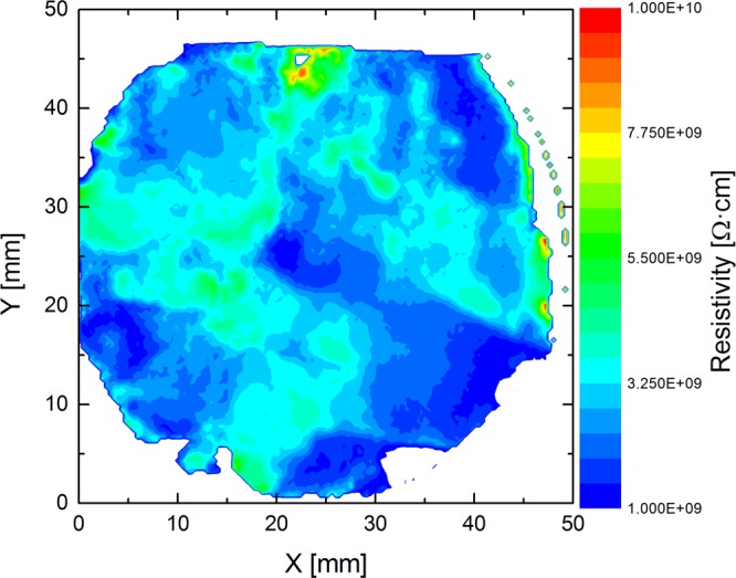
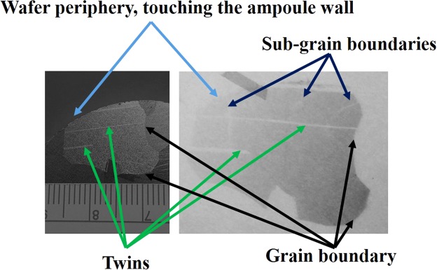
Because of its ideal band gap, high density and high electron mobility-lifetime product, cadmium zinc telluride (CdZnTe or CZT) is currently the best room-temperature compound-semiconductor X- and gamma-ray detector material. However, because of its innate poor thermo-physical properties and above unity segregation coefficient for Zn, the wide spread deployment of this material in large-volume CZT detectors is still limited by the high production cost. The underlying reason for the low yield of high-quality material is that CZT suffers from three major detrimental defects: compositional inhomogeneity, high concentrations of dislocation walls/sub-grain boundary networks and high concentrations of Te inclusions/precipitates. To mitigate all these disadvantages, we report for the first time the effects of the addition of selenium to the CZT matrix. The addition of Se was found to be very effective in arresting the formation of sub-grain boundaries and its networks, significantly reducing Zn segregation, improving compositional homogeneity and resulting in much lower concentrations of Te inclusions/precipitates. Growth of the new quaternary crystal Cd1-xZnxTe1-ySey (CZTS) by the Traveling Heater Method (THM) is reported in this paper. We have demonstrated the production of much higher yield according to its compositional homogeneity, with substantially lower sub-grain boundaries and their network, and a lower concentration of Te inclusions/precipitates.
Conflict of interest statement
The authors declare no competing interests.
Figures

References
-
- Schlesinger TE, et al. Cadmium zinc telluride and its use as a nuclear radiation detector material. Materials Science and Engineering. 2001;R 32:103. doi: 10.1016/S0927-796X(01)00027-4. - DOI
-
- Yang, G. & James, R. B. Physics, Defects, Hetero- and Nano-structures, Crystal Growth, Surfaces and Applications Part II, (EDAX. Triboulet R. et al.) 214 (Elsevier 2009).
-
- Iniewski K. CZT sensors for Computed Tomography: from crystal growth to image quality. J. Instrumentation. 2016;11:C12034. doi: 10.1088/1748-0221/11/12/C12034. - DOI
-
- Kincl V, et al. Cadmium–zinc–telluride SPECT scanners–New perspectives in nuclear cardiology. Cor et Vasa. 2015;57:e214. doi: 10.1016/j.crvasa.2015.01.001. - DOI
-
- Wahl CG, et al. The Polaris-H imaging spectrometer. Nucl. Instrum. Methods A. 2015;784:377. doi: 10.1016/j.nima.2014.12.110. - DOI
LinkOut - more resources
Full Text Sources
Miscellaneous
