Review
. 2019 Apr;15(4):199-219.
doi: 10.1038/s41581-019-0116-9.

Cellular signalling by primary cilia in development, organ function and disease

Affiliations
Review

Cellular signalling by primary cilia in development, organ function and disease

Zeinab Anvarian et al. Nat Rev Nephrol. 2019 Apr.

Abstract

Primary cilia project in a single copy from the surface of most vertebrate cell types; they detect and transmit extracellular cues to regulate diverse cellular processes during development and to maintain tissue homeostasis. The sensory capacity of primary cilia relies on the coordinated trafficking and temporal localization of specific receptors and associated signal transduction modules in the cilium. The canonical Hedgehog (HH) pathway, for example, is a bona fide ciliary signalling system that regulates cell fate and self-renewal in development and tissue homeostasis. Specific receptors and associated signal transduction proteins can also localize to primary cilia in a cell type-dependent manner; available evidence suggests that the ciliary constellation of these proteins can temporally change to allow the cell to adapt to specific developmental and homeostatic cues. Consistent with important roles for primary cilia in signalling, mutations that lead to their dysfunction underlie a pleiotropic group of diseases and syndromic disorders termed ciliopathies, which affect many different tissues and organs of the body. In this Review, we highlight central mechanisms by which primary cilia coordinate HH, G protein-coupled receptor, WNT, receptor tyrosine kinase and transforming growth factor-β (TGFβ)/bone morphogenetic protein (BMP) signalling and illustrate how defects in the balanced output of ciliary signalling events are coupled to developmental disorders and disease progression.


Figures
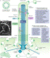
Figure 1.
Figure 1.. Overview of primary cilia, cellular signalling and ciliopathies.
a| The primary cilium is a non-motile organelle that extends as a solitary unit from the centrosomal mother centriole (basal body). a | The cilium comprises a microtubule (MT)-based axoneme containing a ring of nine outer microtubule doublets. Between the basal body and cilium is the ciliary transition zone (TZ), which contains specialized gating structures such as Y-links that along with the basal body transition fibres control the entrance and exit of ciliary proteins, thereby contributing to compartmentalization of the organelle. The intraflagellar transport (IFT) system zips up (anterograde) and down (retrograde) axonemal microtubules to mediate the transport of specific ciliary cargo, such as receptors, into or out of the organelle, whereupon they are degraded or recycled. Cilia can also release ectosomes by shedding off membrane-enclosed material from the surface of the organelle. The function of these extracellular vesicles has been linked to maintenance of ciliary integrity, balancing of intraciliary signalling events and/or in transmission of signals across cells , . b| Image of a primary cilium in a mouse embryonic fibroblast analysed by scanning electron microscopy. c| Selected ciliopathies are caused by dysfunctional primary cilia. d| Overview of diverse sensory capabilities of primary cilia and their associated signalling pathways. BBSome: protein complex of eight Bardet–Biedl syndrome proteins; SDA, sub-distal appendages, TF, transition fibres.
Figure 2.
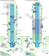
Figure 2.. Overview of canonical hedgehog signalling in primary cilia.
a| In the absence of sonic hedgehog (SHH; that is, under conditions of basal suppression) the receptor patched-1 (PTCH1) is enriched in the ciliary membrane, preventing ciliary accumulation of smoothened (SMO). A transmembrane sterol sensing domain (SSD) in PTCH1 can accommodate cholesterol or cholesterol derivatives. The class A GPCR, GPR161, is targeted to the cilium by tubby-like protein 3 (TULP3) and intraflagellar transport complex A (IFT-A) to activate adenylyl cyclases via G-proteins (Gαs), leading to increased ciliary levels of cAMP. Increased cAMP levels release protein kinase A (PKA) from regulatory subunits (RI), which in conjunction with glycogen synthase kinase 3 beta (GSK3-β) and casein kinases (CK) promote the limited proteolytic cleavage of full-length versions of GLI transcription factors (GLI-FL) into their repressor form (GLI-R) in a cilia-dependent manner. Suppressor of FUSED (SUFU) restrains GLI3 in the cytoplasm and promotes GLI3 processing. b| Binding of SHH to PTCH1 extracellular domains (ECDs) is regulated by cholesterol derivatives. In addition, SMURF1/2-mediated ubiquitination regulates PTCH1 exit from cilia and internalization at the ciliary base. Removal of PTCH1 causes concomitant enrichment and activation of SMO in cilia by cholesterol or derivatives. A longitudinal tunnel in the transmembrane region of SMO (dotted arrow) could move cholesterol from the ciliary membrane to its binding domain in the cysteine rich region (CRD). SHH stimulation results in dissociation of SUFU from GLI transcription factors, formation of full-length activator forms of GLIs (GLI-A), and accumulation of these proteins along with the microtubule-associated atypical kinesin KIF7 at ciliary tips. Downstream effectors for SMO include the EVC–EVC2 complex, which localizes in cilia distal to the transition zone. SHH stimulation further triggers the ciliary exit of GPR161 and ciliary entry of GPR175, which inhibits the production of cAMP. Both GLI-R and GLI-A translocate from the cilium into the nucleus to repress and induce transcriptional activation of HH target genes, respectively. Abbreviations: PI(4,5)P2: phosphatidylinositol 4,5-bisphosphate, Ub: ubiquitination.
Figure 3.
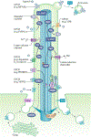
Figure 3.. Overview of ciliary GPCR signalling.
Binding of agonists to various G-protein-coupled receptors (GPCRs) expressed on cilia modulates a variety of signalling pathways on different cell types. For example, binding of endocrine gland-derived vascular endothelial growth factor to prokineticin receptor 1 (PROKR1) on trophoblast cell cilia activates mitogen-activated protein kinase kinase 1/2 (ERK1/2) at the ciliary base, presumably through a Gαq-mediated increase in calcium levels. Dopamine binding to dopamine receptor 5 (D5) on renal epithelial cell cilia facilitates Gβγ subunit dissociation and activation of an L-type calcium channel, resulting in an increase in ciliary calcium levels. ADP binding to the purinergic receptor P2Y12 (P2RY12) on cholangiocyte cilia stimulates Gαi, which inhibits adenylyl cyclase activity and subsequently reduces cAMP levels. Vasopressin binding to the type 2 vasopressin receptor (V2R) on renal epithelial cell cilia results in activation of adenylyl cyclase, increased cAMP levels, activation of PKA, and stimulation of a cation selective channel on the ciliary membrane. Increased cAMP levels and activated PKA can also activate exchange protein directly activated by cAMP (EPAC) and cAMP response element binding protein (CREB) in the cilium, respectively. Somatostatin binding to somatostatin receptor subtype 3 (SSTR3) on neuronal cilia stimulates β-arrestin (ARRB2) recruitment into the cilium where it mediates SSTR3 ciliary export in cooperation with the BBSome and intraflagellar transport (IFT) complex. Neuropeptide Y (NPY) binding to neuropeptide Y receptor subtype 2 (NPY2R) on neuronal cilia causes accumulation of the receptor at the ciliary tip and release through ectosomes.
Figure 4.
Figure 4.
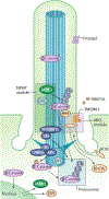
Overview of ciliary transition zone and basal body modulation of WNT signalling. The ciliary transition zone (TZ) is composed of several interacting protein modules or complexes, including the nephronophtisis (NPHP) and Meckel-Gruber Syndrome (MKS) modules that establish ciliary gating and contribute to the regulating of WNT signalling. The NPHP module protein retinitis pigmentosa GTPase regulator-interacting protein 1-like (RPGRIP1L) inhibits canonical WNT signalling through its interacting with a component of the 19S proteasome subunit, PSMD2, thereby promoting ubiquitin (Ub)-mediated proteasomal degradation of β-catenin and DVL. Further, β-catenin is ubiquitinated by the E3 ubiquitin ligase JADE1, which is present in the TZ as well as at the basal body. In the MKS module, transmemembrane protein 67 (TMEM67) recruits receptor tyrosine kinase-like orphan receptor 2 (ROR2) to the TZ to form a receptor complex that binds WNT5A, which is a ligand for non-canonical WNT signalling. In the presence of a cilium, the MKS module component Jouberin (JBN) recruits cytoplasmic β-catenin, which accumulates in response to WNT activation. In the absence of a cilium, JBN facilitates β-catenin nuclear entry. Several key components of the β-catenin destruction complex localize to the basal body, including the scaffold protein adenomatous polyposis coli (APC), GSK3-β glycogen synthase kinase 3 beta (GSK3-β) and casein kinases CK1-α, CK1-μ and CK1-δ –, . The main product of the destruction complex, phosphorylated β-catenin, is also concentrated at the basal body, where it undergoes degradation by the proteasome. Whether β-catenin is actively phosphorylated and ubiquitinated within the cilium, or whether the modified protein is recruited to the basal body, is unknown. Frizzled receptors can accumulate in the primary cilia but the functional relevance of this localization is unknown. RHOA: Ras homolog gene family member A.
Figure 5.
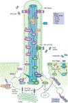
Figure 5.. Overview of ciliary PDGFRα, insulin and IGF-1 signalling.
Targeting of platelet-derived growth factor receptor alpha (PDGFRα) to the cilium relies on intraflagellar transport protein 20 (IFT20) in complex with the E3 ubiquitin ligases c-CBL and CBL-b; activation of the receptor in the cilium activates the MEK1/2–ERK1/2–RSK and PI3K–AKT pathways, which in turn control activation and translocation of the Na+/H+ exchanger 1 (NHE1) to leading edge of the cell for directional migration. AKT might also become activated at the ciliary base in complex with NPHP2 (also known as inversin). Feedback inhibition of PDGFRα signalling might be controlled by CBL-mediated ubiquitination (Ub) of the receptor in the cilium as well as by inositol 1,4,5-trisphosphate 5-phosphatase (INPP5E), which inhibits AKT signalling. PDGFRβ localizes at the plasma membrane to induce aurora kinase A (AURKA)-mediated disassembly of the primary cilium, which promotes cell cycle re-entry. Similarly, expression of the oncogenic mutant PDGFRα Asp842Val, which localizes to the Golgi, promotes ciliary disassembly and cell proliferation via activation of AURKA. Insulin receptor (IR) and insulin-like growth factor 1 receptor (IGF-1R) also localize to primary cilia to balance the regulation of various cellular processes, including adipogenesis, neuronal differentiation, mitogenic signalling and insulin production in islet β-cells of the pancreas. Ciliary IGF-1R activation also induces ciliary resorption and cell cycle entry possibly via IGF-1-mediated recruitment of phosphorylated TCTEX-1 to the ciliary TZ via non-canonical G-protein signalling, PI3K, phosphatidylinositol-4,5-bisphosphate 3-kinase; IRS-1: insulin receptor substrate 1; PLCγ, phospholipase C gamma; PIP2, Phosphatidylinositol 4,5-bisphosphate; IP3, Inositol trisphosphate; HSP90α, heat shock protein 90 alpha.
Figure 6.
Figure 6.. Overview of ciliary TGFβ/BMP signalling.
Receptors of the transforming growth factor beta (TGFβ) and bone morphogenic protein (BMP) family are recruited to the primary cilium to activate receptor (R)–SMAD transcription factors (SMAD2/3 and SMAD1/5/8) partly via internalization of active receptors by clathrin-mediated endocytosis (CME) at the ciliary pocket. Activated R-SMADs form a trimeric complex with the co-SMAD, SMAD4, which translocate to the nucleus for targeted gene expression. TGFβ may activate ERK1/2 in the cilium independently of CME. RAB11-mediated recycling of TGFβ receptors to the primary cilium may be regulated by the subdistal appendage protein, CEP128. Feedback inhibition of ciliary TGFβ/BMP signalling is under the control of the inhibitor SMAD, SMAD7, as well as the E3 ubiquitin ligase, SMURF1. Ciliary TGFβ receptors may further stimulate the hedgehog pathway component, smoothened (SMO), leading to activation of GLI transcription factors. Abbreviations: TGFβ-RI/II, TGFβ receptors I and II; BMP-RI/II, BMP receptors I and II; GLI-A: activator form of GLI transcription factors; ERK1/2: extracellular signal–regulated kinase 1/2; EE: early endosome.

References

    1. Satir P & Christensen ST Overview of structure and function of mammalian cilia. Annu Rev Physiol 69, 377–400 (2007). - PubMed
    1. Ciliopathies: A reference for clinicians. (eds. Kenny TD & Beales PL) 1–282 (Oxford University Press, Oxford, Oxford; 2014).
    1. Heydeck W, Fievet L, Davis EE & Katsanis N The complexity of the cilium: spatiotemporal diversity of an ancient organelle. Current Opinion in Cell Biology 55, 139–149 (2018). - PMC - PubMed
    1. Sorokin S Centrioles and the formation of rudimentary cilia by fibroblasts and smooth muscle cells. J Cell Biol 15, 363–377 (1962). - PMC - PubMed
    1. Sorokin SP Reconstructions of centriole formation and ciliogenesis in mammalian lungs. J Cell Sci 3, 207–230 (1968). - PubMed

Publication types

Substances