Revascularization of AlloDerm Used during Endoscopic Skull Base Surgery
- PMID: 30733900
- PMCID: PMC6365292
- DOI: 10.1055/s-0038-1666851
Revascularization of AlloDerm Used during Endoscopic Skull Base Surgery
Abstract
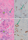
Objectives AlloDerm is an acellular dermal matrix often used for reconstruction throughout the body. AlloDerm has been shown to undergo revascularization when used to reconstruct soft tissue such as in abdominal wall reconstruction. In this study, the authors review the literature on revascularization of AlloDerm and demonstrate the histologic findings of AlloDerm after implantation during skull base reconstruction. Study Design Literature review and case reports. Setting Tertiary Care Institution Participants Patients from a tertiary care institution Main Outcome Measures Histologic slides are evaluated and compared with nonimplanted AlloDerm. Methods The authors review a case of explanted AlloDerm that had been used for skull base reconstruction after endoscopic skull base surgery. Results Upon reviewing the histologic slides of explanted AlloDerm to nonimplanted AlloDerm, we demonstrate revascularization of AlloDerm when used in skull base reconstruction. Representative slides will be included. Conclusions AlloDerm undergoes revascularization when used for skull base reconstruction.
Keywords: AlloDerm; acellular dermal matrix; revascularization; skull base reconstruction; skull base surgery.
Conflict of interest statement
Figures


References
-
- Achauer B M, VanderKam V M, Celikoz B, Jacobson D G. Augmentation of facial soft-tissue defects with Alloderm dermal graft. Ann Plast Surg. 1998;41(05):503–507. - PubMed
-
- Kridel R W, Foda H, Lunde K C. Septal perforation repair with acellular human dermal allograft. Arch Otolaryngol Head Neck Surg. 1998;124(01):73–78. - PubMed
-
- Jones F R, Schwartz B M, Silverstein P. Use of a nonimmunogenic acellular dermal allograft for soft tissue augmentation: a preliminary report. Aesthet Surg J. 1996;16:196–201.
-
- Wainwright D, Madden M, Luterman A et al.Clinical evaluation of an acellular allograft dermal matrix in full-thickness burns. J Burn Care Rehabil. 1996;17(02):124–136. - PubMed
-
- Chaplin J M, Costantino P D, Wolpoe M E, Bederson J B, Griffey E S, Zhang W X. Use of an acellular dermal allograft for dural replacement: an experimental study. Neurosurgery. 1999;45(02):320–327. - PubMed
LinkOut - more resources
Full Text Sources
Other Literature Sources

