. 2019 May;76(10):1987-2002.
doi: 10.1007/s00018-019-03030-7. Epub 2019 Feb 7.

Tight junction proteins at the blood-brain barrier: far more than claudin-5

Affiliations

Tight junction proteins at the blood-brain barrier: far more than claudin-5

Philipp Berndt et al. Cell Mol Life Sci. 2019 May.

Abstract

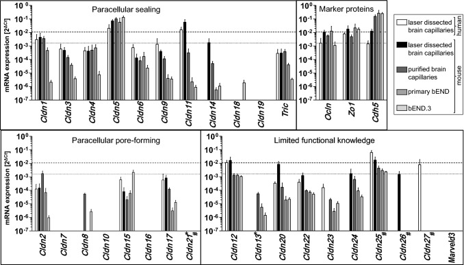
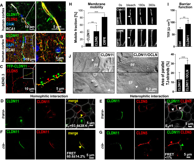
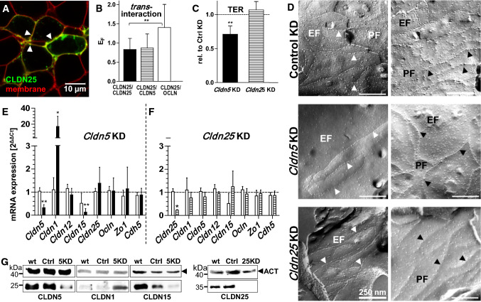
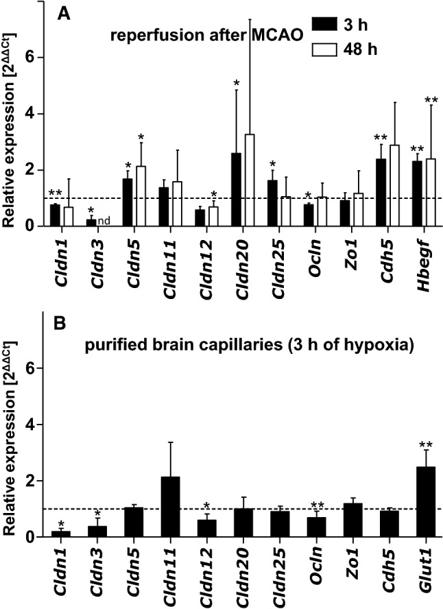
At the blood-brain barrier (BBB), claudin (Cldn)-5 is thought to be the dominant tight junction (TJ) protein, with minor contributions from Cldn3 and -12, and occludin. However, the BBB appears ultrastructurally normal in Cldn5 knock-out mice, suggesting that further Cldns and/or TJ-associated marvel proteins (TAMPs) are involved. Microdissected human and murine brain capillaries, quickly frozen to recapitulate the in vivo situation, showed high transcript expression of Cldn5, -11, -12, and -25, and occludin, but also abundant levels of Cldn1 and -27 in man. Protein levels were quantified by a novel epitope dilution assay and confirmed the respective mRNA data. In contrast to the in vivo situation, Cldn5 dominates BBB expression in vitro, since all other TJ proteins are at comparably low levels or are not expressed. Cldn11 was highly abundant in vivo and contributed to paracellular tightness by homophilic oligomerization, but almost disappeared in vitro. Cldn25, also found at high levels, neither tightened the paracellular barrier nor interconnected opposing cells, but contributed to proper TJ strand morphology. Pathological conditions (in vivo ischemia and in vitro hypoxia) down-regulated Cldn1, -3, and -12, and occludin in cerebral capillaries, which was paralleled by up-regulation of Cldn5 after middle cerebral artery occlusion in rats. Cldn1 expression increased after Cldn5 knock-down. In conclusion, this complete Cldn/TAMP profile demonstrates the presence of up to a dozen TJ proteins in brain capillaries. Mouse and human share a similar and complex TJ profile in vivo, but this complexity is widely lost under in vitro conditions.

Keywords: Brain endothelium; Ischemia; Laser capture microdissection; Neurovasculature; Protein–protein interaction.


Figures

Fig. 1
Fig. 1
Diversity of mRNA expression of tight junction proteins in human and murine brain endothelium. Transcript levels in laser-dissected human and murine brain capillaries, purified murine brain capillaries, primary murine brain capillary endothelial cells and bEND.3, an endothelial cell line of the murine cerebral cortex. mRNA expression normalized to that of β-actin (ΔCt = Cttarget − Ctactin). Cldn claudin, Tric tricellulin, Ocln occludin, Zo1 Zonula occludens protein 1, Cdh5 cadherin-5, Ct cycle threshold. Dotted/dashed line, Ocln expression in laser microdissected human/mouse brain capillaries (used as a benchmark of tight junction protein abundance); missing columns, mRNA not detectable (detection limit for 2ΔCt is 5.75 × 10−7 in primary murine brain capillary endothelial cells). *Cldn13 (human) and Cldn21 (mouse), not determined; #synonyms: human Cldn21, putative Cldn25; Cldn25, Cldnd1; Cldn26, Tmem114; Cldn27, Tmem235
Fig. 2
Fig. 2
Protein levels of tight junction proteins in purified brain capillaries (fmol/µg total protein). a Representative images of western blots to quantify the TJ protein concentration via a dilution series of the purified Cldn epitope recognized by the respective antibody. The endogenous protein in purified brain capillaries is shown on the left. The dilution series was realized by recombinant MBP-tagged epitopes. b Protein concentrations given for Triton X-100 (TX-100)-insoluble and -soluble fraction. n.d. not detectable; mean ± SD; n ≥ 4
Fig. 3
Fig. 3
Claudin (Cldn)-1, -3, -4, -5, -11, -20, and -25 and occludin (Ocln) co-localize to the tight junction (TJ) area of human brain capillaries. Immunofluorescence staining of human brain sections for TJ proteins (green). Microvessels were visualized by RCA1 (Ricinus communis agglutinin 1, white), an endothelial marker. The TJ marker Ocln exclusively localized to cell–cell contacts visualized by the junction marker Zo1 (Zonula occludens protein 1, red). Arrowheads indicate overlap of TJ proteins with Zo1 (yellow-orange); nuclei stained by DAPI (blue)
Fig. 4
Fig. 4
Claudin(Cldn)-11 increases transcellular electrical resistance (TER) through homophilic but not heterophilic cis- and trans-interactions. In brain, Cldn11 localized in a capillaries (marker: RCA1) and in b oligodendrocytes ensheathing neuronal processes (marker: neurofilament, NeuF; red arrow). In capillary tight junctions (TJs), Cldn11 was traced with and without (green arrow) the cerebral TJ marker Cldn5. c After transfection in mouse brain endothelial cells (bEND.3), yellow fluorescent protein (YFP)–Cldn11 alternated with endogenous Cldn5 within TJs (red/green arrowhead). d Co-culture of cells mono-transfected with cyan fluorescent protein (CFP)–Cldn11 or YFP–Cldn11, plasma membrane of living cells visualized by trypan blue (white). Co-localization (yellow arrowhead) indicated strong homophilic trans-interaction quantified by the enrichment factor (EF). e Living cells mono-transfected with CFP–Cldn11 or Cldn5–YFP showed no heterophilic trans-interaction (white arrowhead), EF < 1; plasma membrane (white). Image representative for all other claudins tested. f Cells co-transfected with CFP/YFP–Cldn11 showing strong homophilic cis-interaction (yellow arrowhead) in cell contacts quantified by FRET (fluorescence resonance energy transfer). g No co-localization of CFP–Cldn11 and Cldn5–YFP in co-transfected cells, FRET efficiency < 1% as for all other claudins tested. h CFP–Cldn11 displayed much lower membrane mobility than CFP–Cldn5 or the negative control corticotropin releasing factor receptor (CRFR)–CFP, quantified in cells by fluorescence recovery after photobleaching. Kruskal–Wallis test; n ≥ 9. i Transfection of CFP–Cldn11 in Madin–Darby canine kidney (MDCK-II) cells increased transcellular electrical resistance (TER) compared to non-transfected cells, Mann–Whitney test; n ≥ 9. j Occludin (Ocln) modulated the TJ strands (arrowheads) of claudin-11 visualized by freeze-fracture electron microscopy of cells mono-transfected with CFP–Cldn11 (branched single strands) or co-transfected with CFP–Cldn11/YFP–Ocln (parallel strands). EF/PF, extracellular/protoplasmic fracture face; Mann–Whitney test, n ≥ 6. In dh and j, TJ-/Cldn-free HEK-293 cells were used. Mean ± SD; *p < 0.05; **p < 0.01; ***p < 0.001
Fig. 5
Fig. 5
Loss of claudin (Cldn)-25 and -5 compromises tight junction (TJ) strand morphology, whereas decrease in paracellular tightness and changes in TJ protein expression occur in Cldn5 knock-down (KD) only. a In living HEK (human embryonic kidney)-293 cells, transfected yellow fluorescent protein(YFP)–Cldn25 (green) localized at the plasma membrane (red, trypan blue) but b no homophilic trans-interaction of Cldn25 was found based on calculation of the enrichment factor (EF) quantifying the fluorescence intensity at contacts (arrowheads in a) between transfected cells. However, EF increased between YFP–Cldn25 and YFP–Ocln, suggesting heterophilic trans-interaction; Student’s t test, n ≥ 8. cg experiments in bEND.3 (mouse brain endothelial cell line): c Cldn5 KD but not Cldn25 KD affected transendothelial electrical resistance (TER) compared to control (Ctrl, dashed line); Kruskal–Wallis test; n ≥ 8. d Cldn5 KD weakened the TJ strand network on the extracellular (EF) and protoplasmic (PF) fracture face, i.e. enlarged and more round-shaped meshes. Cldn25 KD led to more unstructured strands and reduced mesh number on PF, i.e. less particles on the strands and larger meshes. e Cldn5 KD (black bars) but not f Cldn25 KD (striped bars) influenced the mRNA expression of other Cldn’s mRNA compared to control KD (white bars), expression levels normalized to wild-type (wt) cells (dashed line). ΔΔCt = (Cttarget − Ctactin)KD − (Cttarget − Ctactin)wt; Ct, cycle threshold. Kruskal–Wallis test; n ≥ 4. g Western blots of wt, Ctrl KD, Cldn5 KD (5KD), Cldn25 KD (25KD), loading control β-actin (ACT). Mean ± SD; n ≥ 4; *p < 0.05; **p < 0.01
Fig. 6
Fig. 6
In cerebral capillaries, mRNA expression of claudin(Cldn)-1, -3, and -12, and occludin (Ocln) is reduced under hypoxic conditions in vitro and under post-ischemic reperfusion in vivo, which in vivo is compensated by enhanced expression of Cldn- 5 and -25. a mRNA expression in laser-dissected mouse brain capillaries after middle cerebral artery occlusion (60 min or 30 min, corresponding reperfusion for 3 h or 48 h), normalized to the contralateral region (dashed line). ΔΔCt = (Cttarget − CtRn28S)ipsilateral − (Cttarget − CtRn28S)contralateral; Ct, cycle threshold. Heparin-binding epithelial growth factor-like growth factor (Hbegf) was used as indicator for ischemic dysfunction (nd, not detected). b mRNA expression in purified mouse brain capillaries exposed to 3 h of hypoxia; dashed line, expression levels under normoxic conditions. ΔΔCt = (Cttarget − Ctactin)hypoxia − (Cttarget − Ctactin)normoxia. Glucose transporter type 1 (Glut1) served as an indicator for hypoxic condition. Mean ± SD, n ≥ 4, Mann–Whitney test; *p < 0.05; **p < 0.01. Rn28S, 28S ribosomal RNA; Zo1, Zonula occludens protein 1; Cdh5, cadherin-5. No other Cldn exhibited significant changes

References

    1. Krause G, Winkler L, Mueller SL, Haseloff RF, Piontek J, Blasig IE. Structure and function of claudins. Biochim Biophys Acta. 2008;1778(3):631–645. doi: 10.1016/j.bbamem.2007.10.018. - DOI - PubMed
    1. Haseloff RF, Dithmer S, Winkler L, Wolburg H, Blasig IE. Transmembrane proteins of the tight junctions at the blood–brain barrier: structural and functional aspects. Semin Cell Dev Biol. 2015;38:16–25. doi: 10.1016/j.semcdb.2014.11.004. - DOI - PubMed
    1. Maher GJ, Hilton EN, Urquhart JE, Davidson AE, Spencer HL, Black GC, Manson FD. The cataract-associated protein TMEM114, and TMEM235, are glycosylated transmembrane proteins that are distinct from claudin family members. FEBS Lett. 2011;585(14):2187–2192. doi: 10.1016/j.febslet.2011.05.060. - DOI - PubMed
    1. Mineta K, Yamamoto Y, Yamazaki Y, Tanaka H, Tada Y, Saito K, Tamura A, Igarashi M, Endo T, Takeuchi K, Tsukita S. Predicted expansion of the claudin multigene family. FEBS Lett. 2011;585(4):606–612. doi: 10.1016/j.febslet.2011.01.028. - DOI - PubMed
    1. Cording J, Berg J, Kading N, Bellmann C, Tscheik C, Westphal JK, Milatz S, Gunzel D, Wolburg H, Piontek J, Huber O, Blasig IE. In tight junctions, claudins regulate the interactions between occludin, tricellulin and marvelD3, which, inversely, modulate claudin oligomerization. J Cell Sci. 2013;126(Pt 2):554–564. doi: 10.1242/jcs.114306. - DOI - PubMed

MeSH terms