. 2019 Jun;75(6):1750-1759.
doi: 10.1002/ps.5372. Epub 2019 Mar 21.

Assessment of neuropeptide binding sites and the impact of biostable kinin and CAP2b analogue treatment on aphid (Myzus persicae and Macrosiphum rosae) stress tolerance

Affiliations

Assessment of neuropeptide binding sites and the impact of biostable kinin and CAP2b analogue treatment on aphid (Myzus persicae and Macrosiphum rosae) stress tolerance

Lucy Alford et al. Pest Manag Sci. 2019 Jun.

Abstract

Background: Neuropeptides are regulators of critical life processes in insects and, due to their high specificity, represent potential targets in the development of greener insecticidal agents. Fundamental to this drive is understanding neuroendocrine pathways that control key physiological processes in pest insects and the screening of potential analogues. The current study investigated neuropeptide binding sites of kinin and CAPA (CAPA-1) in the aphids Myzus persicae and Macrosiphum rosae and the effect of biostable analogues on aphid fitness under conditions of desiccation, starvation and thermal (cold) stress.

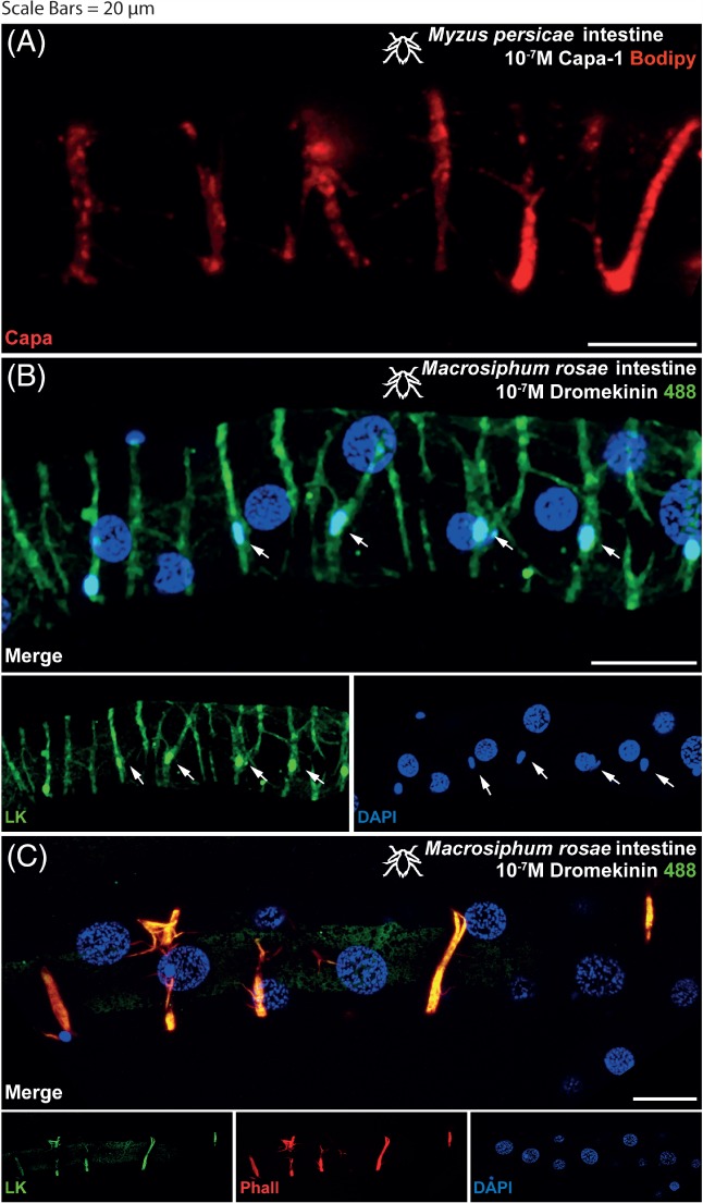
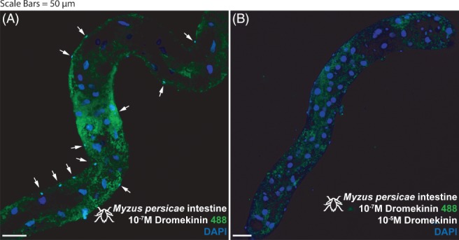
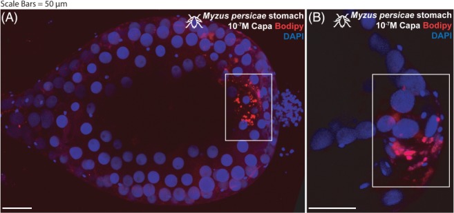
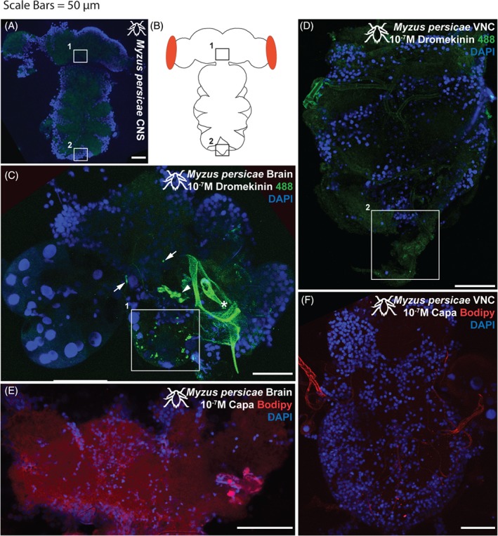
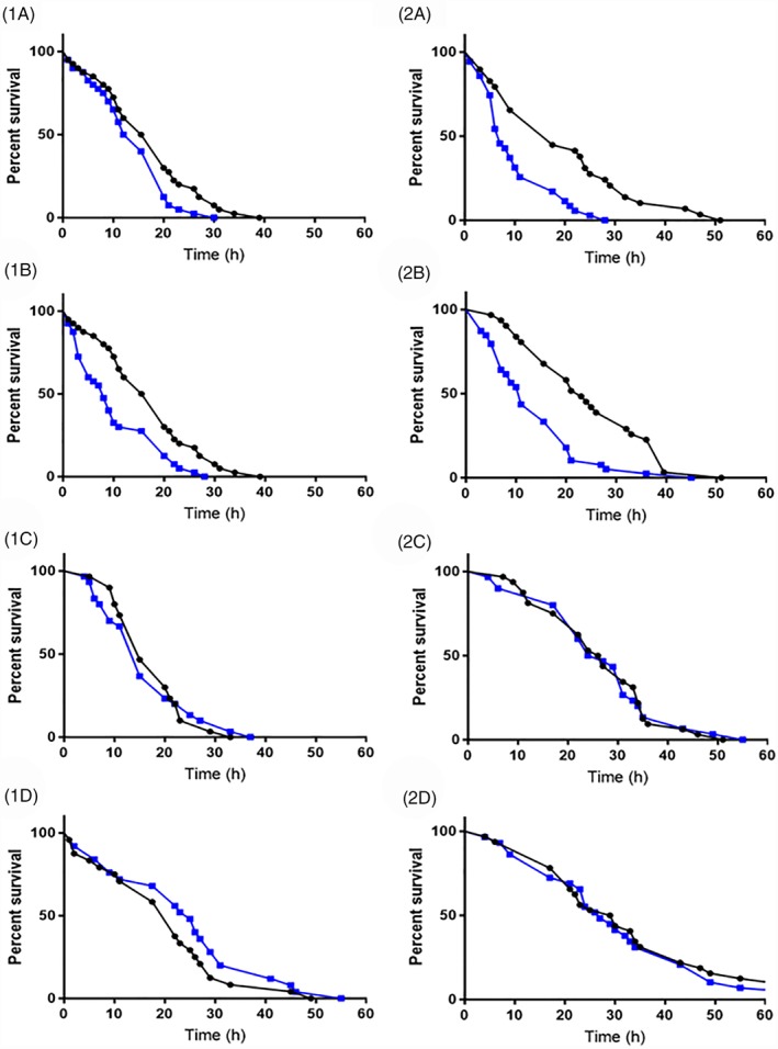
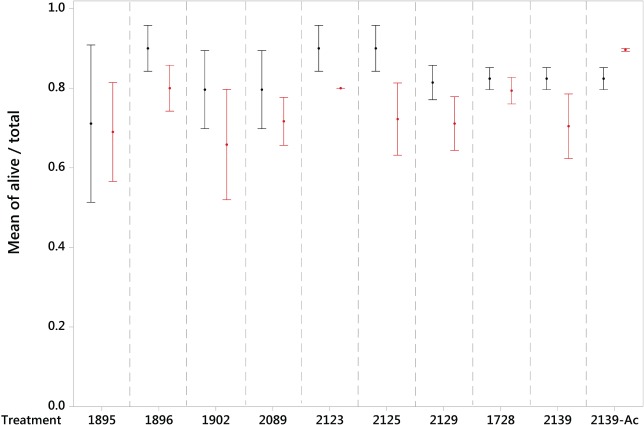
Results: M. persicae and M. rosae displayed identical patterns of neuropeptide receptor mapping along the gut, with the gut musculature representing the main target for kinin and CAPA-1 action. While kinin receptor binding was observed in the brain and VNC of M. persicae, this was not observed in M. rosae. Furthermore, no CAPA-1 receptor binding was observed in the brain and VNC of either species. CAP2b/PK analogues (with CAPA receptor cross-activity) were most effective in reducing aphid fitness under conditions of desiccation and starvation stress, particularly analogues 1895 (2Abf-Suc-FGPRLa) and 2129 (2Abf-Suc-ATPRIa), which expedited aphid mortality. All analogues, with the exception of 2139-Ac, were efficient at reducing aphid survival under cold stress, although were equivalent in the strength of their effect.

Conclusion: In demonstrating the effects of analogues belonging to the CAP2b neuropeptide family and key analogue structures that reduce aphid fitness under stress conditions, this research will feed into the development of second generation analogues and ultimately the development of neuropeptidomimetic-based insecticidal agents. © 2019 The Authors. Pest Management Science published by John Wiley & Sons Ltd on behalf of Society of Chemical Industry.

Keywords: G-protein coupled receptors receptor-mapping; aphicide; cold tolerance; desiccation; ligand-binding; pest control.


Figures

Figure 1
Figure 1
Aphid intestine (distal midgut and proximal hindgut) stained with 10−7 m CAPA‐1‐F labelled with TMR C5‐Maleimide (A) shows receptor localization in the gut muscles (Myzus persicae shown). Excess unlabeled CAPA‐1 (10−5 m) displaces fluorescent signal in a ligand competition assay (not shown), thus confirming the specificity of binding. Aphid intestine stained with 10−7 m kinin labelled with alexafluor488 (B) shows receptor localization in the gut muscles (M. rosae shown). Excess unlabeled kinin (10−5 m) displaces fluorescent signal in a ligand competition assay (not shown), thus confirming the specificity of binding. Staining by kinin‐F was also present in a population of basal cells, characterized by smaller nuclei, as indicated by the white arrows. DAPI was used for nuclear staining (blue). Staining with rhodamine phalloidin labelled with tetramethylrhodamine (TRITC) reaffirms the gut musculature as the site of receptor binding (C). (A and B): Kinin‐F, green; CAPA‐1‐F, red; DAPI, blue. (C) Kinin‐F, green; rhodamine phalloidin, red; DAPI, blue. Scale bars = 20 µm.
Figure 2
Figure 2
Myzus persicae intestine (distal midgut and proximal hindgut) stained with 10−7 m kinin labelled with alexafluor488 (A) and then out‐competed with 10−5 m unlabeled kinin (B). (A) Staining apparent in a population of basal cells, characterized by overtly smaller nuclei (arrows). (B) Staining abrogated in basal (small nuclei) cells (realized by DAPI staining, arrows) during out‐competition with unlabeled 10−5 m kinin. Kinin‐F, green; DAPI, blue. Scale bars = 50 µm.
Figure 3
Figure 3
Myzus persicae stomach (midgut) stained with 10−7 m CAPA‐1‐F labeled with TMR C5‐Maleimide. (A) Staining apparent at junctional area between the fore‐ and midgut (white box). (B) Higher magnification detail of staining associated with this junctional area. Staining is abrogated when outcompeted with unlabeled 10−5 m CAPA‐1 (not shown). CAPA‐1‐F, red; DAPI, blue. Scale bars = 50 µm.
Figure 4
Figure 4
(A) Unstained Myzus persicae CNS, demonstrating baseline autofluorescent levels (488 nm excitation range; green). (B) cartoon schematic. (C) Myzus persicae brain and (D) VNC incubated with 10−7 m kinin‐F labelled with alexafluor488. (C) Staining apparent in a bilateral ‘ladder’ of neurons and a set of more baso‐lateral neurons in the suboesophageal ganglion (1white box). Position of 1white box indicated by 1boxes in (A) and (B). Staining also apparent in symmetrical pairs of neurons/neuronal clusters in the ventro‐ to dorso‐lateral protocerebrum (arrows). Some neurons obscured by cuticular material associated with the aphid feeding stylus (asterisk). (D) Little to no kinin‐F staining apparent in the VNC, although a faint set of cells in the most distal tip of the abdominal ganglion (2white box) are consistently observed. Position of 2white box indicated by 2boxes in (A) and (B). (E) Myzus persicae brain and (F) VNC incubated with 10−7 m CAPA‐1‐F labelled with TMR C5‐Maleimide. No apparent staining with CAPA‐1‐F in either brain or VNC. Kinin‐F, green; CAPA‐1‐F, red; DAPI, blue. Scale bars = 50 µm.
Figure 5
Figure 5
Effect of CAP2b and kinin analogue treatment on the survival of Myzus persicae (1) and M. rosae (2) under conditions of desiccation and starvation stress. Control aphids are indicated by the black line and analogue‐treated aphids by the blue line. CAP2b analogues 1895 (a) and 2129 (b) were administered to a final concentration of ×10−5 m via microinjection and acted to significantly increase mortality relative to the control. CAP2b analogue 2125 (c) and kinin analogue 2139 (d) are presented to illustrate non‐significant survival curves.
Figure 6
Figure 6
Survival curve calculated via Probit analysis for Myzus persicae pre‐reproductive adults following a 1 h exposure at the desired temperature. Raw data values are indicated by black circles.
Figure 7
Figure 7
Mean ± standard error proportion survival of M. persicae when treated with biostable peptide analogues (CAP2b/PK: 1895, 1896, 1902, 2089, 2123, 2125, 2129; kinin: 1728, 2139, 2139‐Ac) via microinjection and subjected to a discriminating temperature for a 1 h exposure. Control groups are shown in black and peptide treatment groups in red.

Similar articles

Cited by

References

    1. Pimentel D, Acquay H, Biltonen M, Rice P, Silva M, Nelson J et al., Environmental and economic costs of pesticide use. Bioscience 42:750–760 (1992).
    1. Wilson C and Tisdell C, Why farmers continue to use pesticides despite environmental, health and sustainability costs. Ecol Econ 39:449–462 (2001).
    1. Altstein M and Nässel DR, Neuropeptide signaling in insects. Adv Exp Med Biol 692:155–165 (2010). - PubMed
    1. Nachman RJ and Pietrantonio PV, Interaction of mimetic analogs of insect kinin neuropeptides with arthropod receptors. Adv Exp Med Biol 692:27–48 (2010). - PubMed
    1. Audsley N and Down RE, G protein coupled receptors as targets for next generation pesticides. Insect Biochem Molec 67:27–37 (2015). - PubMed

MeSH terms