

Self-digitization chip for quantitative detection of human papillomavirus gene using digital LAMP
- PMID: 30734822
- PMCID: PMC6420227
- DOI: 10.1039/c8lc01223g
Self-digitization chip for quantitative detection of human papillomavirus gene using digital LAMP
Abstract
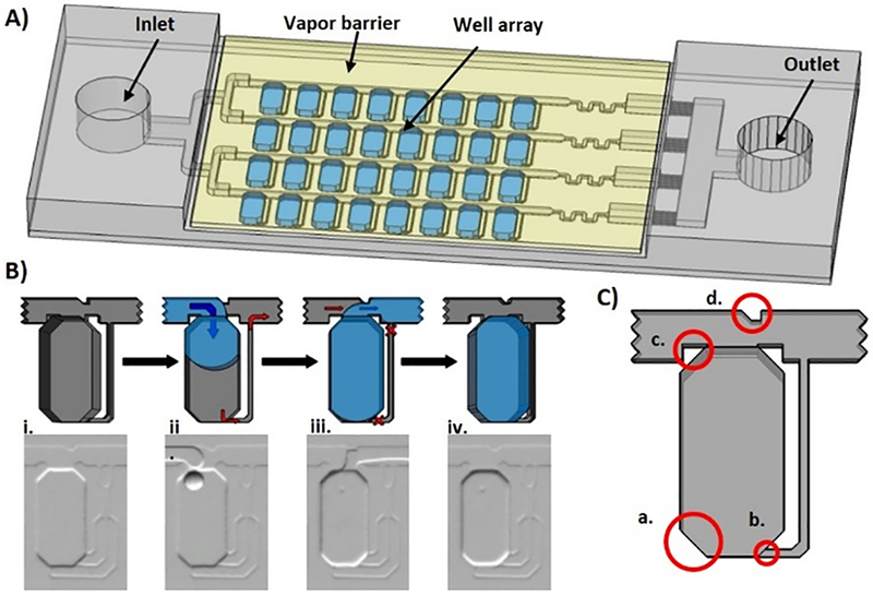
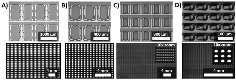
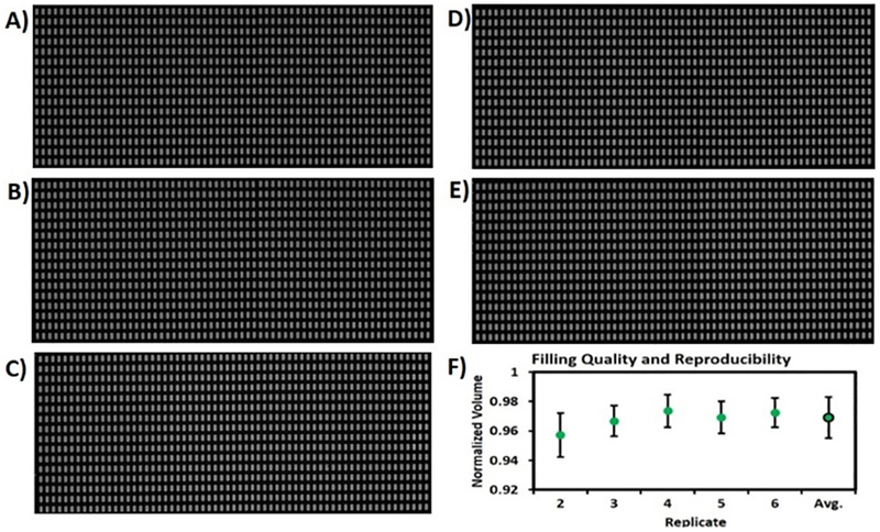
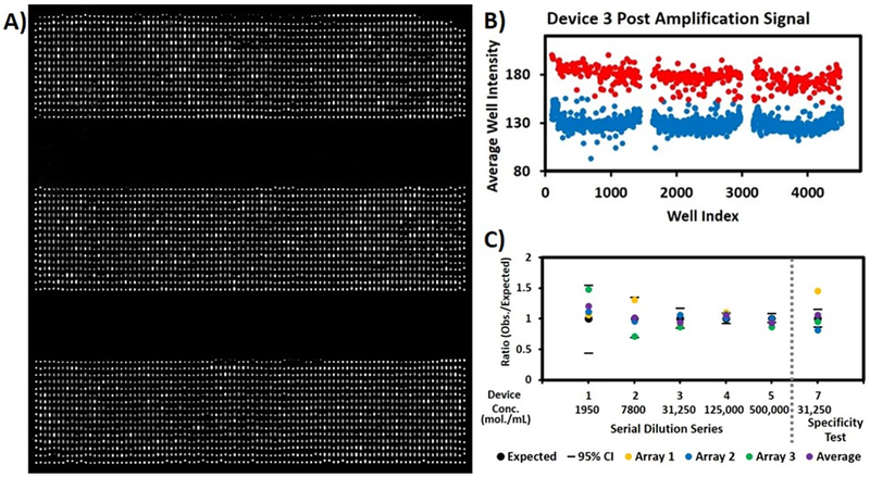
Digital nucleic acid amplification and detection methods provide excellent sensitivity and specificity and allow absolute quantification of target nucleic acids. Isothermal methods such as digital loop-mediated isothermal amplification (digital LAMP) have potential for use in rapid disease diagnosis in low-resource settings due to their speed and lack of thermal cycling. We previously developed a self-digitization (SD) chip, a simple microfluidics device that automatically digitizes a sample into an array of nanoliter wells, for use in digital LAMP. In this work, we improve the SD chip design to increase sample loading efficiency, speed, and completeness, and test a range of well volumes and numbers. We demonstrate the diagnostic capability of this platform by applying it to quantifying human papillomavirus 18 gene.
Figures
Similar articles
-
Digital LAMP in a sample self-digitization (SD) chip.Lab Chip. 2012 Jun 21;12(12):2247-54. doi: 10.1039/c2lc21247a. Epub 2012 Mar 7. Lab Chip. 2012. PMID: 22399016 Free PMC article.
-
Digital Quantification of Human Viral RNA and DNA Using a Self-Digitization Chip.Methods Mol Biol. 2022;2393:279-295. doi: 10.1007/978-1-0716-1803-5_15. Methods Mol Biol. 2022. PMID: 34837185
-
Optimizing the performance of digital loop-mediated isothermal amplification.Anal Biochem. 2021 Oct 15;631:114371. doi: 10.1016/j.ab.2021.114371. Epub 2021 Sep 10. Anal Biochem. 2021. PMID: 34509442
-
Integration of isothermal amplification methods in microfluidic devices: Recent advances.Biosens Bioelectron. 2017 Apr 15;90:174-186. doi: 10.1016/j.bios.2016.11.045. Epub 2016 Nov 19. Biosens Bioelectron. 2017. PMID: 27888686 Review.
-
[Analysis of Common Issues and Research Progress in Loop Mediated Isothermal Amplification].Bing Du Xue Bao. 2016 Sep;32(5):659-65. Bing Du Xue Bao. 2016. PMID: 30004194 Review. Chinese.
Cited by
-
Recent Advances in Digital Biosensing Technology.Biosensors (Basel). 2022 Aug 23;12(9):673. doi: 10.3390/bios12090673. Biosensors (Basel). 2022. PMID: 36140058 Free PMC article. Review.
-
Droplet microfluidic-based loop-mediated isothermal amplification (dLAMP) for simultaneous quantification of multiple targets.STAR Protoc. 2022 Apr 18;3(2):101335. doi: 10.1016/j.xpro.2022.101335. eCollection 2022 Jun 17. STAR Protoc. 2022. PMID: 35496787 Free PMC article.
-
Application of a clamshell isothermal nucleic acid amplification analyzer in the detection of lower respiratory tract bacteria.Pract Lab Med. 2024 Apr 16;40:e00394. doi: 10.1016/j.plabm.2024.e00394. eCollection 2024 May. Pract Lab Med. 2024. PMID: 38680549 Free PMC article.
-
An LED-Driven AuNPs-PDMS Microfluidic Chip and Integrated Device for the Detection of Digital Loop-Mediated Isothermal DNA Amplification.Micromachines (Basel). 2020 Feb 8;11(2):177. doi: 10.3390/mi11020177. Micromachines (Basel). 2020. PMID: 32046315 Free PMC article.
-
Nucleic Acid Target Sensing Using a Vibrating Sharp-Tip Capillary and Digital Droplet Loop-Mediated Isothermal Amplification (ddLAMP).Sensors (Basel). 2024 Jun 30;24(13):4266. doi: 10.3390/s24134266. Sensors (Basel). 2024. PMID: 39001045 Free PMC article.
References
-
- Dietel M, Jöhrens K, Laffert M, Hummel M, Bläker H, Müller BM, Lehmann A, Denkert C, Heppner FL, Koch A, Sers C and Anagnostopoulos I, Cancer Gene Ther, 2013, 20, 211. - PubMed
-
- Petralia S and Conoci S, ACS Sensors, 2017, 2, 876–891. - PubMed
-
- Nolan T, Hands RE and Bustin SA, Nat Protocols, 2006, 1, 1559. - PubMed
-
- Nagamine K, Watanabe K, Ohtsuka K, Hase T and Notomi T, Clin Chem, 2001, 47, 1742–1743. - PubMed
Publication types
MeSH terms
Substances
Grants and funding
LinkOut - more resources
Full Text Sources
Other Literature Sources