. 2019 Apr 1;316(4):H889-H899.
doi: 10.1152/ajpheart.00106.2018. Epub 2019 Feb 8.

TRPA1 channel contributes to myocardial ischemia-reperfusion injury

Affiliations

TRPA1 channel contributes to myocardial ischemia-reperfusion injury

Daniel J Conklin et al. Am J Physiol Heart Circ Physiol. .

Abstract

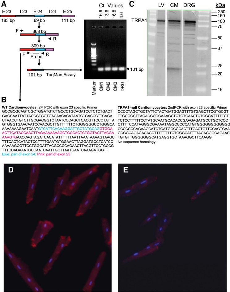
Myocardial ischemia-reperfusion (I/R) results in the generation of free radicals, accumulation of lipid peroxidation-derived unsaturated aldehydes, variable angina (pain), and infarction. The transient receptor potential ankyrin 1 (TRPA1) mediates pain signaling and is activated by unsaturated aldehydes, including acrolein and 4-hydroxynonenal. The contribution of TRPA1 (a Ca2+-permeable channel) to I/R-induced myocardial injury is unknown. We tested the hypothesis that cardiac TRPA1 confers myocyte sensitivity to aldehyde accumulation and promotes I/R injury. Although basal cardiovascular function in TRPA1-null mice was similar to that in wild-type (WT) mice, infarct size was significantly smaller in TRPA1-null mice than in WT mice (34.1 ± 9.3 vs. 14.3 ± 9.9% of the risk region, n = 8 and 7, respectively, P < 0.05), despite a similar I/R-induced area at risk (40.3 ±8.4% and 42.2 ± 11.3% for WT and TRPA1-null mice, respectively) after myocardial I/R (30 min of ischemia followed by 24 h of reperfusion) in situ. Positive TRPA1 immunofluorescence was present in murine and human hearts and was colocalized with connexin43 at intercalated disks in isolated murine cardiomyocytes. Cardiomyocyte TRPA1 was confirmed by quantitative RT-PCR, DNA sequencing, Western blot analysis, and electrophysiology. A role of TRPA1 in cardiomyocyte toxicity was demonstrated in isolated cardiomyocytes exposed to acrolein, an I/R-associated toxin that induces Ca2+ accumulation and hypercontraction, effects significantly blunted by HC-030031, a TRPA1 antagonist. Protection induced by HC-030031 was quantitatively equivalent to that induced by SN-6, a Na+/Ca2+ exchange inhibitor, further supporting a role of Ca2+ overload in acrolein-induced cardiomyocyte toxicity. These data indicate that cardiac TRPA1 activation likely contributes to I/R injury and, thus, that TRPA1 may be a novel therapeutic target for decreasing myocardial I/R injury. NEW & NOTEWORTHY Transient receptor potential ankyrin 1 (TRPA1) activation mediates increased blood flow, edema, and pain reception, yet its role in myocardial ischemia-reperfusion (I/R) injury is unknown. Genetic ablation of TRPA1 significantly decreased myocardial infarction after I/R in mice. Functional TRPA1 in cardiomyocytes was enriched in intercalated disks and contributed to acrolein-induced Ca2+ overload and hypercontraction. These data indicate that I/R activation of TRPA1 worsens myocardial infarction; TRPA1 may be a potential target to mitigate I/R injury.

Keywords: acrolein; cardiomyocytes; lipid peroxidation; myocardial infarction; transient receptor potential ankyrin 1 channel; unsaturated aldehydes.


Conflict of interest statement

No conflicts of interest, financial or otherwise, are declared by the authors.

Figures

Fig. 1.
Fig. 1.
Effects of ischemia and reperfusion on Langendorff-perfused hearts from male wild-type (WT) and transient receptor potential ankyrin 1 (TRPA1)-null mice. Isolated hearts were perfused for 30 min and then subjected to 30 min of ischemia followed by 45 min of reperfusion. Cardiac parameters, including heart rate (HR, beats/min; A), left ventricular (LV) developed pressure (LVDP; B), maximum and minimum rate of rise and fall of LV developed pressure (dP/dt; C), LV pressure minimum (LVPmin; D), and coronary flow (E), were monitored in real time by a LV implanted balloon and an in-line flow probe, respectively. Values are means ± SE. *P < 0.05, TRPA1-null vs. WT mice.
Fig. 2.
Fig. 2.
Transient receptor potential ankyrin 1 (TRPA1) contributes to ischemia-reperfusion injury in mouse heart. A: representative images of triphenyltetrazolium chloride (TTC)- and phthalocyanine blue-stained midventricular cross sections of hearts from male wild-type (WT; i) and TRPA1-null (ii) mice 24 h after myocardial ischemia (30 min). The unstained (white) area in the risk region (TTC, red) represents the infarcted myocardium. B: summary data of the risk region [% of the left ventricle (LV); left y-axis) and infarct size (% of the risk region; right y-axis). Values are means ± SE. *P < 0.05, TRPA1-null vs. WT mice.
Fig. 3.
Fig. 3.
Transient receptor potential ankyrin 1 (TRPA1) in the healthy mouse heart and failing human heart. A–C: mid left ventricular cross sections of male wild-type mouse stained with hematoxylin and eosin (A), fluorescently labeled TRPA1 antibody (red) and DAPI [blue (nuclear stain)] (B), and fluorescently labeled TRPA1 antibody, specific TRPA1 blocking peptide, and DAPI (C). D–F: ventricular sections of the uninfarcted failing human myocardium stained with hematoxylin and eosin (D), fluorescently labeled TRPA1 antibody (green) and DAPI (E), and fluorescently labeled TRPA1 antibody, specific TRPA1 blocking peptide, isolectin B4 [red (stains capillaries)], and DAPI (F). Cardiomyocytes and the coronary vasculature stained positive with TRPA1 antibody.
Fig. 4.
Fig. 4.
Molecular evidence of transient receptor potential ankyrin 1 (TRPA1) in cardiomyocytes. A: mRNA of wild-type (WT) cardiomyocytes was probed for the specific TRPA1 sequence bridging exons (E) 23 and 24 (101-bp product), run on gel electrophoresis, and quantified by RT-PCR. CM, cardiomyocytes; Ct, cycle threshold; I, intron; F, forward; R, reverse. B: sequencing of cDNA from WT and TRPA1-null cardiomyocytes. The WT group matched the mTRPA1 sequence, whereas the TRPA1-null group (as expected) did not. C: Western blot for TRPA1 protein (relative molecular mass ≈ 100,000) in lysates of the left ventricle (LV), isolated cardiomyocyte (CM), and dorsal root ganglion (DRG; positive control) of WT mice. D and E: representative epifluorescence micrographs of isolated WT (D) and TRPA1-null (E) cardiomyocytes stained for TRPA1.
Fig. 5.
Fig. 5.
Transient receptor potential ankyrin 1 (TRPA1) in cardiomyocyte membranes colocalized to the intercalated disk. A–E: isolated male wild-type (WT) mouse cardiomyocytes stained with DAPI [blue (nuclear stain); A], fluorescently labeled TRPA1 antibody (green; B), connexin 43 antibody (red; C), and combined antibodies without (D; white arrows indicate colocalization) or with (E) TRPA1-blocking peptide. F: representative cell-attached patch recordings (holding potential: −40 mV) of single channel events at the intercalated disk region of an isolated adult cardiomyocyte in the absence [control (i, ii, and iii)] and presence of cinnamaldehyde (50 µM; iv, v, and vi) and HC-030031 (10 µM; vii, viii, and ix). Open probabilities (nPo) are shown for each condition. Recordings are representative of two independent experiments.
Fig. 6.
Fig. 6.
Acrolein-induced transient receptor potential ankyrin 1 (TRPA1)-dependent Ca2+ entry and cardiomyocyte hypercontracture. Cardiomyocytes isolated from hearts of male wild-type (WT) mice were plated overnight and superfused with acrolein (10 or 25 µM), and hypercontraction was monitored over 60 min. A: time course of acrolein-induced changes in intracellular Ca2+ [fluorescence at 488 nm (F488)] in isolated fluo 8-loaded cardiomyocytes. Acrolein (25 µM) was added at time indicated by arrow in the absence or presence of HC-030031. B: representative images of cardiomyocytes at time 0 or 60 min after acrolein exposure (rounded myocyte in the bottom left corner). C: isolated cardiomyocyte survival after superfusion with acrolein alone or in the presence of TRPA1 antagonist (HC-030031; 10 µM), SN-6 (10 µM), or HC-030031 + SN-6 over 60 min. Values are means ± SE; n = 2–4 separate experiments. *P < 0.05 vs. acrolein alone.

References

    1. Andrè E, Campi B, Materazzi S, Trevisani M, Amadesi S, Massi D, Creminon C, Vaksman N, Nassini R, Civelli M, Baraldi PG, Poole DP, Bunnett NW, Geppetti P, Patacchini R. Cigarette smoke-induced neurogenic inflammation is mediated by α,β-unsaturated aldehydes and the TRPA1 receptor in rodents. J Clin Invest 118: 2574–2582, 2008. doi:10.1172/JCI34886. - DOI - PMC - PubMed
    1. Andrei SR, Ghosh M, Sinharoy P, Dey S, Bratz IN, Damron DS. TRPA1 ion channel stimulation enhances cardiomyocyte contractile function via a CaMKII-dependent pathway. Channels (Austin) 11: 587–603, 2017. doi:10.1080/19336950.2017.1365206. - DOI - PMC - PubMed
    1. Andrei SR, Sinharoy P, Bratz IN, Damron DS. TRPA1 is functionally co-expressed with TRPV1 in cardiac muscle: co-localization at z-discs, costameres and intercalated discs. Channels (Austin) 10: 395–409, 2016. doi:10.1080/19336950.2016.1185579. - DOI - PMC - PubMed
    1. Aubdool AA, Graepel R, Kodji X, Alawi KM, Bodkin JV, Srivastava S, Gentry C, Heads R, Grant AD, Fernandes ES, Bevan S, Brain SD. TRPA1 is essential for the vascular response to environmental cold exposure. Nat Commun 5: 5732, 2014. doi:10.1038/ncomms6732. - DOI - PMC - PubMed
    1. Baines CP, Song CX, Zheng YT, Wang GW, Zhang J, Wang OL, Guo Y, Bolli R, Cardwell EM, Ping P. Protein kinase Cε interacts with and inhibits the permeability transition pore in cardiac mitochondria. Circ Res 92: 873–880, 2003. doi:10.1161/01.RES.0000069215.36389.8D. - DOI - PMC - PubMed

Publication types

LinkOut - more resources