

Review
doi: 10.1016/j.stem.2019.01.001.
Stem Cell Quiescence: Dynamism, Restraint, and Cellular Idling
Affiliations
- PMID: 30735649
- PMCID: PMC6413865
- DOI: 10.1016/j.stem.2019.01.001
Item in Clipboard
Review
Stem Cell Quiescence: Dynamism, Restraint, and Cellular Idling
Cell Stem Cell.
.
Abstract
Stem cells can reside in a state of reversible growth arrest, or quiescence, for prolonged periods of time. Although quiescence has long been viewed as a dormant, low-activity state, increasing evidence suggests that quiescence represents states of poised potential and active restraint, as stem cells "idle" in anticipation of activation, proliferation, and differentiation. Improved understanding of quiescent stem cell dynamics is leading to novel approaches to enhance maintenance and repair of aged or diseased tissues. In this Review, we discuss recent advances in our understanding of stem cell quiescence and techniques enabling more refined analyses of quiescence in vivo.
Published by Elsevier Inc.
Figures
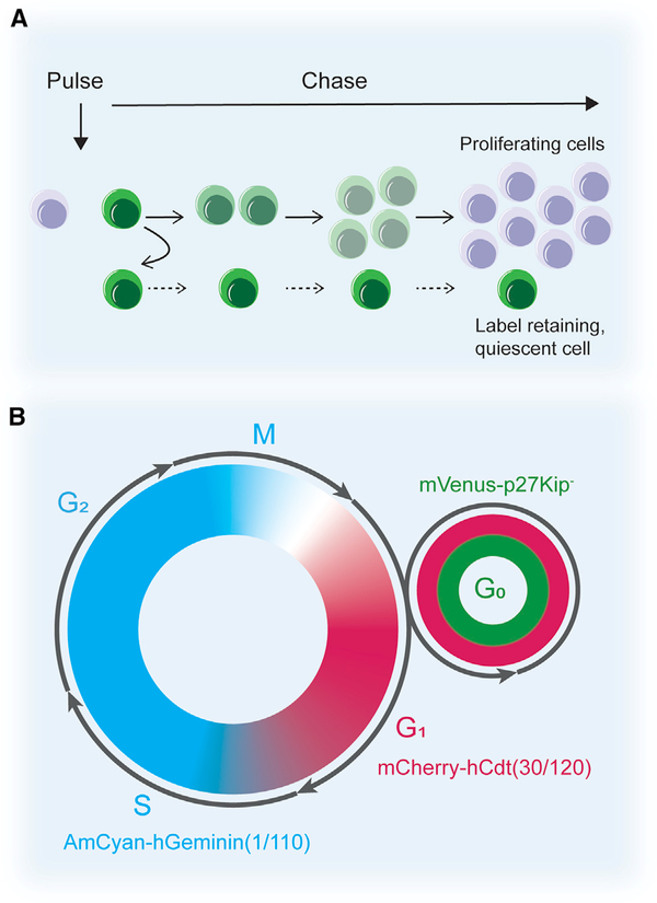
(A) Label retention assays. Cells are labeled when they divide during the pulse and, as they further divide, the label is diluted among the daughter cells where it eventually becomes undetectable. Slowly dividing cells (i.e., self-renewed quiescent stem cells) retain the label over time. (B) The fluorescent ubiquitination-based cell cycle indicator (Fucci) system allows for analysis of the different cell cycle phases. The system uses the fusion of fluorescent proteins to cell-cycle specific proteins, geminin, Cdt1, and p27, to visualize specific cell cycle phases. Based upon the ubiquitination and degradation of these proteins in the various cell cycle phases, nuclei in G1 are labeled red, nuclei in S/G2/M are labeled cyan, and nuclei in G0 are labeled red and green.
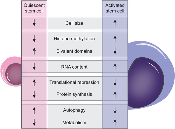
Quiescent stem cells are characterized by tight regulation of all cellular processes. Some of the key processes that have been studied in the context of the dynamics of cellular quiescence and the maintenance of the quiescent state are illustrated here, with an indication of whether they are upregulated or downregulated compared to fully activated stem cells.
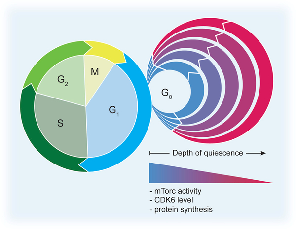
The cell cycle is divided into specific phases, i.e., G1, S, G2, and M. Cells can exit this proliferative cycle in G1 and enter the quiescent state, G0. Recent studies have shown that different levels of quiescence depth exist. Examples of quiescent cells in relatively “shallow quiescent” states, termed “GAlert” or “primed” cells, can enter G1 and the cell cycle faster than more “deeply quiescent” cells. Signaling through mTORC1 has been shown to control the transition from G0 to GAlert in MuSCs, whereas the primed state in HSCs has been shown to be regulated by the level of CDK6 (Rodgers et al., 2014; Laurenti et al., 2015). Undoubtedly, stem cells from different lineages can persist in comparable alternative quiescent states, controlled by other signaling pathways.
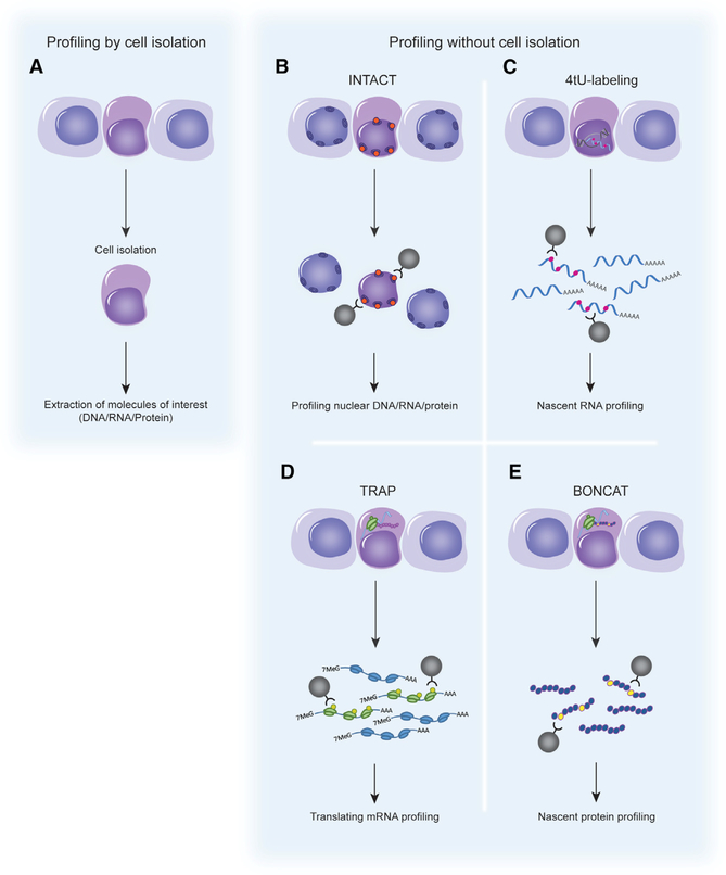
(A) Conventionally, quiescent stem cells are identified and isolated from the tissue by methods such as FACS. Total DNA, RNA, and/or protein is extracted and analyzed. (B–E) Profiling of quiescent stem cells without cell isolation. In each case, a labeling construct is expressed in a cell-specific manner in vivo. The organelles/molecules of interest are labeled only in these cells. Labeled molecules can be purified from whole tissue homogenates without the need for tissues dissociation and cell purification. (B) Isolation of nuclei tagged in specific cell types (INTACT). A nuclear targeting fusion (NTF) protein carrying the biotin ligase recognition peptide and the biotin ligase, BirA, are co-expressed in cells of interest. The NTF protein is targeted to the nuclear membrane where it acts as a substrate for BirA. After tissue homogenization, biotinylated nuclei can be recovered. (C) 4tU tagging to label nascent RNA. The uracil phosphoribosyltransferase (UPRT) enzyme is expressed in a cell-specific manner. 4tU is administered, converted by UPRT to 4-thiouridine, and incorporated into nascent transcripts. The tissue is homogenized and thio-RNAs are extracted. (D) Translating ribosome affinity purification (TRAP) to isolate actively translated transcripts. Tagged ribosome subunits are expressed in a cell-specific manner. After homogenization of the tissue, mRNAs associated with tagged ribosome/polysome complexes can be extracted. (E) Bio-orthogonal non-canonical amino acid tagging (BONCAT) to label nascent proteins. A single mutation (L274G) in the amino acid binding site of methionyl-tRNA synthetase (MetRS) enables the loading of the non-canonical amino acid azidonorleucine (ANL) onto methionine tRNA. Nascent proteins can be labeled with ANL in cells that express MetRS L274G.
References
-
- Alvarez-Castelao B, Schanzenbächer CT, Hanus C, Glock C, Tom Dieck S, Dörrbaum AR, Bartnik I, Nassim-Assir B, Ciirdaeva E, Mueller A, et al. (2017). Cell-type-specific metabolic labeling of nascent proteomes in vivo. Nat. Biotechnol 35, 1196–1201. - PubMed
-
- Benecke BJ, Ben-Ze’ev A, and Penman S (1978). The control of mRNA production, translation and turnover in suspended and reattached anchorage-dependent fibroblasts. Cell 14, 931–939. - PubMed
-
- Bernstein BE, Mikkelsen TS, Xie X, Kamal M, Huebert DJ, Cuff J, Fry B, Meissner A, Wernig M, Plath K, et al. (2006). A bivalent chromatin structure marks key developmental genes in embryonic stem cells. Cell 125, 315–326. - PubMed
Publication types
MeSH terms
Substances
Grants and funding
LinkOut - more resources
Full Text Sources
Medical
