. 2019 Apr 5;64(8):085004.
doi: 10.1088/1361-6560/ab056d.

Semi-automated IGRT QA using a cone-shaped scintillator screen detector for proton pencil beam scanning treatments

Affiliations

Semi-automated IGRT QA using a cone-shaped scintillator screen detector for proton pencil beam scanning treatments

Weixing Cai et al. Phys Med Biol. .

Abstract

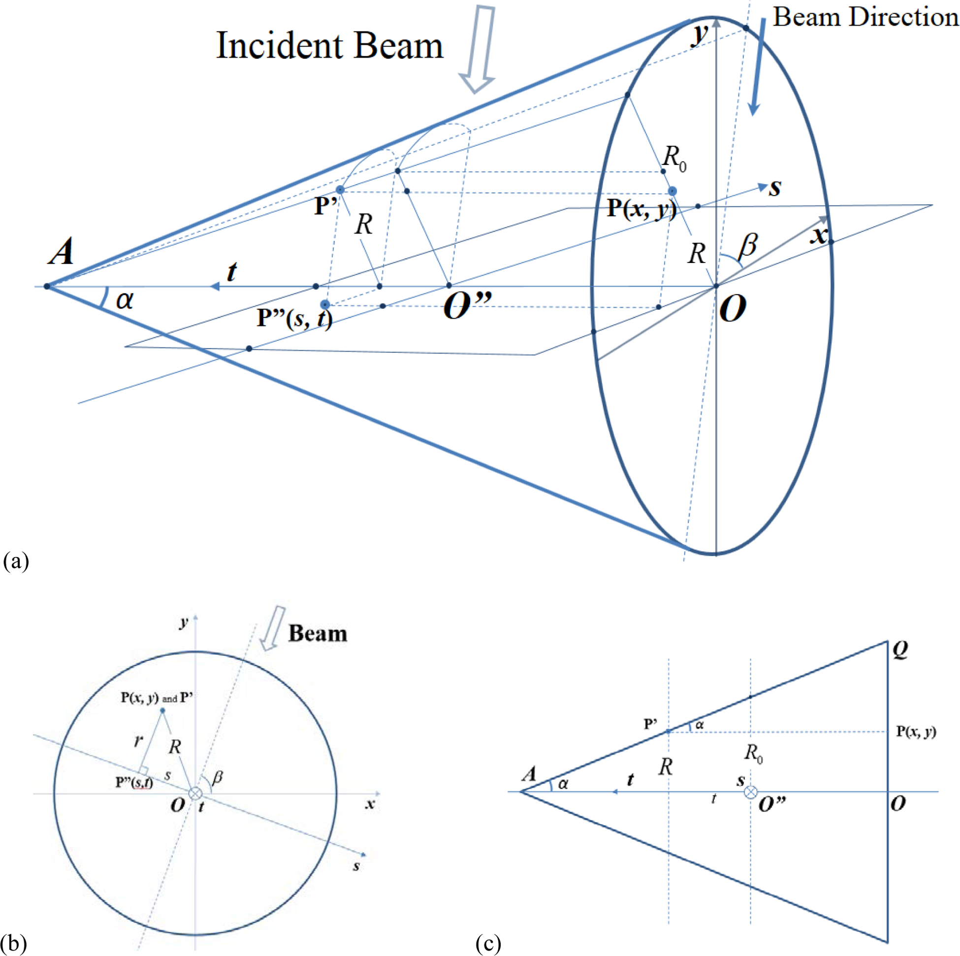
To promote accurate image-guided radiotherapy (IGRT) for a proton pencil beam scanning (PBS) system, a new quality assurance (QA) procedure employing a cone-shaped scintillator detector has been developed for multiple QA tasks in a semi-automatic manner. The cone-shaped scintillator detector (XRV-124, Logos Systems, CA) is sensitive to both x-ray and proton beams. It records scintillation on the cone surface as a 2D image, from which the geometry of the radiation field that enters and exits the cone can be extracted. Utilizing this feature, QA parameters that are essential to PBS IGRT treatment were measured and analyzed. The first applications provided coincidence checks of laser, imaging and radiation isocenters, and dependencies on gantry angle and beam energies. The analysis of the Winston-Lutz test was made available by combining the centricity measurements of the x-ray beam and the pencil beam. The accuracy of the gantry angle was validated against console readings provided by the digital encoder and an agreement of less than 0.2° was found. The accuracy of the position measurement was assessed with a robotic patient positioning system (PPS) and an agreement of less than 0.5 mm was obtained. The centricity of the two onboard x-ray imaging systems agreed well with that from the routinely used Digital Imaging Positioning System (DIPS), up to a consistent small shift of (-0.5 mm, 0.0 mm, -0.3 mm). The pencil beam spot size, in terms of σ of Gaussian fitting, agreed within 0.2 mm for most energies when compared to the conventional measurements by a 2D ion-chamber array (MatriXX-PT, IBA Dosimetry, Belgium). The cone-shaped scintillator system showed advantages in making multi-purpose measurements with a single setup. The in-house algorithms were successfully implemented to measure and analyze key QA parameters in a semi-automatic manner. This study presents an alternative and more efficient approach for IGRT QA for PBS and potentially for linear accelerators.


Figures

Figure 1.
Figure 1.
Picture of the Logos XRV-124 device. (a) The XRV device is composed of a cone-shaped scintillator screen and a CCD camera viewing from the base of the cone (Logos Systems 2014); (b) The XRV system is sitting on the patient position system (PPS) and is aligned at the isocenter of the PBS gantry. Three coordinate systems are involved in the study and are illustrated in the image. Note that BEV-Y is defined as the same direction as IEC-Y, and BEV-X remains in the plane defined by IEC-X and IEC-Z as the gantry rotates.
Figure 2.
Figure 2.
Geometric correction of the XRV-124 system. (a) shows the known equally spaced hole pattern on the cone surface (Logos Systems 2015), (b) shows the raw XRV image of the hole pattern, which is not equally spaced due to optical distortion. Using the information from (a) and (b), a correction function was established to map image distance (pixels) to physical distance (mm), based on which a raw XRV image can be corrected into an orthogonal projection, viewed along the cone axis from the base. As an example, image (b) is corrected into image (d) using the calibration function. The hole pattern in (d) is now equally spaced and matches the orthogonal projection of (a).
Figure 3.
Figure 3.
(a) shows the 3D geometry of this transform. (x, y) is the coordinate system on the orthogonal projection, the radiation beam makes an angle β with x-axis, and the BEV plane (s, t) is perpendicular to the beam. For any point P(x, y) in the orthogonal projection, the algorithm finds the imaged point P’ on the cone surface, and then its counterpart P”(s, t) in the (s, t) plane (BEV). (b) and (c) are the bottom view and the side view, respectively.
Figure 4.
Figure 4.
Typical orthogonal projection images and BEV images. The first row shows x-ray cross-hair images: (a) orthogonal projection, (b) BEV of entry beam and (c) BEV of exit beam. The second row shows pencil beam spot images in the same order. Beam direction is calculated in the orthogonal projection images and is indicated by the blue line. The beam center is calculated in the BEV images and is shown against the coordinate center (big blue cross).
Figure 5.
Figure 5.
Cross-hair center measurements for both beamline and orthogonal x-ray systems. Deviations were calculated from the origin of the IEC coordinates.
Figure 6.
Figure 6.
Pencil beam spot size measurements for various proton energies. Error bars on the MatriXX reference curve is +/−10%, which is our clinical tolerance for routine QA.
Figure 7.
Figure 7.
Winston-Lutz test at different gantry angles. The insert is a fused view of the x-ray cross-hair image and the pencil beam spot image, showing the relative position of the two centers (red for cross hair center and blue for pencil beam spot center).

Similar articles

Cited by

References

    1. Bissonnette JP et al. 2012. Quality assurance for image-guided radiation therapy utilizing CT-based technologies: A report of the AAPM TG-179 Med. Phys 39(4) 1946–1963 - PubMed
    1. Bujold A, Craig T, Jaffray D and Dawson L 2012. Image-guided radiotherapy: Has it influenced patient outcomes? Semin. Radiat. Oncol 22 50–61 - PubMed
    1. Chang Z, Bowsher J, Cai J, Yoo S, Wang Z, Adamson J, Ren L and Y FF 2012. Imaging system QA of a medical accelerator, Novalis Tx, for IGRT per TG 142: our 1 year experience J. Appl. Clin. Med. Phys 13(4) 113–140 - PMC - PubMed
    1. Dhanesar S et al. 2013. Quality assurance of proton beams using a multilayer ionization chamber system Med. Phys 40(9) 092102. - PubMed
    1. Kooy HM et al. 2010. A case study in proton pencil-beam scanning delivery Int. J. Radiat. Oncol. Biol. Phys 76(2) 624–630 - PubMed

Publication types

MeSH terms

LinkOut - more resources