Review
. 2019 Feb 6;20(3):705.
doi: 10.3390/ijms20030705.

The Role of Osteoprotegerin and Its Ligands in Vascular Function

Affiliations
Review

The Role of Osteoprotegerin and Its Ligands in Vascular Function

Luc Rochette et al. Int J Mol Sci. .

Abstract

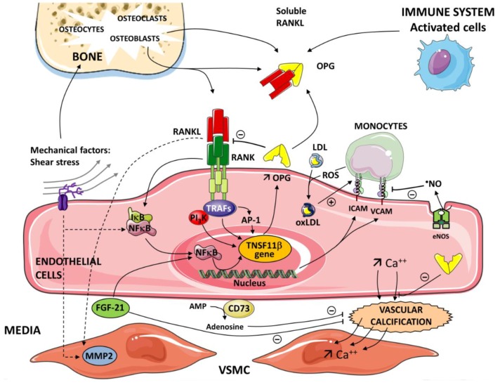
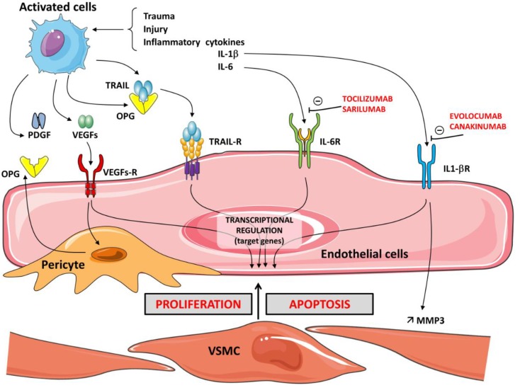
The superfamily of tumor necrosis factor (TNF) receptors includes osteoprotegerin (OPG) and its ligands, which are receptor activators of nuclear factor kappa-B ligand (RANKL) and TNF-related apoptosis-inducing ligand (TRAIL). The OPG/RANKL/RANK system plays an active role in pathological angiogenesis and inflammation as well as cell survival. It has been demonstrated that there is crosstalk between endothelial cells and osteoblasts during osteogenesis, thus establishing a connection between angiogenesis and osteogenesis. This OPG/RANKL/RANK/TRAIL system acts on specific cell surface receptors, which are then able to transmit their signals to other intracellular components and modify gene expression. Cytokine production and activation of their receptors induce mechanisms to recruit monocytes and neutrophils as well as endothelial cells. Data support the role of an increased OPG/RANKL ratio as a possible marker of progression of endothelial dysfunction in metabolic disorders in relationship with inflammatory marker levels. We review the role of the OPG/RANKL/RANK triad in vascular function as well as molecular mechanisms related to the etiology of vascular diseases. The potential therapeutic strategies may be very promising in the future.

Keywords: OPG/RANKL/RANK; endothelium; osteoprotegerin; vascular disease.


Conflict of interest statement

The authors declare no conflict of interest.

Figures

Figure 1
Figure 1
Critical role of the nuclear factor kappa-B/nuclear factor kappa-B ligand/osteoprotegerin (RANK/RANKL/OPG) axis in the pathogenesis of inflammatory processes and vascular calcification. OPG is produced by different cells—activated cells (immune system), osteoblasts in bone. The inflammatory cells and immune cells up-regulate expression of receptor activator of the RANKL. A soluble form of RANKL, sRANKL, also circulates in the blood. The interaction between RANK and RANKL initiates a signaling and gene expression cascade, activating the transcription factor NF-κB. OPG binds to RANKL and prevents the RANKL/RANK interaction. Tumor necrosis factor (TNF) receptor-associated factors (TRAFs 2,5,6) to specific sites are present in the cytoplasmic domain of RANK. Subendothelial retention of low-density lipoprotein (LDL) and its oxidative modification (OxLDL) represent the initial event in atherogenesis. Reactive oxygen species (ROS) generated by monocytes contribute to the level of oxidation of LDL. OxLDLs induce endothelial cell (EC) expression of adhesion molecules intercellular adhesion molecule-1 (ICAM-1) and vascular adhesion molecule-1 (VCAM-1). Nitric oxide (NO) generated in the endothelium by the catalytic action of the enzyme nitric oxide synthase (eNOS) reduces the endothelial expression of ICAM-1 and VCAM-1. In the nucleus of ECs, via NF-κB and AP -1, OPG induces the expression of ICAM-1 and VCAM-1 and promotes leukocyte adhesion, an early step in ECs dysfunction. Various pathways and mediators are involved in vascular calcification depending on the etiology of the atherosclerosis. Vascular calcification is an active cell-regulated process of mineralization implicating matrix mineral metabolism. Sensors and effectors associated with shear stress regulate cellular functions and gene expression via the activation of NF-κB target genes. Osteogenic differentiation of vascular smooth muscle cells (VSMC) plays a pivotal role in the progression of vascular calcification. RANK-RANKL-OPG and other regulatory proteins are major pathways in the progression of vascular calcification. Fibroblast growth factor21 (FGF21) and Ecto-5’-nucleotidase (CD73) contribute to the regulation of this calcification. FGF21 protects the vascular system by limiting VSMC calcification. CD73 hydrolyses extracellular AMP to adenosine. Adenosine has been shown to play a protective role against calcification.
Figure 2
Figure 2
Schema illustrating the relationship between the OPG/TRAIL/TRAIL-R system, pericytes, growth factors, and the cytokines IL-1 and IL-6 on the balance between proliferation and apoptosis of vascular smooth muscle cells (VSMC). In the presence of inflammatory cytokines IL-1β or IL-6 and trauma or injury, activated cells express OPG. Activation of cytokine receptors IL-1R and IL-6R induces the recruitment of monocytes and neutrophils. The growth factor system, which includes vascular endothelial growth factors (VEGFs) and PDGF, influences the proliferation (angiogenesis) and OPG expression in vascular cells. Associated with the microvasculature, pericytes secrete elevated amounts of OPG. OPG also acts as a receptor for TRAIL. TRAIL binds to its receptors, TRAIL-Rs. The cellular actions of TRAIL are tightly regulated in a balance between the apoptosis and proliferation of vascular cells. TRAIL and RANKL increase matrix metalloproteinase (MMP) activity, leading to degradation of the extracellular matrix. The cellular actions promoted by IL-1β induce MMP-3 production. MMP-3 is constitutively expressed in EC and VSMC. The human monoclonal antibodies canakinumab and evolocumab, which target IL-1β, have anti-inflammatory effects. Sarilumab and tocilizumab are human monoclonal antibodies against the IL-6 receptor-α. Intracellular signaling of IL-6 in response to receptor activation is complex—anti-inflammatory protective but pro-inflammatory for the immune response.

References

    1. Walsh M.C., Choi Y. Biology of the RANKL-RANK-OPG System in Immunity, Bone, and Beyond. Front. Immunol. 2014;5:511. doi: 10.3389/fimmu.2014.00511. - DOI - PMC - PubMed
    1. Rochette L., Meloux A., Rigal E., Zeller M., Cottin Y., Vergely C. The role of osteoprotegerin in the crosstalk between vessels and bone: Its potential utility as a marker of cardiometabolic diseases. Pharm. Ther. 2018;182:115–132. doi: 10.1016/j.pharmthera.2017.08.015. - DOI - PubMed
    1. Barbu C.G., Arsene A.L., Florea S., Albu A., Sirbu A., Martin S., Nicolae A.C., Burcea-Dragomiroiu G.T.A., Popa D.E., Velescu B.S., et al. Cardiovascular risk assessment in osteoporotic patients using osteoprotegerin as a reliable predictive biochemical marker. Mol. Med. Rep. 2017;16:6059–6067. doi: 10.3892/mmr.2017.7376. - DOI - PMC - PubMed
    1. Harper E., Forde H., Davenport C., Rochfort K.D., Smith D., Cummins P.M. Vascular calcification in type-2 diabetes and cardiovascular disease: Integrative roles for OPG, RANKL and TRAIL. Vasc. Pharm. 2016;82:30–40. doi: 10.1016/j.vph.2016.02.003. - DOI - PubMed
    1. Kuroda Y., Matsuo K. Molecular mechanisms of triggering, amplifying and targeting RANK signaling in osteoclasts. World J. Orthop. 2012;3:167–174. doi: 10.5312/wjo.v3.i11.167. - DOI - PMC - PubMed

MeSH terms

LinkOut - more resources