. 2019 Feb 7;20(3):711.
doi: 10.3390/ijms20030711.

CGRP Induces Differential Regulation of Cytokines from Satellite Glial Cells in Trigeminal Ganglia and Orofacial Nociception

Affiliations

CGRP Induces Differential Regulation of Cytokines from Satellite Glial Cells in Trigeminal Ganglia and Orofacial Nociception

Shaista Afroz et al. Int J Mol Sci. .

Abstract

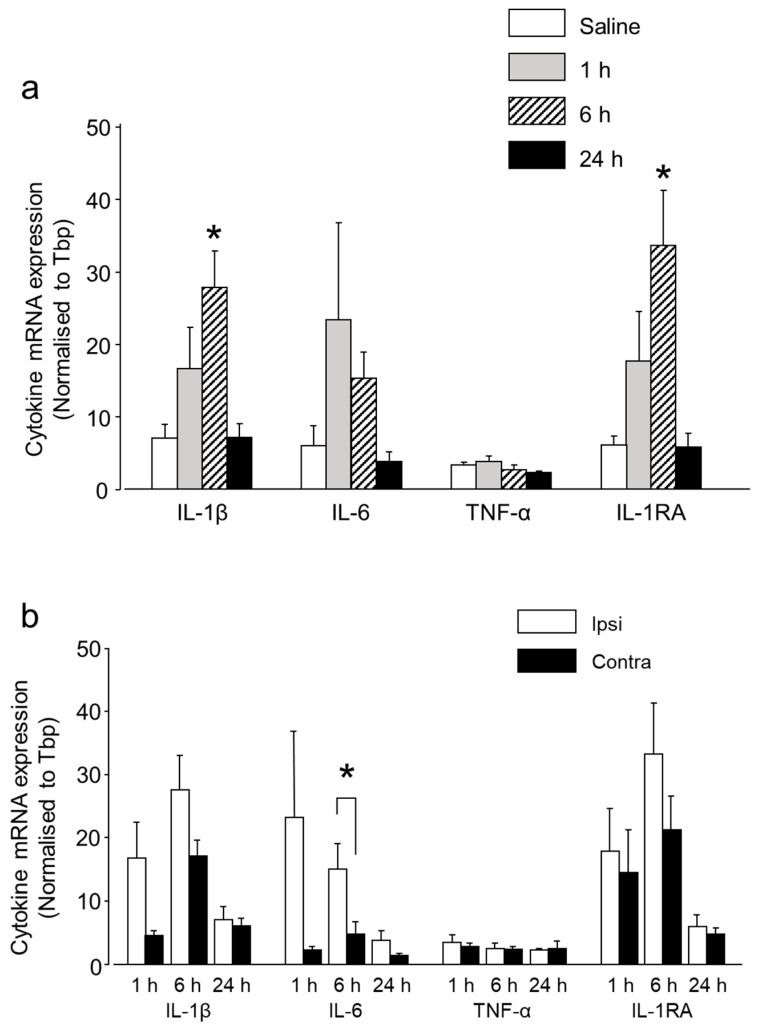
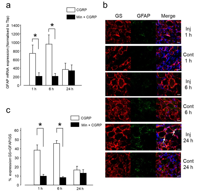
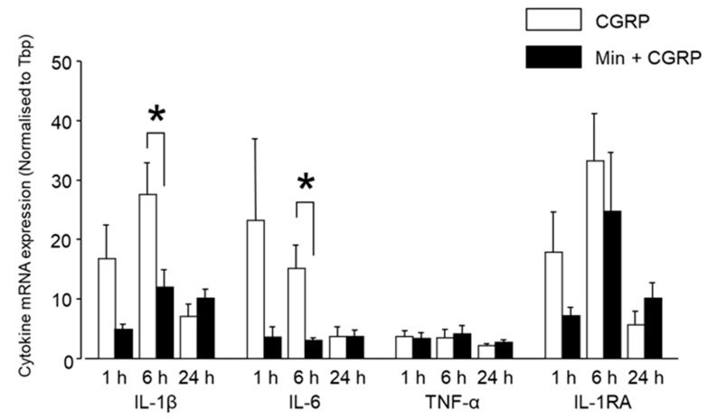
Neuron-glia interactions contribute to pain initiation and sustainment. Intra-ganglionic (IG) secretion of calcitonin gene-related peptide (CGRP) in the trigeminal ganglion (TG) modulates pain transmission through neuron-glia signaling, contributing to various orofacial pain conditions. The present study aimed to investigate the role of satellite glial cells (SGC) in TG in causing cytokine-related orofacial nociception in response to IG administration of CGRP. For that purpose, CGRP alone (10 μL of 10-5 M), Minocycline (5 μL containing 10 μg) followed by CGRP with one hour gap (Min + CGRP) were administered directly inside the TG in independent experiments. Rats were evaluated for thermal hyperalgesia at 6 and 24 h post-injection using an operant orofacial pain assessment device (OPAD) at three temperatures (37, 45 and 10 °C). Quantitative real-time PCR was performed to evaluate the mRNA expression of IL-1β, IL-6, TNF-α, IL-1 receptor antagonist (IL-1RA), sodium channel 1.7 (NaV 1.7, for assessment of neuronal activation) and glial fibrillary acidic protein (GFAP, a marker of glial activation). The cytokines released in culture media from purified glial cells were evaluated using antibody cytokine array. IG CGRP caused heat hyperalgesia between 6⁻24 h (paired-t test, p < 0.05). Between 1 to 6 h the mRNA and protein expressions of GFAP was increased in parallel with an increase in the mRNA expression of pro-inflammatory cytokines IL-1β and anti-inflammatory cytokine IL-1RA and NaV1.7 (one-way ANOVA followed by Dunnett's post hoc test, p < 0.05). To investigate whether glial inhibition is useful to prevent nociception symptoms, Minocycline (glial inhibitor) was administered IG 1 h before CGRP injection. Minocycline reversed CGRP-induced thermal nociception, glial activity, and down-regulated IL-1β and IL-6 cytokines significantly at 6 h (t-test, p < 0.05). Purified glial cells in culture showed an increase in release of 20 cytokines after stimulation with CGRP. Our findings demonstrate that SGCs in the sensory ganglia contribute to the occurrence of pain via cytokine expression and that glial inhibition can effectively control the development of nociception.

Keywords: calcitonin gene related peptide; cytokine; satellite glial cells; thermal hyperalgesia; trigeminal ganglion.


Conflict of interest statement

The authors declare no conflict of interest.

Figures

Figure 1
Figure 1
Behavioral outcome of intra-ganglionic (IG) calcitonin gene-related peptide (CGRP) administration. (a) The reward-licking events/face-contact events (L/F) ratio (mean ± standard error of the mean (SEM)) was significantly reduced at 45 °C, both at 6 and 24 h after IG CGRP administration. (b) The stimulus duration/face-contact (s) (Mean ± SEM) was significantly reduced at 45 °C, at 6 h after IG CGRP administration. These results indicate that CGRP decreases the tolerance to heat between 6 and 24 h. Results are presented as percent change from untreated baseline at the respective temperature. *: p < 0.05, **: p < 0.01 with paired-t test. n = 7 rats were assigned to each group.
Figure 2
Figure 2
IG CGRP induced satellite glial cells (SGCs) activation. (a) The mRNA expression of glial fibrillary acidic protein (GFAP) in the trigeminal ganglion (TG) was significantly increased at 1 and 6 h after IG CGRP administration. Results are presented as Mean ± SEM of the relative expression. *: p < 0.05, **: p < 0.01 with one-way analysis of variance (ANOVA) followed by the Dunnett t test. n = 5 rats were assigned to each group. (b) Confocal images of immunofluorescent staining of TG sections with glutamine synthetase (GS, red), GFAP (green), and 4’,6-Diamidino-2-phenylindole dihydrochloride (DAPI, blue) at 1, 6 and 24 h after IG CGRP administration and contralateral TGs. Colocalization of GS and GFAP in the SGCs is denoted by white arrow. Scale bar: 20 µm. (c) IG CGRP administration increased the GFAP protein expression on the injected side compared to the contralateral side both at 1 and 6 h. *: p < 0.05, with t-test. n = 3 rats were assigned to each group and data were acquired from three independent sections (i.e., examined in three non-overlapping views).
Figure 3
Figure 3
Differential mRNA expression of cytokines after IG CGRP administration. (a) IG CGRP increased the expression of IL-1β, IL-6 and IL-1RA at 1 and 6 h after administration compared to the saline-injected group. However, the statistically significant result was observed only for IL-1β and IL-1RA after 6 h. After 24 h, the expression of IL-1β, IL-6 and IL-1RA decreased. TNF-α expression was similar in all the groups. #: p < 0.05 with one-way ANOVA followed by the Dunnett t test for comparison with saline injection. n = 5 rats were assigned to each group. (b) A comparison between ipsilateral and contralateral side showed increased expression on the injected side at 1 and 6 h for IL-1β, IL-6 and IL-1RA. However, statistically significant result was observed only for IL-6 at 6 h using one-way ANOVA, *: p < 0.05, n = 5 in each group.
Figure 4
Figure 4
Cytokine array membrane. Protein profiling in cell culture supernate in stimulated and control condition. Overnight stimulation of glial-rich culture with 1 μM CGRP causes differential expression of cytokines compared to control condition.
Figure 5
Figure 5
Behavioral outcome to IG CGRP and Minocycline (Min) + CGRP administration. (a) The L/F ratio (Mean ± SEM) was significantly increased at 45 °C, 6 h after administration in the Min + CGRP injected group. (b) The stimulus duration/face-contact (s) (Mean ± SEM) were significantly increased at 45 °C, 6 h after administration in the Min + CGRP injected group. Results are presented as percent change from untreated baseline at the respective temperature. *: p < 0.05 with t-test. n = 7 rats were assigned to each group.
Figure 6
Figure 6
Effect of injecting Min 1 h before CGRP on glial inhibition. (a) mRNA expression of GFAP in TG was significantly decreased 1 and 6 h after administration in the Min + CGRP group compared to CGRP alone group. Results are presented as Mean ± SEM of relative expression. *: p < 0.05 with t-test. n = 5 rats were assigned to each group. (b) Confocal images of immunofluorescent staining of TG sections with GS (red), GFAP (green), and DAPI (blue) after 1, 6 and 24 h of IG Min + CGRP administration and contralateral TG. Colocalization of GS and GFAP in the SGCs is denoted by white arrow. Scale bar: 20 µm. (c) IG Min 1 h before CGRP administration decreased the GFAP protein expression compared to only CGRP injection at 1 and 6 h. *: p < 0.05 with t test. n = 3 rats were assigned to each group and data were acquired from three independent sections (i.e., examined in three non-overlapping views).
Figure 7
Figure 7
Differential mRNA expression of cytokines after IG CGRP and Min + CGRP administration. IG Min administered 1 h before CGRP decreased the expression of IL-1β, IL-6 and IL-1RA after 1 and 6 h, compared to CGRP alone. After 24 h, there was a decrease in the expression of IL-1β, IL-6 and IL-1RA in bothe the groups. Min administration had no effect on TNF-α expression. *: p < 0.05 with t-test. n = 5 rats were assigned to each group.
Figure 8
Figure 8
Effect of IG CGRP and Min + CGRP on NaV1.7 expression. CGRP increased the mRNA expression of NaV1.7 after 6 h and Min reduced the CGRP induced upregulation of NaV1.7 expression. *: p < 0.05 with t test and #: p < 0.05 with one-way ANOVA followed by the Dunnett t test for comparison with saline injection. n = 5 rats were assigned to each group.
Figure 9
Figure 9
Schematic representation of the effect of exogenously administered CGRP on neurons and SGCs. CGRP receptors are present on the SGCs and neurons [namely, receptor activity-modifying protein 1 (RAMP1), and calcitonin receptor-like receptor (CLR)]. Injected CGRP causes its activity by engaging these receptors and causing activation of SGCs as demonstrated by an increased expression of GFAP. Pro-inflammatory cytokines IL-1β and IL-6 are released from the SGCs in the TG and these cytokines cause neuronal activation as shown by upregulation of NaV 1.7. Hypothetically, once initiated these effects are self-sustaining because of the formation of a feedback loop due to the secretion of endogenous CGRP and responsible for hyperalgesia as observed at 6 h in the present experiment. Injecting Min (a glial inhibitor) reduced the effect of CGRP, leading to an alleviation of hyperalgesia.
Figure 10
Figure 10
Glial-rich culture, SGCs are identified based on: (a) morphology using light microscopy, (b) and immunoreactivity as glutamine synthetase (marker of SGC) and DAPI-positive cells using confocal microscopy. Arrow points to the SGCs. Scale bar: 20 µm.

References

    1. Basbaum A.I., Bautista D.M., Scherrer G., Julius D. Cellular and molecular mechanisms of pain. Cell. 2009;139:267–284. doi: 10.1016/j.cell.2009.09.028. - DOI - PMC - PubMed
    1. Takeda M., Takahashi M., Matsumoto S. Contribution of the activation of satellite glia in sensory ganglia to pathological pain. Neurosci. Biobehav. Rev. 2009;33:784–792. doi: 10.1016/j.neubiorev.2008.12.005. - DOI - PubMed
    1. Seal R.P. Illuminating the Gap: neuronal cross-talk within sensory ganglia and persistent pain. Neuron. 2016;91:950–951. doi: 10.1016/j.neuron.2016.08.030. - DOI - PubMed
    1. Matsuka Y., Neubert J.K., Maidment N.T., Spigelman I. Concurrent release of ATP and substance P within guinea pig trigeminal ganglia in vivo. Brain Res. 2001;915:248–255. doi: 10.1016/S0006-8993(01)02888-8. - DOI - PubMed
    1. Matsuka Y., Ono T., Iwase H., Mitrirattanakul S., Omoto K.S., Cho T., Lam Y.Y., Snyder B., Spigelman I. Altered ATP release and metabolism in dorsal root ganglia of neuropathic rats. Mol. Pain. 2008;4:66. doi: 10.1186/1744-8069-4-66. - DOI - PMC - PubMed

Substances