. 2019 Feb 7;8(2):213.
doi: 10.3390/jcm8020213.

Collagen Fiber Array of Peritumoral Stroma Influences Epithelial-to-Mesenchymal Transition and Invasive Potential of Mammary Cancer Cells

Affiliations

Collagen Fiber Array of Peritumoral Stroma Influences Epithelial-to-Mesenchymal Transition and Invasive Potential of Mammary Cancer Cells

Marco Franchi et al. J Clin Med. .

Abstract

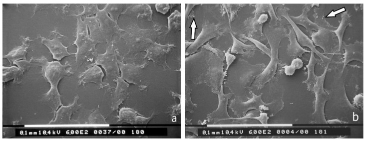
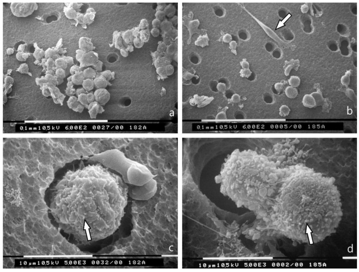
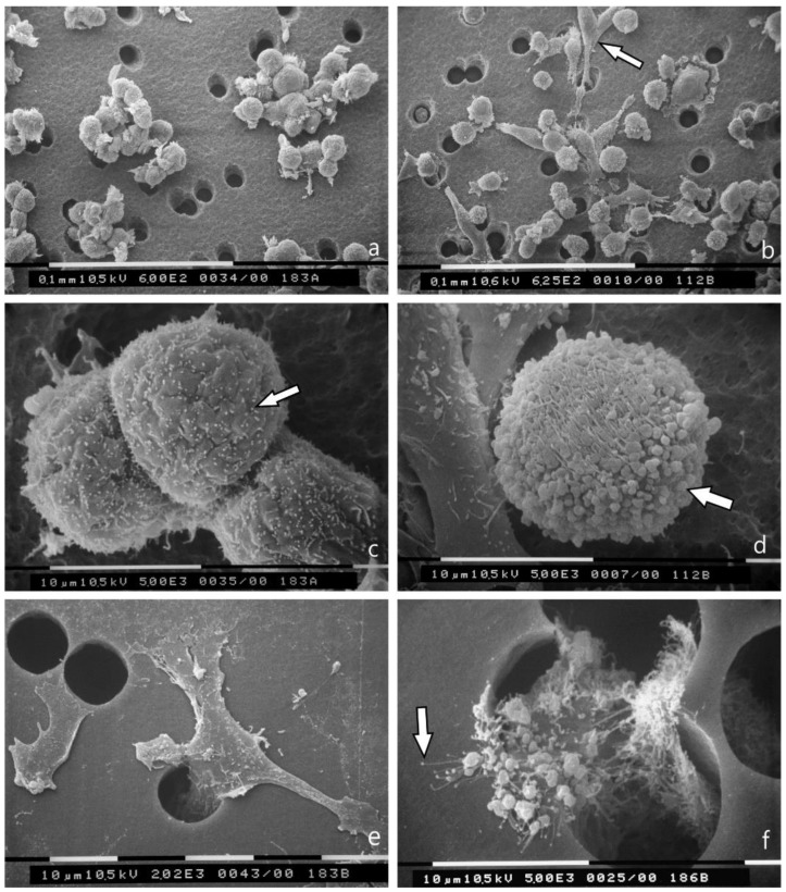
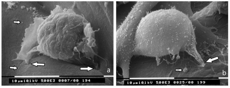
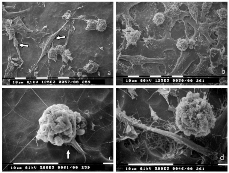
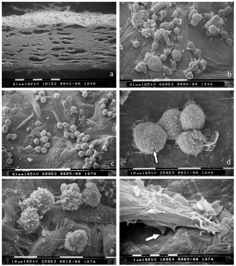
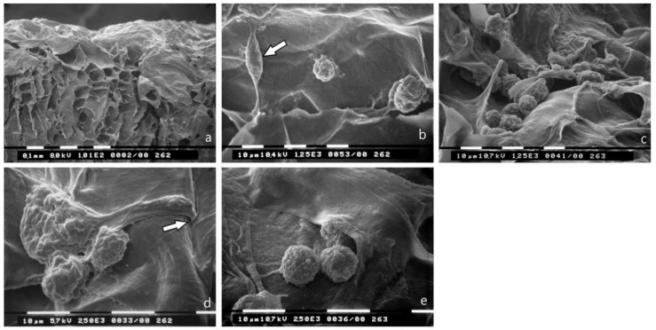
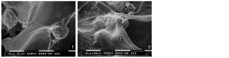
: Interactions of cancer cells with matrix macromolecules of the surrounding tumor stroma are critical to mediate invasion and metastasis. In this study, we reproduced the collagen mechanical barriers in vitro (i.e., basement membrane, lamina propria under basement membrane, and deeper bundled collagen fibers with different array). These were used in 3D cell cultures to define their effects on morphology and behavior of breast cancer cells with different metastatic potential (MCF-7 and MDA-MB-231) using scanning electron microscope (SEM). We demonstrated that breast cancer cells cultured in 2D and 3D cultures on different collagen substrates show different morphologies: i) a globular/spherical shape, ii) a flattened polygonal shape, and iii) elongated/fusiform and spindle-like shapes. The distribution of different cell shapes changed with the distinct collagen fiber/fibril physical array and size. Dense collagen fibers, parallel to the culture plane, do not allow the invasion of MCF-7 and MDA-MB-231 cells, which, however, show increases of microvilli and microvesicles, respectively. These novel data highlight the regulatory role of different fibrillar collagen arrays in modifying breast cancer cell shape, inducing epithelial-to-mesenchymal transition, changing matrix composition and modulating the production of extracellular vesicles. Further investigation utilizing this in vitro model will help to demonstrate the biological roles of matrix macromolecules in cancer cell invasion in vivo.

Keywords: breast cancer; collagen type I; epithelial-to-mesenchymal transition; extracellular matrix; scanning electron microscope.


Conflict of interest statement

The authors declare no conflict of interest.

Figures

Figure 1
Figure 1
2D cultures in polystyrene flasks of breast cancer cells observed at SEM. (a) Most of the MCF-7 cells appear grouped with many cell-cell contacts and show a flattened, smoothly polygonal shape with very few cytoplasmic microvilli and rare cytoplasmic vesicles. Bar 100 μm; (b) MDA-MB-231 cells are presented as isolated cells with very few cell-cell contacts. These cells appear as globular/spherical cells, flattened elongated cells and spindle-like cells with a few cytoplasmic vesicles (arrows). Bar 100 μm.
Figure 2
Figure 2
3D Millipore filter cultures observed at SEM. (a) MCF-7 cells assemble around the holes of the Millipore filter with many cell-cell contacts and show a globular/spherical morphology with a diameter of about 10 μm. Bar 100 μm; (b) Most of the MDA-MB-231 cells have a globular/spherical shape with a mean diameter of 10 μm and evident cytoplasmic microvesicles. An elongated and spindle-like cell (arrow) with very few microvilli and microvesicles is also visible. Bar 100 μm; (c) A globular/spherical cell is passing through a hole of Millipore filter and exhibits few microvilli (arrow) but no microvesicles. Bar 10 μm; (d) Two MDA-MB-231 globular cells with microvesicles (arrow) are crossing the holes of the Millipore filter. Bar 10 μm.
Figure 3
Figure 3
3D Matrigel (0.18 μg/mL) cell cultures as observed at SEM. (a) MCF-7 cells cultured on Matrigel are grouped and pass through the holes of Millipore filter. Bar 100 μm; (b) Most of the MDA-MB-231 cells show a globular/spherical shape and are rich in superficial microvesicles but elongated /spindle-like cells are also visible (arrow). Bar 100 μm; (c) MCF-7 cells show globular/spherical shape with cell-cell contacts and microvilli (arrow) on the cytoplasmic surface. Bar 100 μm; (d) A globular/spherical MDA-MB-231cell shows microvilli and many microvesicles (arrow). On the left, an elongated, flattened cell with few microvilli and microvesicles is detectable. Bar 10 μm; (e) In the lower side of the Millipore filter, MCF-7 cells passed through the holes do not show microvesicles but few microvilli. Bar 10 μm; (f) In the lower surface of the Millipore filter one MDA-MB-231 cell is passing through a filter hole and shows many microvesicles and microvilli generating exosomes (arrow). Bar 10 μm.
Figure 4
Figure 4
3D Matrigel (3.0 μg/mL) cell cultures analyzed by SEM. (a) MCF-7 cell lying on a thick layer of Matrigel shows very few microvilli and no microvesicles. Two invadopodia penetrating the Matrigel develop from the ventral side of the cell (large arrows). A few exosomes and microvesicles are shed on the Matrigel surface (small arrows). Bar 10 μm; (b) An MDA-MB-231 cell with few microvilli and microvesicles shows invadopodia (large arrow) penetrating into the thickness of Matrigel. Two adjacent elongated cells with few microvesicles are also visible. Few exosomes and microvesicles (small arrow) shed from MDA-MB-231 cells are present on the Matrigel surface next to the cells. Bar 10 μm.
Figure 5
Figure 5
Breast cancer cells cultured on type I collagen fibrils (lamina propria under basement membrane) observed at SEM. The randomly arranged collagen fibrils completely occlude the holes of the Millipore filter. (a) Several MCF-7 cells showing both globular/spherical and flattened elongated shapes appear isolated or relatively grouped. A few spindle-like cells are also detectable (arrows). Bar 10 μm; (b) MDA-MB-231 breast cancer cells show both globular/spherical and flattened elongated shapes. Bar 10 μm; (c) A globular/spherical MCF-7 cell shows cytoplasmic convolutions, very few microvilli and no microvesicles. Invadosomes from the cell ventral surface are also visible (arrow). Bar 10 μm; (d) A globular/spherical MDA-MB-231 cell in proximity of a Millipore hole covered by sparse collagen fibrils shows microvesicles. Bar 10 μm.
Figure 6
Figure 6
Breast cancer cells cultured on collagen type I fibers bundled parallel to the culture plane (TACS II). (a) Section of the collagen membrane of Achilles tendon including collagen fibers parallel to the culture plane used as substrate for culture of breast cancer cells. Bar 100 μm; (b) MCF-7 cells on collagen membrane appear as grouped globular/spherical cells with many microvilli but no microvesicles. Bar 100 μm; (c) Grouped MDA-MB-231 cells on the same collagen membrane show globular/spherical shape and elongated and spindle-like ones. Bar 100 μm; (d) At a higher enlargement, four MCF-7 cells show cell-cell contacts and many microvilli on their cytoplasmic surface. Bar 10 μm; (e) At higher enlargement the globular/spherical MDA-MB-231 cells appear covered by many microvesicles, which are very few on elongated or spindle-like cells. Bar 10 μm; (f) A spindle-like MDA-MB-231 cell with microvilli shows two invadopodia (arrow) degrading the collagen fibrils (easily recognizable in the lower portion of the picture). Bar 1 μm; (g) MDA-MB-231 cells are visible on the collagen membrane but not in the thickness of the same substrate. However, many exosomes and microvesicles (arrows) shed by MDA-MB-231 cells penetrated the superficial thickness of the collagen membrane and are visible in the inter fiber spaces. Bar 100 μm; (h) At higher enlargement, exosomes (small arrow) and microvesicles (large arrow) lying on collagen fibrils are clearly visible in the thickness of the collagen membrane. Bar 1 μm.
Figure 6
Figure 6
Breast cancer cells cultured on collagen type I fibers bundled parallel to the culture plane (TACS II). (a) Section of the collagen membrane of Achilles tendon including collagen fibers parallel to the culture plane used as substrate for culture of breast cancer cells. Bar 100 μm; (b) MCF-7 cells on collagen membrane appear as grouped globular/spherical cells with many microvilli but no microvesicles. Bar 100 μm; (c) Grouped MDA-MB-231 cells on the same collagen membrane show globular/spherical shape and elongated and spindle-like ones. Bar 100 μm; (d) At a higher enlargement, four MCF-7 cells show cell-cell contacts and many microvilli on their cytoplasmic surface. Bar 10 μm; (e) At higher enlargement the globular/spherical MDA-MB-231 cells appear covered by many microvesicles, which are very few on elongated or spindle-like cells. Bar 10 μm; (f) A spindle-like MDA-MB-231 cell with microvilli shows two invadopodia (arrow) degrading the collagen fibrils (easily recognizable in the lower portion of the picture). Bar 1 μm; (g) MDA-MB-231 cells are visible on the collagen membrane but not in the thickness of the same substrate. However, many exosomes and microvesicles (arrows) shed by MDA-MB-231 cells penetrated the superficial thickness of the collagen membrane and are visible in the inter fiber spaces. Bar 100 μm; (h) At higher enlargement, exosomes (small arrow) and microvesicles (large arrow) lying on collagen fibrils are clearly visible in the thickness of the collagen membrane. Bar 1 μm.
Figure 7
Figure 7
Breast cancer cells cultured on type I collagen fibers bundled orthogonal to the culture plane (TACS III). (a) Section of the collagen membrane with collagen fibers orthogonal to the culture plane used as substrate for cultures of breast cancer cells. Inter fiber spaces and channels are available for breast cancer invasion. (b) MCF-7 cells lying on the collagen membrane surface mainly show a globular/spherical shape with cytoplasmic convolutions and no microvilli (in the center and on the right), but also some spindle-like cells (arrow). Bar 10 μm. (c) MDA-MB-231 cells on the collagen membrane show a globular/spherical shape with microvesicles on the cell surface. Bar 10 μm. (d) Two globular MCF-7 cells and a flattened/elongated cell showing invadopodia penetrating the collagen membrane surface (arrow) are visible. Cytoplasmic convolutions with no microvilli are detectable on the cell surfaces. Bar 10 μm. (e) MDA-MB-231 cells on the collagen membrane show a globular/spherical shape with few microvesicles. Bar 10 μm. (f) Globular/spherical MCF-7 cells penetrated the thickness of the collagen membrane through inter fibers wide channels. Bar 10 μm. (g) Globular/spherical cells with few microvesicles (stars) partially or completely invaginated a superficial sheet of the collagen membrane. Bar 10 μm.
Figure 7
Figure 7
Breast cancer cells cultured on type I collagen fibers bundled orthogonal to the culture plane (TACS III). (a) Section of the collagen membrane with collagen fibers orthogonal to the culture plane used as substrate for cultures of breast cancer cells. Inter fiber spaces and channels are available for breast cancer invasion. (b) MCF-7 cells lying on the collagen membrane surface mainly show a globular/spherical shape with cytoplasmic convolutions and no microvilli (in the center and on the right), but also some spindle-like cells (arrow). Bar 10 μm. (c) MDA-MB-231 cells on the collagen membrane show a globular/spherical shape with microvesicles on the cell surface. Bar 10 μm. (d) Two globular MCF-7 cells and a flattened/elongated cell showing invadopodia penetrating the collagen membrane surface (arrow) are visible. Cytoplasmic convolutions with no microvilli are detectable on the cell surfaces. Bar 10 μm. (e) MDA-MB-231 cells on the collagen membrane show a globular/spherical shape with few microvesicles. Bar 10 μm. (f) Globular/spherical MCF-7 cells penetrated the thickness of the collagen membrane through inter fibers wide channels. Bar 10 μm. (g) Globular/spherical cells with few microvesicles (stars) partially or completely invaginated a superficial sheet of the collagen membrane. Bar 10 μm.
Figure 8
Figure 8
Migration of breast cancer cells cultured on different substrates. MDA-MB-231 and MCF-7 cells were seeded on Millipore filters or Millipore filters covered with Matrigel (0.18 μg/mL) or collagen type I (Coll I) and incubated for 5 h. All assays were performed as described in Materials and Methods. Asterisks (**) indicate statistically significant differences (p ≤ 0.01).
Figure 9
Figure 9
Type I collagen induces striking changes in EMT markers and significant alterations in the expression of ECM components of breast cancer cells. Real-time PCR analysis of a-SMA, E-cadherin, vimentin, fibronectin, MMP-2 and MMP-9 on MDA-MB-231 and MCF-7 cells seeded on uncoated Millipore filter or filter covered with collagen type I and incubated for 24 hours. Expression was normalized to GAPDH expression. Asterisks (*), (**) indicate statistically significant differences (p ≤ 0.05 and p ≤ 0.01, respectively).

Similar articles

Cited by

References

    1. Page-McCaw A., Ewald A.J., Werb Z. Matrix metalloproteinases and the regulation of tissue remodelling. Nat. Rev. Mol. Cell Biol. 2007;8:221–233. doi: 10.1038/nrm2125. - DOI - PMC - PubMed
    1. Garcia M.G., Bayo J., Bolontrade M.F., Sganga L., Malvicini M., Alaniz L., Aquino J.B., Fiore E., Rizzo M.M., Rodriguez A., et al. Hepatocellular carcinoma cells and their fibrotic microenvironment modulate bone marrow-derived mesenchymal stromal cell migration in vitro and in vivo. Mol. Pharm. 2011;8:1538–1548. doi: 10.1021/mp200137c. - DOI - PubMed
    1. Jaafar H., Sharif S.E., Murtey M.D. Distinctive features of advancing breast cancer cells and interactions with surrounding stroma observed under the scanning electron microscope. Asian Pac. J. Cancer Prev. 2012;13:1305–1310. doi: 10.7314/APJCP.2012.13.4.1305. - DOI - PubMed
    1. Theocharis A.D., Skandalis S.S., Gialeli C., Karamanos N.K. Extracellular matrix structure. Adv. Drug. Deliv. Rev. 2016;97:4–27. doi: 10.1016/j.addr.2015.11.001. - DOI - PubMed
    1. Karamanos N.K., Piperigkou Z., Theocharis A.D., Watanabe H., Franchi M., Baud S., Brezillon S., Gotte M., Passi A., Vigetti D., et al. Proteoglycan Chemical Diversity Drives Multifunctional Cell Regulation and Therapeutics. Chem. Rev. 2018 doi: 10.1021/acs.chemrev.8b00354. - DOI - PubMed

LinkOut - more resources